New Images of RSV may Expose Weak Points in the Stubborn Virus

Photo by Andrea Piacquadio on Pexels

The complex shape of respiratory syncytial virus is one hurdle limiting the development of treatments for an infection that leads to hospitalisation or worse. New images of the virus published in Nature from researchers at the University of Wisconsin–Madison may hold the key to preventing or slowing RSV infections.

RSV is of greatest concern in young children, the elderly and adults at high risk for respiratory complications. Yet unlike the flu and other common, communicable respiratory illnesses that annually sweep through schools, there are few options for fighting RSV. In the US, prophylactic treatments are available for young children, and existing vaccines are approved only for pregnant women and the elderly.

The virus’s structure, which consists of tiny, bending filaments, have eluded researchers. This has made it difficult to identify key drug targets, including viral components that are conserved across related viruses.

RSV F proteins, shown in this image created by University of Wisconsin–Madison researchers using a technique called cryo-electron tomography, may make RSV more potent by keeping it from infecting cells prematurely. Image by Wright Lab, UW–Madison

“There are a number of viruses related to RSV that are also significant human pathogens, including measles,” says Elizabeth Wright, a UW–Madison biochemistry professor. “What we know about related viruses gives us clues about RSV protein structures, but to identify drug targets we need a closer look at RSV proteins that are intimately associated with the membranes of host cells.”

Using an imaging technique called cryo-electron tomography, Wright and her team have now revealed details of molecules and structures essential to RSV’s form and function. They published their findings recently in Nature.

Cryo-ET freezes viral particles or other molecules at ultracold temperatures, stopping biological processes in action. This allows researchers to examine the structures of organisms, cells and organelles, and viruses and capture small-scale images of structures frozen in time. Flash-freeze many RSV particles, and cryo-ET imaging will capture (nearly) all the virus’s possible configurations from many different angles. These 2D images are combined to produce a representation of the virus’s 3D structures at high resolutions – even at the level of individual atoms.

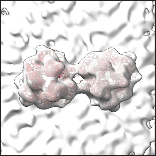
Wright’s recent study produced high-resolution images detailing the structure of two RSV proteins, RSV M protein and RSV F protein, that are crucial to the interaction between the virus and the host cell membrane. Both proteins are also present in related viruses.

RSV M protein interacts with host cell membranes, holding together the virus’s filamentous structure and coordinating viral components and other proteins – including RSV F proteins. RSV F proteins sit on the viral surface, ready to engage with host cell receptors and regulate the virus’s fusion and entry into the host cell. The scientists’ images reveal that in RSV, two F proteins come together to form a more stable unit. Wright says that this association may prevent the F proteins from prematurely infecting the host cell.

“Our primary findings reveal structural details that allow us to better understand not only how the protein regulates assembly of viral particles, but also the coordination of proteins that enable the virus to be infectious,” says Wright.

The scientists believe that F protein pairs may be a key to destabilising the virus before it is ready to infect its next host, making pairs of F proteins a possible target for future drug development. They will continue to explore how RSV proteins interact with each other to cause infection.

Source: University of Wisconsin-Madison

Airborne Levels of Chemicals Released by Plastics Shock Researchers

Photo by Ryan Zazueta on Unsplash

A new study documents how people in California are chronically being exposed to toxic airborne chemicals called plasticisers, including one banned from children’s items and beauty products. 

Plasticizers are chemical compounds that make materials more flexible. They are used in a wide variety of products ranging from lunchboxes and shower curtains to garden hoses and upholstery. 

“It’s not just for drinking straws and grocery bags,” said David Volz, environmental sciences professor at UC Riverside, and corresponding author of the study published in the journal Environmental Research

Previous California monitoring programs focused on plasticisers called ortho-phthalates, some of which were phased out of manufacturing processes due to health and environmental concerns. Less research has focused on the health effects of their replacements, called non-ortho-phthalates. This study revealed the presence of both types of plasticisers in the air throughout Southern California.

“The levels of these compounds are through the roof,” Volz said. “We weren’t expecting that. As a result, we felt it was important for people to learn about this study.”

The National Institute of Environmental Health Sciences also wants to increase the visibility of this study, one of only a few to document the phthalates’ presence in the air of urban environments. The institute’s monthly newsletter, Environmental Factor, highlights the study in their October 2024 issue.  

The researchers tracked two groups of UCR undergraduate students commuting from different parts of Southern California. Both groups wore silicone wristbands designed to collect data on chemical exposures in the air. 

The first group wore their wristbands for five days in 2019, and the second group wore two different wristbands for five days each in 2020. Both groups wore the bands continuously, all day, as they went about their activities. At the end of the data collection period, the researchers chopped the wristbands into pieces, then analysed the chemicals they contained. 

In a previous paper, the team focused on TDCIPP, a flame-retardant and known carcinogen, picked up in the wristbands. They saw that the longer a student’s commute, the higher their exposure to TDCIPP. 

Unlike TDCIPP, which most likely migrates out of commuters’ car seats into dust, the team cannot pinpoint the origin of the plasticisers. Because they are airborne, rather than bound to dust, the wristbands could have picked them up anywhere, even outside the students’ cars.

For every gram of chopped-up wristband, the team found between 100 000 and 1 million nanograms of three phthalates, DiNP, DEHP, and DEHT. Ten total plasticizers were measured, but the levels of these three stood out.

Both DiNP and DEHP are included on California’s Proposition 65 list, which contains chemicals known to cause cancer, birth defects, or other reproductive harm. DEHT was introduced as an alternative, but its effects on human health have not been well studied. 

This study suggests that introducing DEHT also has not done much to reduce the public’s level of exposure to DiNP or DEHP. Levels of all three chemicals found by Volz and his team were similar to those found by researchers in unrelated studies conducted on the East Coast. 

Despite differences in climate, the air on both coasts is likely carrying similar levels of phthalates.

“No matter who you are, or where you are, your daily level of exposure to these plasticizer chemicals is high and persistent,” Volz said. “They are ubiquitous.”

To Volz, studies like this one amplify the need to find alternatives to plastic. As plastics degrade, these compounds and others like them are leaching out into the environment and into the body. 

“The only way to decrease the concentration of plasticisers in the air is to decrease our production and consumption of materials containing plasticisers,” he said. 

Source: University of California – Riverside

Antigens in Foods Suppress Gut Tumours by Activating Immune Cells

Photo by Pixabay on Pexels

Researchers led by Hiroshi Ohno at the RIKEN Center for Integrative medical sciences (IMS) in Japan have discovered that food antigens like milk proteins help keep tumours from growing in our guts, specifically the small intestines. Experiments revealed how these proteins trigger the intestinal immune system, allowing it to effectively stop the birth of new tumours. The study was published in the scientific journal Frontiers in Immunology.

Food antigens get a lot of negative press because they are the source of allergic reactions to foods such as peanuts, shellfish, bread, eggs, and milk. Even if not allergenic, these antigens, along with the many others found in plants and beans, are still considered foreign objects that need to be checked out by the immune system. Ohno and his team have previously reported that food antigens activate immune cells in the small intestines, but not the large intestines. At the same time, some immune cells activated by gut bacteria are known to suppress tumours in the gut. In the new study, the RIKEN IMS researchers bring these two lines of thought together and tested whether food antigens suppress tumours in the small intestines.

The team began with a mouse model with a mutated tumour-suppression gene. Like people with familial adenomatous polyposis, when this gene malfunctions, the mice develop tumours throughout the small and large intestines. The first experiment was fairly simple. They fed these mice normal food or antigen-free food and found that the ones that got normal food had fewer tumours in the small intestines, but the same amount in the large intestines.

Next, they added a common representative antigen called albumin – which can be found in meat and was not in the normal food – to the antigen-free diet, making sure that the total amount of the protein equalled the amount of protein in the normal diet. When the mice were given this diet, tumours in the small intestine were suppressed just as they has been with normal food. This means that tumour suppression was directly related to the presence of antigen, not the nutritional value of the food or any specific antigen.

Mice that got the plain antigen-free diet had many fewer T cells than those that got the normal food or the antigen-free food with milk protein. Further experiments revealed the biological process that makes this possible.

These findings have clinical implications. Similar to antigen-free diets, clinical elemental diets include simple amino acids, but not proteins. This reduces digestive work and can help people with severe gastrointestinal conditions, such as Crohn’s disease or irritable bowel syndrome. According to Ohno, “small intestinal tumours are much rarer than those in the colon, but the risk is higher in cases of familial adenomatous polyposis, and therefore the clinical use of elemental diets to treat inflammatory bowel disease or other gastrointestinal conditions in these patients should be considered very carefully.”

Elemental diets are sometimes adopted by people without severe gastrointestinal conditions or allergies as a healthy way to lose weight or reduce bloating and inflammation. The new findings suggest that this could be risky and emphasises that these kinds of diets should not be used without a doctor’s recommendation.

Source: RIKEN

A New Heart Failure Treatment Targets Abnormal Hormone Activity

Right side heart failure. Credit: Scientific Animations CC4.0

Scientists have discovered a potential new treatment for heart failure with preserved ejection fraction (HFpEF), a type of heart disease that is notoriously difficult to treat. The diseased heart cells were found to have high levels of glucagon activity, a pancreatic hormone that raises blood glucose levels. The scientists then demonstrated that a drug that blocks the hormone’s activity can significantly improve heart function.

In heart failure, which is considered a global pandemic, the heart can no longer pump blood effectively. Globally, an estimated 64 million people live with this condition with HFpEF accounting for around half of the cases.

In HFpEF, the heart can pump normally but its muscles are too stiff to relax to re-fill the chambers with blood properly. It is often seen in older adults and people with multiple risk factors including high blood pressure (hypertension), obesity and diabetes. They typically have symptoms such as shortness of breath, fatigue and reduced ability to exercise. This is unlike heart failure with reduced ejection fraction (HFrEF), where heart muscle is weakened and pumping volume reduced.

There have been studies on how the heart is stressed by hypertension and metabolic diseases associated with obesity, such as diabetes, but these have been done in isolation of each other. This latest study, which was published in Circulation Researchaddresses this gap by taking into account both stressors, revealing for the first time, the molecular pathway that contributes to HFpEF progression.

In pre-clinical studies, the team of scientists, which included collaborators from the University of Cincinnati College of Medicine, University of California Los Angeles, University of Toronto and University of North Carolina School of Medicine, investigated how stress from hypertension affected lean hearts versus diabetic/obese ones. In their findings, the lean models developed heart failure with reduced ejection fraction (HFrEF), typically observed in hypertensive patients. The obese models however, developed heart failure with preserved ejection fraction (HFpEF), proving that a combination of stressors give rise to the disease and providing a good model for further studies.

Using advanced single-cell RNA-sequencing technologies, the scientists were then able to study the expression of every detected gene in every single heart cell, allowing them to uncover specific genetic variations in cells associated with HFpEF. The scientists found that in the obese models, the most active genes were the ones driving the activity of glucagon.

Professor Wang Yibin, Director of the Cardiovascular & Metabolic Disorders Programme at Duke-NUS and senior author of the study, said:

“Under stress conditions such as high blood pressure and metabolic disorders like obesity and diabetes, we found that glucagon signalling becomes excessively active in heart cells. This heightened activity contributes to the development of heart failure with preserved ejection fraction (HFpEF) by increasing heart stiffness and impairing its ability to relax and fill with blood.”

The team then tested a drug that blocks the glucagon receptor in a pre-clinical model of HFpEF and found significant improvements in heart function, including reduced heart stiffness, enhanced relaxation, improved blood filling capacity and overall better heart performance.

Assistant Professor Chen Gao from the Department of Pharmacology, Physiology and Neurobiology at the University of Cincinnati College of Medicine; and the study’s first author, said:

“Our study shows strong evidence that a glucagon receptor blocker could work well to treat HFpEF. Repurposing this drug, which is already being tested in clinical trials for diabetes, could bypass the lengthy drug development process and provide quicker and more effective relief to millions of heart patients.”

Professor Patrick Tan, Senior Vice-Dean for Research at Duke-NUS, commented:

“With our ageing population, there will likely be more patients with multiple conditions, including heart failure, diabetes and hypertension, presenting a significant challenge to health systems. Uncovering the synergistic impact of such illnesses and their underlying mechanisms is key to better understanding the complex process of heart failure and developing an effective treatment for the disease.”  

The researchers hope to work with clinical partners to conduct clinical trials to test the glucagon receptor blocker in humans with HFpEF. If these succeed, it could become one of the first effective treatments for this challenging condition, significantly improving the quality of life for millions worldwide.

Source: Duke University

Hand-held Medical Scanner could Transform Cancer and Arthritis Diagnosis

PAT images of wrist vasculature acquired in high-resolution scan mode. Wrist region, (i) x-y and (ii) x-z depth-to-colour encoded MIPs, (iii) x-z and (iv) y-z greyscale MIP slices of regions indicated by dashed red and blue rectangles in (i) showing fine dermal microvasculature (DM), radial artery (RA) and large wrist veins. Inset: x-z greyscale MIP showing cross-sectional view of the radial artery and adjacent veins in the plane indicated by the dashed yellow line in (iv). Huynh et al., Nature Communications, 2024.

A new hand-held scanner developed by UCL researchers and tested in a series of clinical trials on UCLH patients can generate highly detailed 3D photoacoustic images in just seconds, paving the way for their use in a clinical setting for the first time and offering the potential for earlier disease diagnosis.

In the study, published in Nature Biomedical Engineering, the UCL and UCLH team show their technology can deliver photoacoustic tomography (PAT) imaging scans to doctors in real time, providing them with accurate and intricate images of blood vessels, helping inform patient care.

Photoacoustic tomography imaging uses laser-generated ultrasound waves to visualise subtle changes (an early marker of disease) in the sub-millimetre-scale veins and arteries up to 15mm deep in human tissues.

However, up until now, existing PAT technology has been too slow to produce high-enough quality 3D images for use by clinicians.

The older PAT scanners took more than five minutes to take an image – by reducing that time to a few seconds or less, image quality is much improved and far more suitable for people who are frail or poorly.

The researchers say the new scanner could help to diagnose cancer, cardiovascular disease and arthritis in three to five years’ time, subject to further testing.

In this study, the team tested the scanner during pre-clinical tests on 10 UCLH patients with type-2 diabetes, rheumatoid arthritis or breast cancer, along with seven healthy volunteers. They also compared the PAT scans to regular clinical scans taken at UCLH. Larger scale trials of the device are ongoing at UCLH and UCL.

In three patients with type-2 diabetes, the scanner was able to produce detailed 3D images of the microvasculature in the feet, highlighting deformities and structural changes in the vessels. The scanner was also used to visualise the skin inflammation linked to breast cancer.

UCLH consultant radiologist Andrew Plumb, a senior author of the study and Chief Investigator of the clinical PAT studies, said: “One of the complications often suffered by people with diabetes is low blood flow in the extremities, such as the feet and lower legs, due to damage to the tiny blood vessels in these areas. But until now we haven’t been able to see exactly what is happening to cause this damage or characterise how it develops.

“In one of our patients, we could see smooth, uniform vessels in the left foot and deformed, squiggly vessels in the same region of the right foot, indicative of problems that may lead to tissue damage in future. Photoacoustic imaging could give us much more detailed information to facilitate early diagnosis, as well as better understand disease progression more generally.” Dr Plumb is also Associate Professor of Medical Imaging at UCL.

Patients were identified and recruited from a number of clinics at UCLH, including consultant rheumatologist Madhura Castelino, consultant interventional radiologist Conrad von Stempel and research staff Katerina Soteriou and Antonia Yeung who co-ordinated safe, timely scanning at UCLH and UCL on the new PAT scanner.

UCL Professor of Biomedical Photoacoustics Paul Beard, corresponding author, said: “We’ve come a long way with photoacoustic imaging in recent years, but there were still barriers to using it in the clinic.

“The breakthrough in this study is the acceleration in the time it takes to acquire images, which is between 100 and 1000 times faster than previous scanners.

“This speed avoids motion-induced blurring, providing highly-detailed images of a quality that no other scanner can provide. It also means that rather than taking five minutes or longer, images can be acquired in real time, making it possible to visualise dynamic physiological events.

“These technical advances make the system suitable for clinical use for the first time, allowing us to look at aspects of human biology and disease that we haven’t been able to before.

“Now more research is needed with larger groups of patients to confirm our findings.”

Professor Beard added that a key potential use for the new scanner was to assess inflammatory arthritis, which requires scanning all 20 finger joints in both hands. With the new scanner, this can be done in a few minutes – older PAT scanners take nearly an hour, which is too long for elderly, frail patients, he said.

Source: University College London Hospitals

Artificial Spider Silk: A Next-generation Medical Material

Photo by Anthony Levlev on Unsplash

It’s almost time to dust off the Halloween decorations and adorn the house with all manner of spooky things, including the classic polyester spider webs. Scientists report in ACS Nano that they have made their own version of fake spider silk, but this one consists of proteins and heals wounds instead of haunting hallways. The artificial silk is strong enough to be woven into bandages that helped treat joint injuries and skin lesions in mice.

Spider silk is one of the strongest materials on Earth, technically stronger than steel for a material of its size. But it’s tough to obtain – spiders are too territorial (and cannibalistic!) to breed them like silkworms, leading scientists to turn to artificial options. Teaching microbes to produce the spider silk proteins through genetic engineering is one such option, but this has proved challenging because the proteins tend to stick together, reducing the silk’s yield. So, Bingbing Gao and colleagues wanted to modify the natural protein sequence to design an easily spinnable, yet still stable, spider silk using microbes.

The team first used these microbes to produce the silk proteins, adding extra peptides as well. The new peptides, following a pattern found in the protein sequence of amyloid polypeptides, helped the artificial silk proteins form an orderly structure when folded and prevented them from sticking together in solution, increasing their yield. Then, using an array of tiny, hollow needles attached to the nozzle of a 3D printer, the researchers drew the protein solution into thin strands in the air and spun them together into a thicker fibre. This setup acted like a giant artificial spider spinning its web.

They then wove their artificial silk fibres into prototype wound dressings that they applied on mice with osteoarthritis (a degenerative joint disease) and chronic wounds caused by diabetes. Drug treatments were easily added to the dressings, and the team found these modified dressings boosted wound healing better than traditional bandages. Compared with a control group with neutral dressings, mice with osteoarthritis showed decreased swelling and repaired tissue structure after 2 weeks of treatment, while diabetic mice with skin lesions treated with a similar dressing showed significant wound healing after 16 days of treatment. The new silken bandages are biocompatible and biodegradable, and the researchers say that they show promise for future applications in medicine.

Source: American Chemical Society

Over 100 Key Alzheimer’s Papers Found To Have Suspicious Data

Photo by National Cancer Institute on Unsplash

An investigation by Science has shown that over 100 key papers on Alzheimer’s research have used falsified data. The papers all have a common author – veteran neuropathologist Eliezer Masliah, a key researcher at the National Institute on Aging (NIA), typically as first or last author.

The investigation has found that scores of Masliah’s lab studies at the University of California San Diego (UCSD) and NIA are riddled with apparently falsified Western blots (images used to show the presence of proteins) and micrographs of brain tissue. Numerous images seem to have been inappropriately reused within and across papers, sometimes published years apart in different journals, under supposedly different experimental conditions.

At UCSD, Masliah had amassed decades of experience researching Alzheimer’s and Parkinson’s disease, amassing 800 papers. Some important topics in them, such as alpha-synuclein (a protein linked to both diseases), continue to have great influence. The US Congress had released a flood of funding for Alzheimer’s research, US$2.6 billion for last year’s budget, far outstripping that for the rest of the NIA, and Masliah was an ideal choice for its neuroscience division director. This was a position which was enormously influential for Alzheimer’s research in the US as well as internationally, allowing him to fund selected research over and above others with better scores form peer-review.

One of the drugs being developed based on his work is prasinezumab, which failed to show benefit over placebo in a trial of 316 Parkinson’s patients – but resulting in a host of adverse effects, though none serious. The drug was based on an idea by Masliah and another scientist (whose papers were also seemingly doctored) that a vaccine-like approach could cause the body to create antibodies against harmful precursors in both Parkinson’s and Alzheimer’s.

Questions began to be raised about his research two years ago. These were assessed by a team of forensic analysts and a neuroscientist, who concluded, “In our opinion, this pattern of anomalous data raises a credible concern for research misconduct and calls into question a remarkably large body of scientific work.” They acknowledge that accidental duplication is a possibility, and that images can acquire artefacts resembling improper manipulation during the publication process.

Columbia University neurobiologist Mu Yang used specialised software to detect similarities and alterations in images. She had previously worked with the team investigating manipulation in Alzheimer’s and stroke data. In her analysis, duplicated sections in certain Western blots that had been “seamlessly blended” quickly floated into view, she said. “It tells me someone put a lot of thought and effort into the image … and usually indicates something is very wrong.”

A team of 11 neuroscientists was less charitable when they viewed the images. Samuel Gandy, a prominent neurologist at the Mount Sinai Alzheimer’s Disease Research Center said that he was “floored” by what he saw, noting that even a “bus driver” could see that two images of a mitochondrion published two years apart were identical. “Hundreds of images,” he said in a video interview. “There had to have been ongoing manipulation for years.”

In response to this latest dossier, the NIH issued a statement stating that there was a finding of “research misconduct” for Masliah over reuse of figures in two papers, further stating that Masliah no longer serves as NIA’s neuroscience division director. The NIH stated that it had started its own investigation in 2023.

Source: Science

Oestrogens are Implicated in More than Just Breast Cancers

Photo by National Cancer Institute on Unsplash

Oestrogens are known to drive tumour growth in breast cancer cells that carry its receptors, but a new study by Duke Cancer Institute researchers unexpectedly finds that oestrogens play a role in fuelling the growth of breast cancers without the receptors, as well as numerous other cancers.

Writing in the journal Science Advances, the researchers describe how oestrogens not only decrease the ability of the immune system to attack tumours, but also reduce the effectiveness of immunotherapies that are used to treat many cancers, notably triple-negative breast cancers. Triple-negative breast cancers are an aggressive form of disease that are negative for oestrogen, progesterone, and the HER2 receptor proteins.

Informed by retrospective analysis of patient data and experiments in mice, the researchers found that anti-oestrogen drugs reversed the effects of oestrogens, restoring potency to immunotherapies.

“The treatment for triple-negative breast cancer has been greatly improved with the advent of immunotherapy,” said senior author Donald McDonnell, PhD, professor at Duke University School of Medicine.

“Developing ways to increase the anti-cancer activity of immunotherapies is a primary goal of our research,” McDonnell said. “Here we have found a simple way bolster the effectiveness of immunotherapy for this type of breast cancer and the benefit was even seen in other cancers, including melanoma and colon cancers.”

McDonnell and colleagues, including lead author Sandeep Artham, a postdoctoral associate in the McDonnell lab, focused on a type of white blood cell called eosinophils, which are typically activated during allergic reactions and inflammatory diseases.

Eosinophils have recently been identified as important in tumours, and a phenomenon called tumour associated tissue eosinophilia, or TATE, is associated with better outcomes among patients with multiple types of cancer, including colon, oesophageal, gastric, oral, melanoma and liver cancers.

In their studies, the Duke team described how oestrogens decrease the number of eosinophils and TATE in mice. The hormone contributes to increased tumour growth in oestrogen receptor-negative breast cancer tumours and in melanoma tumours, which do not rely on oestrogen receptors for tumour growth.

Conversely, anti-oestrogen therapies inhibited oestrogen receptor signalling and enhanced the efficacy of immunotherapies, slowing tumour growth.

“These findings highlight the importance of oestrogen-receptor signalling as a regulator of eosinophil biology and TATE and highlight the potential near-term clinical application of anti-oestrogen drugs to increase the benefits of immunotherapies in multiple tumour types,” McDonnell said.

He said clinical trials are being planned using an investigational anti-oestrogen drug called lasofoxifene among patients with triple-negative breast cancers.

Source: Duke University Medical Center

Breast Cancer Knows no Gender: the Rising Need for Awareness, Early Detection, and Financial Preparedness

Breast cancer cells. Image by National Cancer Institute

Breast cancer is a significant health issue in South Africa, being the most common cancer among women, with a lifetime risk of 1 in 27. However, while breast cancer predominantly affects women, it is crucial to acknowledge that men can also develop the disease, and awareness needs to span genders. Early detection is key in improving outcomes, but the financial implications of treatment can be significant, as many medical aid schemes do not fully cover the extensive costs associated with treatment, including surgeries, chemotherapy, and follow-up care. Having gap cover in place can significantly ease the burden of out-of-pocket expenses, providing peace of mind for patients and their families.

Incidence on the rise

The latest statistics from the National Cancer Registry (NCR) indicate that breast cancer remains the most prevalent cancer among women in South Africa. According to the 2022 NCR report, breast cancer accounted for 20.4% of all cancers diagnosed in females, with a significant increase in incidence rates over the years. Although not very common, men also get breast cancer; approximately 1% of all breast cancer cases occur in men, and this number is also increasing.

Steve Kelly, a male breast cancer survivor, has been cancer-free for five years. “In December 2018, my partner spotted a lump in my right breast. It was painless, and I did not feel ill. It was diagnosed as stage 3 grade 3 breast cancer. I had surgery the following week, followed by six months of chemotherapy and six weeks of radiation therapy,” he explains.

While Kelly is one of the lucky ones, the reality is that many men who receive a diagnosis of breast cancer are not, because it is typically diagnosed late, which increases the mortality rate and also means that treatment has to be more aggressive. The increasing prevalence of breast cancer, along with the challenges of late-stage diagnoses, underscores the importance of early detection and education. Initiatives aimed at promoting regular screenings and self-examinations are vital for improving outcomes for all individuals affected by breast cancer in South Africa, including men.

Awareness is crucial

“Men do not scan and are generally poor at self-examination. More significantly, research shows that up to 33% of men would not seek medical attention if they found a painless lump in their breast. Because of embarrassment or ignorance, men would often present later with a more advanced breast cancer and a worse prognosis,” Kelly says.

Awareness campaigns need to evolve to become more inclusive. However, they also need to evolve to effectively target women, given the growing prevalence of breast cancer as well as the fact that it is increasingly affecting women at younger ages. Regular self-examination is a critical element in the early detection of breast cancers in both women and men, and having appropriate testing in place is essential.

Joanne Stroebel is another breast cancer survivor, and she credits her early diagnosis and successful treatment to her healthy lifestyle and her regular self-screenings. “Have your screenings done regularly and make sure to self-examine at least once a month. Once you have been diagnosed, involve your medical aid broker (or get one that knows the systems) and let them help you with the claims. The healthcare system can be very daunting when you have a new diagnosis, and extra stress is the last thing you need,” she recommends.

Easing the financial strain

Having medical aid is important in covering the cost of breast cancer treatment, but the reality is that many medical aid schemes do not fully fund treatments. There are many areas where you could potentially incur out-of-pocket expenses. Surgery is typically involved, which often comes with shortfalls on doctors’ accounts, such as surgeons and anaesthetists.

Prophylactic bilateral mastectomy (the preventative removal of both breasts) is generally not covered, and neither is reconstruction. Making use of a doctor who is not in a Designated Service Provider (DSP) network means additional shortfalls and co-payments. Medical aids also cover cancer in one of two ways: they either have an annual limit for cancer treatment, and once this is depleted you will only have access to Prescribed Minimum Benefits (PMBs); or they will cover you up to a certain Rand value, and once this is depleted you will incur a 20% co-payment on anything related to oncology treatment as well as the treatment itself.

Gap cover can go a long way toward alleviating the financial burden of breast cancer treatment. If your medical aid pays a lump sum, once this is depleted, then gap cover can assist with funding ongoing treatment, including in-hospital as well as outpatient treatment, pathology, and biological drugs, if these were covered by your medical aid. Gap cover can also help to pay the 20% co-payment, which can add up to significant sums, especially around biological drugs.

“Nearly a quarter (23.3%) of all Turnberry cancer claims are for breast cancer, and the highest individual claim we have seen is in excess of R80 000 resulting in a total treatment cost of more than R170 000. This is not an outlying number either – individual claims are frequently in the tens of thousands of Rands, and total treatment cost is usually over R100 000,” says Brian Harris, GM: Operations at Turnberry Management Risk Solutions.

Stroebel concludes, “Being a medical aid specialist, I was fortunate that I had the best cover available for cancer treatment. I never thought that I would need to try and raise funds for treatment, as I was confident that my medical aid and gap cover would cover any shortfalls, which was absolutely the case. I also had a dread disease policy that paid out, and being financially secure meant I never had unnecessary stress. Talk to your broker to make sure you have the best cover to suit your needs.”

Meth and PCP Cognitive Deficits Stem from a Common Neurotransmitter Switch

Photo by Inzmam Khan

The effects of sustained drug abuse can manifest in many ways. Loss of memory and reduced cognitive functions are some of the effects that can persist for years. Neurobiologists at the University of California San Diego have now identified a mechanism in the brain that generates drug-induced cognitive impairments.

The researchers investigated how methamphetamine and phencyclidine (PCP or “angel dust”), which take effect by activating different targets in the brain, induce a similar reduction in cognitive ability. How could the same difficulties in memory emerge in response to drugs that trigger different actions in the brain?

The results of this investigation, led by Assistant Project Scientist Marta Pratelli in Professor Nicholas Spitzer’s laboratory, appear in Nature Communications. They showed that meth and PCP caused neurons to change the way they communicate through a process known as neurotransmitter switching.

Neurotransmitter switching is a form of brain plasticity, an evolving area of research investigating how the brain changes function and structure in response to experience. In recent years, Spitzer and his colleagues have also identified roles for neurotransmitter switching in autism spectrum disorderpost-traumatic stress disorder and in exercise.

Examining the cerebral cortex of mice, the investigators found that meth and PCP each caused a switch from the excitatory neurotransmitter glutamate to the inhibitory neurotransmitter GABA (gamma-aminobutyric acid) in the same neurons in the prelimbic region, an area of the frontal cortex involved in executive functions. This switch was linked to a decrease in memory task performance since drug-treated mice performed well in the tasks when the expression of GABA was blocked.

Further experiments showed that even after repeated exposure to the drugs, the researchers were able to reverse this neurotransmitter switch using molecular tools to locally decrease the brain’s electrical activity or using clozapine, an antipsychotic drug. Each of these treatments reversed the memory loss, restoring the performance of mice in the cognitive tasks.

“These results suggest that targeted manipulation of neuronal activity may be used to ameliorate some of the negative effects of repeated drug abuse,” said Pratelli.  

In this new study, the researchers found that a drug-induced increase in the release of dopamine, a neurotransmitter involved in reward, and an increase in the electrical activity of neurons in the cerebral cortex, were required to produce the neurotransmitter switch.

“This study reveals a shared and reversible mechanism that regulates the appearance of cognitive deficits upon exposure to different drugs,” said Spitzer.

The researchers note in their paper that a deeper understanding of brain mechanisms tied to loss of memory from drug use could boost prospects for new treatments, not only resulting in therapy for meth and PCP consumption, but for other disorders as well.

Source: University of San Diego California