Bird Flu Viruses Are Resistant to Fever – Making Them a Major Threat to Humans
Bird flu viruses are a particular threat to humans because they can replicate at temperatures higher than a typical fever, one of the body’s ways of stopping viruses in their tracks, according to new research led by the universities of Cambridge and Glasgow.
In a study published in Science, the team identified a gene that plays an important role in setting the temperature sensitivity of a virus. In the deadly pandemics of 1957 and 1968, this gene transferred into human flu viruses, and the resulting virus thrived.
Human flu viruses cause millions of infections every year. The most common types of these viruses, which cause seasonal flu, are known as influenza A viruses. They tend to thrive in the upper respiratory tract, where the temperature is around 33°C, rather than deep in the lungs in the lower respiratory tract, where the temperature is around 37°C.
Unchecked, a virus will replicate and spread throughout the body, where it can cause illness, occasionally severe. One of the body’s self-defence mechanisms is fever, which can cause our body temperature to reach as high as 41°C, though until now it has not been clear how fever stops viruses – and why some viruses can survive.
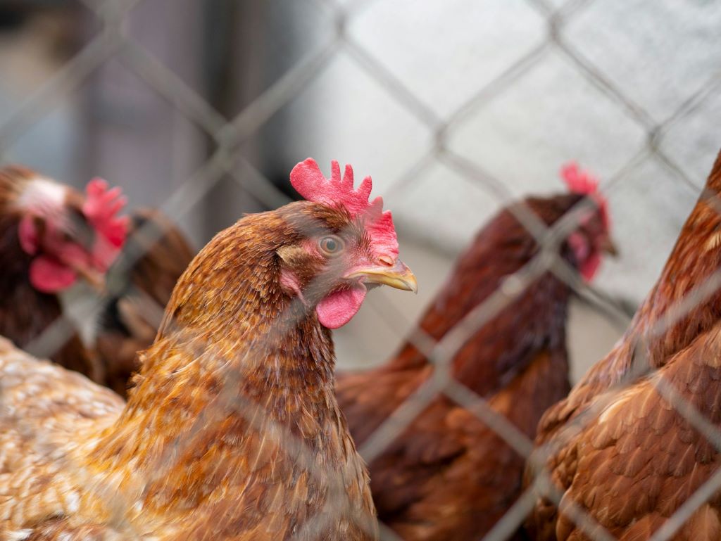
Unlike human flu viruses, avian influenza viruses tend to thrive in the lower respiratory tract. In fact, in their natural hosts, which include ducks and seagulls, the virus often infects the gut, where temperatures can be as high as 40-42C.
In previous studies using cultured cells, scientists have shown that avian influenza viruses appear more resistant to temperatures typically seen in fever in humans. Today’s study uses in vivo models – mice infected with influenza viruses – to help explain how fever protects us and why it may not be enough to protect us against avian influenza.
An international team led by scientists in Cambridge and Glasgow simulated in mice what happens during a fever in response to influenza infections. To carry out the research, they used a laboratory-adapted influenza virus of human origin, known as PR8, which does not pose a risk to humans.
Although mice do not typically develop fever in response to influenza A viruses, the researchers were able to mimic its effect on the virus by raising the ambient temperature where the mice were housed (elevating the body temperature of the mice).
The researchers showed that raising body temperature to fever levels is effective at stopping human-origin flu viruses from replicating, but it is unlikely to stop avian flu viruses. Fever protected against severe infection from human-origin flu viruses, with just a 2C increase in body temperature enough to turn a lethal infection into a mild disease.
The research also revealed that the PB1 gene of the virus, important in the replication of the virus genome inside infected cells, plays a key role in setting the temperature-sensitivity. Viruses carrying an avian-like PB1 gene were able to withstand the high temperatures associated with fever, and caused severe illness in the mice. This is important, because human and bird flu viruses can ‘swap’ their genes when they co-infect a host at the same time, for example when both viruses infect pigs.
Dr Matt Turnbull, the first author of the study, from the Medical Research Council Centre for Virus Research at the University of Glasgow said: “The ability of viruses to swap genes is a continued source of threat for emerging flu viruses. We’ve seen it happen before during previous pandemics, such as in 1957 and 1968, where a human virus swapped its PB1 gene with that from an avian strain. This may help explain why these pandemics caused serious illness in people.
“It’s crucial that we monitor bird flu strains to help us prepare for potential outbreaks. Testing potential spillover viruses for how resistant they are likely to be to fever may help us identify more virulent strains.”
Senior author Professor Sam Wilson, from the Cambridge Institute of Therapeutic Immunology and Infectious Disease at the University of Cambridge, said: “Thankfully, humans don’t tend to get infected by bird flu viruses very frequently, but we still see dozens of human cases a year. Bird flu fatality rates in humans have traditionally been worryingly high, such as in historic H5N1 infections that caused more than 40% mortality.
“Understanding what makes bird flu viruses cause serious illness in humans is crucial for surveillance and pandemic preparedness efforts. This is especially important because of the pandemic threat posed by avian H5N1 viruses.”
The findings may have implications for the treatment of infections, though the team stresses that more research is needed before changes are considered for treatment guidelines. Fever is often treated with antipyretic medication, which include ibuprofen and aspirin. However, there is clinical evidence that treating fever may not always be beneficial to the patient and may even promote transmission of influenza A viruses in humans.
Professor Wendy Barclay, Chair of the Medical Research Council (MRC) Infections and Immunity Board, said: “This elegant study builds on the very simple observation that different animals have different body temperatures, and shows how this may impact the way that viruses replicate in new hosts as they cross species barriers. The authors show that replication of human-adapted influenza virus is attenuated when temperatures are increased, such as in a fever. But avian influenza viruses, whose natural hosts have higher body temperatures, are not controlled by the fever response when they cross into mammals.
“They link their findings to one particular gene of the virus, called PB1, which is often carried over from birds when a new pandemic virus emerges. These findings have important implications for when and how to use drugs to control the fever that is associated with an influenza infection, and may also help us to understand why the disease from some influenza outbreaks is more severe.”
The research was funded primarily by the Medical Research Council, with additional funding from the Wellcome Trust, Biotechnology and Biological Sciences Research Council, European Research Council, European Union Horizon 2020, UK Department for Environment, Food & Rural Affairs, and US Department of Agriculture.
Reference
Turnbull, ML et al. Avian-origin influenza A viruses tolerate elevated pyrexic temperatures in mammals. Science; 27 Nov 2025; DOI: 10.1126/science.adq4691
Republished from Cambridge University under a Creative Commons Attribution-NonCommercial-ShareAlike 4.0 International License.
Read the original article.