Category: Uncategorized

Peer Teasing a Major Driver of Teen Eating Disorders

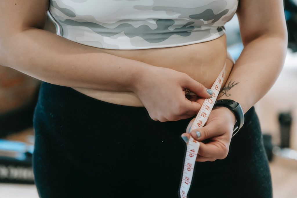
Photo from Freepik.

Weight-related peer teasing is a strong predictor of disordered eating behaviours in adolescents, particularly those at higher genetic risk, new research shows.

Focusing on teenagers, the Flinders University study found that specific environmental factors including peer pressure and conflict with parents can increase the risk of sometimes dangerous disordered eating behaviours – particularly among those with higher genetic risk.

“This study demonstrates that genetic risk for anorexia nervosa exerts a stronger influence on disordered eating behaviours in adolescent girls when combined with adverse parental and peer relationships. These social factors – including parental expectations, criticism, and care, as well as weight-related peer teasing – are pivotal in shaping vulnerability to disordered eating,” says Professor Sarah Cohen-Woods, senior author of the article in the Journal of Child Psychology and Psychiatry.

“It appears that modifiable factors, such as positive peer relationships, and parental behaviour and relationships with their children, may be leveraged to reduce the impact of increased genetic risk for disordered eating behaviours.”

Preventative efforts should target environmental influences such as weight-related peer teasing, and parental interactions and relationships with their children, particularly in those that are genetically at risk.

“For example, high parental expectations or criticism may increase genetic risk for self-induced vomiting and avoidance of eating.

“Weight-related peer teasing was found to increase the influence of genetic factors on disordered eating behaviours, such as objective bulimic episodes.”

The study of 383 Australian teenage twin girls aged between 14 and 19 collected responses to capturing their self-reported experiences of parental expectations, parental criticism, parental care, weight-related peer teasing and parental conflict.

Professor Cohen-Woods, from the Flinders Institute for Mental Health and Wellbeing, says research into the complex interplay between genetic predisposition and environmental factors can in the long term inform and improve clinical and non-clinical solutions to eating disorders.

“The research into anorexia nervosa polygenic risk scores in context with other known environmental risk factors highlights that a person’s genetic risk is not an isolated factor but can be significantly influenced by their social and family environment.”

The research, led by the Blackbird Initiative at the Flinders Institute for Mental Health and Wellbeing, bridges a gap between psychosocial research and molecular genetics, demonstrating a gene-environment interaction using polygenic risk scores and existing parental and peer teasing data.

Researchers, however, say future research is needed with larger cohorts and a broader range of environmental measures to further investigate these complex relationships to guide support and prevention efforts for young people, benefiting everyday families and communities in the future.

Source: Flinders University

Innovative Soft Robotic Intubation Device Could Save Lives

Credit: Pixabay CC0

Endotracheal intubation is a difficult task for highly trained individuals and under the best of circumstances. In the field and in the ER, where seconds matter, emergency medical personnel face many unknowns and wildly challenging conditions which lower their chances of success.

But what if successful endotracheal intubation could be less reliant on ideal conditions and years of specialised training? In a paper published in Science Translational Medicine, UC Santa Barbara researchers  David Haggerty, Elliot Hawkes and collaborators demonstrate a non-electronic soft robotic device that quickly and autonomously guides a soft tube into the trachea. Initial device testing with highly trained users yielded a 100% success rate, and a 96% overall success rate with prehospital medical providers (EMTs and paramedics).

“Current intubation tools require extensive anatomical knowledge, training, skill and ideal conditions to be highly successful,” said recent UC Santa Barbara Ph.D. graduate David Haggerty, a former researcher in the lab of mechanical engineering professor Elliot Hawkes. Current technology calls for the rescuer to first visualize the tracheal opening then manually direct a tube through the serpentine anatomy of the airway into the trachea. The challenge of this procedure increases in prehospital settings due to various factors including inadequate light and nonideal body position in addition to potential injuries and fluid in the airway.

This project is supported in part with funds from the National Science Foundation

From rigid tools to soft robotics

One of the main challenges to successful intubation is the body itself, and the mechanisms it has evolved to prevent food and foreign bodies from entering the lungs. The epiglottis is a small fleshy flap that closes over the trachea and guides food and liquid into the adjacent esophagus with each swallow. Conventional practice typically requires the rescuer to push a metal laryngoscope into the back of the mouth behind the tongue to lift the epiglottis out of the way in order to make room for the breathing tube.

But even with the epiglottis out of the way, the path the endotracheal tube must take is a twisted one, as it has to bend toward the front of the neck where the trachea is located, otherwise air could be delivered to the stomach via the esophagus, instead of to the lungs.

“Traditional tools, which you push from the base, are fundamentally limited in navigating delicate, tortuous anatomy,” Hawkes explained. “They must be relatively stiff so you can push them, and can only get around bends by deflecting off the sensitive tissue.”

The researchers’ device upends that paradigm with a soft, inflatable tube that everts from its tip. Called the soft robotic intubation system (SRIS), it consists of a curved “introducer” that slides into place at the back of the throat and stops at the esophagus. With that in place, a soft, pre-inflated tube is threaded through the introducer, emerging near its tip at the opening of the trachea. As the user advances the tube, it everts from its tip, carrying inside it a soft breathing tube as it enters the trachea. “So instead of trying to push this tube and bend it to get into this complex configuration, we can just mechanically create that complex configuration as we go,” Haggerty said. Once the endotracheal tube is at its destination, the user can inflate a cuff at its distal end to seal the opening and begin ventilation. The introducer can be removed, leaving the breathing tube in place.

Introducing a soft, growing tube eliminates friction with the surrounding tissue and minimizes injury due to excessive or misplaced force. It also automatically conforms to its environment, one of the major benefits of soft robotics. “This growing paradigm naturally accounts for minor variations in anatomical placement, size, shape or configuration,” Haggerty said, and because of this, users need not have extensive skill or a perfect understanding of the environment in order to navigate it.

In tests with mannequins and cadavers, the SRIS proved itself both effective, and, importantly, rapid, with a 100% success rate at a procedure duration of just seconds for expert users. For nonexpert users – the primary target of this technology – a five-minute training session was all that was needed to deliver an 87% success rate for first-pass attempts, translating into an 96% overall success rate, with a significantly lower procedure duration – 21 seconds versus 44 seconds – than state-of-the-art video laryngoscope intubation.

The next step for the researchers is to conduct clinical trials in order to get approvals from the Food and Drug Administration for clinical use. “We have good reason to believe it’s efficacious based on the data, but cadavers don’t say ‘ow’,” Haggerty said. They need to complete more testing to ascertain the device’s safety and effectiveness in a variety of airways and external conditions, he added. If successful, this device could benefit the millions of emergency intubations that occur each year in the US, and find application in military medical care. That’s in addition to its potential to save lives in global populations that have poor or no access to essential health services.

Source: University of California – Santa Barbara

Research Finds that Morning Coffee Really Does Lift Mood

Photo by Porapak Apichodilok on Pexels

People who regularly consume caffeine are usually in a better mood after a cup of coffee or another caffeinated drink – an effect that is much more pronounced in the morning than later in the day. This finding comes from a new study by researchers at the University of Warwick and Bielefeld University, published in the journal Scientific Reports. Participants reported feeling significantly happier and more enthusiastic on those mornings compared with other days at the same hour when they had not yet had coffee.

Links to negative moods such as sadness and feeling upset were also observed, though these effects were less pronounced: after drinking coffee, participants felt slightly less sad or upset than on comparable days without coffee. Unlike the boost in positive emotions, this effect was not dependent on the time of day.

In total, 236 young adults in Germany were studied over a period of up to four weeks. Participants completed short questionnaires on their smartphones seven times a day, indicating their current mood and whether they had consumed a caffeinated drink in the preceding 90 minutes. Accordingly, the study focused on caffeine consumption in everyday life, not just in artificial laboratory settings.

Perceived effect regardless of consumption habits

The researchers also investigated whether coffee has different effects on different individuals. Justin Hachenberger from the Bielefeld University in Germany says, ‘We were somewhat surprised to find no differences between individuals with varying levels of caffeine consumption or differing degrees of depressive symptoms, anxiety, or sleep problems. The links between caffeine intake and positive or negative emotions were fairly consistent across all groups.’

The researchers expected that people with higher levels of anxiety would experience negative mood changes, such as increased nervousness, after consuming caffeine. However, Hachenberger emphasises that people who react to caffeine in a bad way may avoid it and that the study did not include people who completely abstain from caffeine.

Role of caffeine withdrawal symptoms remains unclear

The researchers explain the mood-boosting effect of caffeine on morning mood with its ability to block adenosine receptors, which promotes wakefulness and makes people feel more energetic. ‘Caffeine works by blocking adenosine receptors, which can increase dopamine activity in key brain regions – an effect that studies have linked to improved mood and greater alertness’, says Professor Anu Realo from the University of Warwick.

Yet, it remains unclear whether these effects are related to a reduction in withdrawal symptoms after a night’s sleep. ‘Even people with moderate caffeine consumption can experience mild withdrawal symptoms that disappear with the first cup of coffee or tea in the morning’, says Anu Realo.

Caffeine consumption: a universal habit

‘Around 80 per cent of adults worldwide consume caffeinated beverages, and the use of such stimulating substances dates far back in human history’, says Professor Sakari Lemola from Bielefeld University, the study’s senior author. ‘Even wild animals consume caffeine; bees and bumblebees prefer nectar from plants that contain caffeine.’

Still, the study authors caution that caffeine can lead to dependence. Excessive intake is linked to various health risks, and consuming it later in the day can lead to sleep problems.

Source: Bielefeld University

A New LSD Analogue with Potential for Treating Schizophrenia

A cortical neuron treated with JRT, a synthetic molecule similar to the psychedelic drug LSD. Drugs like JRT might enable new treatments for conditions such as schizophrenia, without the hallucinations and other side effects of psychedelics. (Photo credit: Lee E. Dunlap, Institute for Psychedelics and Neurotherapeutics, UC Davis)

University of California, Davis, researchers have developed a new, neuroplasticity-promoting drug closely related to LSD that harnesses the psychedelic’s therapeutic power with reduced hallucinogenic potential.

The research, published in PNAS, highlights the new drug’s potential as a treatment option for conditions like schizophrenia, where psychedelics are not prescribed for safety reasons. The compound also may be useful for treating other neuropsychiatric and neurodegenerative diseases characterised by synaptic loss and brain atrophy.

To design the drug, dubbed JRT, researchers flipped the position of just two atoms in LSD’s molecular structure. The chemical flip reduced JRT’s hallucinogenic potential while maintaining its neurotherapeutic properties, including its ability to spur neuronal growth and repair damaged neuronal connections that are often observed in the brains of those with neuropsychiatric and neurodegenerative diseases.

“Basically, what we did here is a tire rotation,” said corresponding author David E. Olson, director of the Institute for Psychedelics and Neurotherapeutics and a professor of chemistry, and biochemistry and molecular medicine at UC Davis. “By just transposing two atoms in LSD, we significantly improved JRT’s selectivity profile and reduced its hallucinogenic potential.”

JRT exhibited powerful neuroplastic effects and improved measures in mice relevant to the negative and cognitive symptoms of schizophrenia, without exacerbating behaviours and gene expression associated with psychosis.

“No one really wants to give a hallucinogenic molecule like LSD to a patient with schizophrenia,” said Olson, who is also co-founder and chief innovation officer of Delix Therapeutics, a company that aims to bring neuroplastogens to the market. “The development of JRT emphasises that we can use psychedelics like LSD as starting points to make better medicines. We may be able to create medications that can be used in patient populations where psychedelic use is precluded.”

Testing JRT’s potential

Olson said that it took his team nearly five years to complete the 12-step synthesis process to produce JRT. The molecule was named after Jeremy R. Tuck, a former graduate student in Olson’s laboratory, who was the first to synthesise it and is a co-first author of the study along with Lee E. Dunlap, another former graduate student in Olson’s laboratory.

Following JRT’s successful synthesis, the researchers conducted a battery of cellular and mouse assays that demonstrated the drug’s neuroplastic effects and improved safety profile relative to LSD.

Key findings included:

  • JRT and LSD have the exact same molecular weight and overall shape, but distinct pharmacological properties.
  • JRT is very potent and highly selective for binding to serotonin receptors, specifically 5-HT2A receptors, the activation of which are key to promoting cortical neuron growth.
  • JRT promoted neuroplasticity, or growth between cellular connections in the brain, leading to a 46% increase in dendritic spine density and an 18% increase in synapse density in the prefrontal cortex.
  • JRT did not produce hallucinogenic-like behaviors that are typically seen when mice are dosed with LSD.
  • JRT did not promote gene expression associated with schizophrenia. Such gene expression is typically amplified with LSD use.
  • JRT produced robust anti-depressant effects, with it being around 100-fold more potent than ketamine, the state-of-the-art fast-acting anti-depressant.
  • JRT promoted cognitive flexibility, successfully addressing deficits in reversal learning that are associated with schizophrenia.

“JRT has extremely high therapeutic potential. Right now, we are testing it in other disease models, improving its synthesis, and creating new analogues of JRT that might be even better,” Olson said.

A more effective treatment for schizophrenia

Olson emphasised JRT’s potential for treating the negative and cognitive symptoms of schizophrenia, as most current treatments produce limited effects on anhedonia — the inability to feel pleasure — and cognitive function. Clozapine is the one exception, but it has side effects, and is not first-line drug of choice for people with severe schizophrenia.

Olson and his team are currently testing JRT’s potential against other neurodegenerative and neuropsychiatric diseases.

Source: University of California – Davis

We Found a Germ that ‘Feeds’ on Hospital Plastic – New Study

Photo by Marcelo Leal on Unsplash

Ronan McCarthy, Brunel University of London and Rubén de Dios, Brunel University of London

Plastic pollution is one of the defining environmental challenges of our time – and some of nature’s tiniest organisms may offer a surprising way out.

In recent years, microbiologists have discovered bacteria capable of breaking down various types of plastic, hinting at a more sustainable path forward.

These “plastic-eating” microbes could one day help shrink the mountains of waste clogging landfills and oceans. But they are not always a perfect fix. In the wrong environment, they could cause serious problems.

Plastics are widely used in hospitals in things such as sutures (especially the dissolving type), wound dressings and implants. So might the bacteria found in hospitals break down and feed on plastic?


Get your news from actual experts, straight to your inbox. Sign up to our daily newsletter to receive all The Conversation UK’s latest coverage of news and research, from politics and business to the arts and sciences.


To find out, we studied the genomes of known hospital pathogens (harmful bacteria) to see if they had the same plastic-degrading enzymes found in some bacteria in the environment.

Pseudomonas bacteria. Source: Wikimedia CCO

We were surprised to find that some hospital germs, such as Pseudomonas aeruginosa, might be able to break down plastic.

P aeruginosa is associated with about 559,000 deaths globally each year. And many of the infections are picked up in hospitals.

Patients on ventilators or with open wounds from surgery or burns are at particular risk of a P aeruginosa infection. As are those who have catheters.

We decided to move forward from our computational search of bacterial databases to test the plastic-eating ability of P aeruginosa in the laboratory.

We focused on one specific strain of this bacterium that had a gene for making a plastic-eating enzyme. It had been isolated from a patient with a wound infection. We discovered that not only could it break down plastic, it could use the plastic as food to grow. This ability comes from an enzyme we named Pap1.

Biofilms

P aeruginosa is considered a high-priority pathogen by the World Health Organization. It can form tough layers called biofilms that protect it from the immune system and antibiotics, which makes it very hard to treat.

Our group has previously shown that when environmental bacteria form biofilms, they can break down plastic faster. So we wondered whether having a plastic-degrading enzyme might help P aeruginosa to be a pathogen. Strikingly, it does. This enzyme made the strain more harmful and helped it build bigger biofilms.

To understand how P aeruginosa was building a bigger biofilm when it was on plastic, we broke the biofilm apart. Then we analysed what the biofilm was made of and found that this pathogen was producing bigger biofilms by including the degraded plastic in this slimy shield – or “matrix”, as it is formally known. P aeruginosa was using the plastic as cement to build a stronger bacterial community.

Pathogens like P aeruginosa can survive for a long time in hospitals, where plastics are everywhere. Could this persistence in hospitals be due to the pathogens’ ability to eat plastics? We think this is a real possibility.

Many medical treatments involve plastics, such as orthopaedic implants, catheters, dental implants and hydrogel pads for treating burns. Our study suggests that a pathogen that can degrade the plastic in these devices could become a serious issue. This can make the treatment fail or make the patient’s condition worse.

Thankfully, scientists are working on solutions, such as adding antimicrobial substances to medical plastics to stop germs from feeding on them. But now that we know that some germs can break down plastic, we’ll need to consider that when choosing materials for future medical use.

Ronan McCarthy, Professor in Biomedical Sciences, Brunel University of London and Rubén de Dios, Postdoctoral Research Fellow, Biotechnology, Brunel University of London

This article is republished from The Conversation under a Creative Commons license. Read the original article.

Adolescents Who Sleep Longer Perform Better at Cognitive Tasks

Photo by Eren Li

Adolescents who sleep for longer – and from an earlier bedtime – than their peers tend to have improved brain function and perform better at cognitive tests, researchers from the UK and China have shown.

But the study of adolescents in the US also showed that even those with better sleeping habits were not reaching the amount of sleep recommended for their age group.

Sleep plays an important role in helping our bodies function. It is thought that while we are asleep, toxins that have built up in our brains are cleared out, and brain connections are consolidated and pruned, enhancing memory, learning, and problem-solving skills. Sleep has also been shown to boost our immune systems and improve our mental health.

During adolescence, our sleep patterns change. We tend to start going to bed later and sleeping less, which affects our body clocks. All of this coincides with a period of important development in our brain function and cognitive development. The American Academy of Sleep Medicine says that the ideal amount of sleep during this period is between eight- and 10-hours’ sleep.

Professor Barbara Sahakian from the Department of Psychiatry at the University of Cambridge said: “Regularly getting a good night’s sleep is important in helping us function properly, but while we know a lot about sleep in adulthood and later life, we know surprisingly little about sleep in adolescence, even though this is a crucial time in our development. How long do young people sleep for, for example, and what impact does this have on their brain function and cognitive performance?”

Studies looking at how much sleep adolescents get usually rely on self-reporting, which can be inaccurate. To get around this, a team led by researchers at Fudan University, Shanghai, and the University of Cambridge turned to data from the Adolescent Brain Cognitive Development (ABCD) Study, the largest long-term study of brain development and child health in the United States.

As part of the ABCD Study, more than 3200 adolescents aged 11-12 years old had been given FitBits, allowing the researchers to look at objective data on their sleep patterns and to compare it against brain scans and results from cognitive tests. The team double-checked their results against two additional groups of 13-14 years old, totalling around 1190 participants. The results are published today in Cell Reports.

The team found that the adolescents could be divided broadly into one of three groups:

Group One, accounting for around 39% of participants, slept an average (mean) of 7 hours 10 mins. They tended to go to bed and fall asleep the latest and wake up the earliest.

Group Two, accounting for 24% of participants, slept an average of 7 hours 21 mins. They had average levels across all sleep characteristics.

Group Three, accounting for 37% of participants, slept an average of 7 hours 25 mins. They tended to go to bed and fall asleep the earliest and had lower heart rates during sleep.

Although the researchers found no significant differences in school achievement between the groups, when it came to cognitive tests looking at aspects such as vocabulary, reading, problem solving and focus, Group Three performed better than Group Two, which in turn performed better than Group One.

Group Three also had the largest brain volume and best brain functions, with Group One the smallest volume and poorest brain functions.

Professor Sahakian said: “Even though the differences in the amount of sleep that each group got was relatively small, at just over a quarter-of-an-hour between the best and worst sleepers, we could still see differences in brain structure and activity and in how well they did at tasks. This drives home to us just how important it is to have a good night’s sleep at this important time in life.”

First author Dr Qing Ma from Fudan University said: “Although our study can’t answer conclusively whether young people have better brain function and perform better at tests because they sleep better, there are a number of studies that would support this idea. For example, research has shown the benefits of sleep on memory, especially on memory consolidation, which is important for learning.”

The researchers also assessed the participants’ heart rates, finding that Group Three had the lowest heart rates across the sleep states and Group One the highest. Lower heart rates are usually a sign of better health, whereas higher rates often accompany poor sleep quality like restless sleep, frequent awakenings and excessive daytime sleepiness.

Because the ABCD Study is a longitudinal study – that is, one that follows its participants over time – the team was able to show that the differences in sleep patterns, brain structure and function, and cognitive performance, tended be present two years before and two years after the snapshot that they looked at.

Senior author Dr Wei Cheng from Fudan University added: “Given the importance of sleep, we now need to look at why some children go to bed later and sleep less than others. Is it because of playing videogames or smartphones, for example, or is it just that their body clocks do not tell them it’s time to sleep until later?”

The research was supported by the National Key R&D Program of China, National Natural Science Foundation of China, National Postdoctoral Foundation of China and Shanghai Postdoctoral Excellence Program. The ABCD Study is supported by the National Institutes of Health.

Reference

Ma, Q et al. Neural correlates of device-based sleep characteristics in adolescents. Cell Reports; 22 Apr 2025; DOI: 10.1016/j.celrep.2025.115565


Republished under a Creative Commons Attribution-NonCommercial-ShareAlike 4.0 International License.

Read the original article.

Not Just for Respiration: Lungs Also Produce Blood Cells

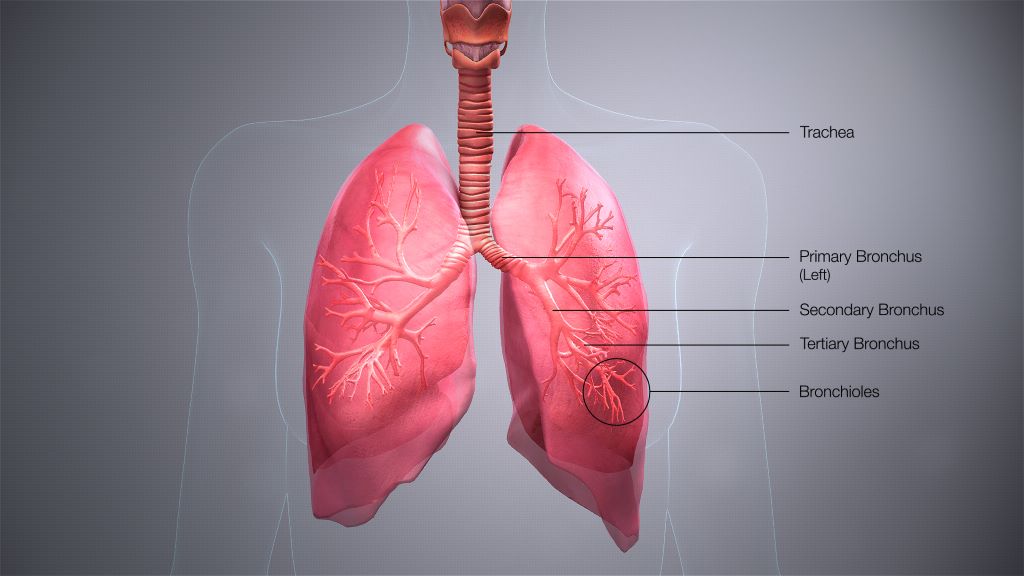
Credit: Scientific Animations CC4.0

For many years, scientists assumed that blood production took place in the bone marrow, providing the 200 billion blood cells needed per day. But now, researchers at UCSF are showing it’s also happening in the lungs. 

They found haematopoietic stem cells (HSCs) in human lung tissue that make red blood cells, as well as megakaryocytes, which produce the platelets that form blood clots. The findings appear in the journal Blood.

The work, which was supported by the National Heart, Lung, and Blood Institute (NHLBI) of the National Institutes of Health (NIH), suggests the lungs could be a potent source for life-saving stem cell transplants.

“For decades, bone marrow transplants have been a lynchpin in the treatment of cancers like leukemia,” said Mark Looney, MD, professor of medicine and laboratory medicine at UCSF and senior author of the paper. “The lung HSCs could prove to be a second and significant reservoir of these precious stem cells.”

From mouse to human

In 2017, the UCSF team found cells in the mouse lung making 50% of the mouse’s platelets

They also discovered lung stem cells in mice that made all the constituents of blood, including red blood cells, megakaryocytes and several types of immune cells.

Looney’s group wanted to prove this was also happening in people. So, they obtained donated samples of lung, bone marrow and blood, and compared what they found in each tissue.

Screening a golf-ball-sized volume of lung tissue, the scientists found stem cells in the lung that strongly resembled the well-known HSCs of bone marrow. Surprisingly, the HSCs were found at similar rates in both lung and bone marrow. 

“The lung HSCs weren’t one-offs – they were a reliable presence in the lungs,” said Catharina Conrad, MD, PhD, postdoctoral scholar in Looney’s lab and first author of the paper. “But we still needed to know that they were actually capable of making blood.”

So, the scientists coaxed lung and bone marrow HSCs to mature in petri dishes and found the lung HSCs were productive just like the bone marrow HSCs.

“Both types of HSCs thrived in our gold-standard stem cell experiment, but the lung HSC colonies made more red blood cells and megakaryocytes, while the bone marrow colonies tended to make more immune cells,” Looney said.

The human lung HSCs also could restore bone marrow in HSC-deficient mice. The discovery confirmed Looney’s earlier discovery that the mouse lung and bone marrow complemented one another in producing blood, even sending stem cells to restore one another.

“We think these HSCs could be a reservoir of haematopoiesis in a particular organ, in this case the lung, that gets activated whenever the body needs more of any part of the blood, whether it’s platelets, red blood cells or immune cells,” Looney said.

Getting to know the new HSC in town

To show that the lung HSCs truly resided in the lung, and weren’t just escapees from the bone marrow, Conrad and Looney looked for the HSCs in human lung tissue samples.

They found them between blood vessels in an arrangement that was reminiscent of what’s seen in bone marrow.

“They really seem to live there and aren’t just passing through,” Conrad said. 

Lastly, the team analysed the output of routine bone marrow transplants, which today begin with a blood draw from a donor followed by a screen for stem cells. 

Remarkably, nearly a fifth of the stem cells isolated for bone marrow transplant carried the signature of lung HSCs – suggesting that cells in “bone marrow transplants” aren’t only from bone marrow.

There’s a lot more to learn about the lung HSCs. Could the different pools of HSCs serve different therapeutic roles in medicine? Why do the lungs themselves need to make blood?

“The lungs are critical to blood circulation, so it’s tantalising to see the lung HSCs as an emergency reservoir for red blood cell and platelet production,” Looney said. “Now that we know they exist, it opens up a lot of new opportunities for a therapy, hematopoietic stem cell transplantation, that is very commonly used for patients with the need.”

Source: EurekAlert!

Type 1 Diabetes: Hybrid Closed-loop and Open-loop Systems Compared

Photo by Photomix Company on Pexels

People with type 1 diabetes require continuous insulin treatment and must regularly measure their glucose levels. With open-loop therapies*, insulin administration is manually controlled, while hybrid closed-loop systems* automatically regulate insulin delivery. A study with the involvement of the German Center for Diabetes Research showed that hybrid closed-loop systems offer improved long-term blood sugar values (HbA1c levels) and a lower risk of hypoglycaemic coma, but lead to a higher rate of diabetic ketoacidosis. The results were published in The Lancet Diabetes & Endocrinology.

Despite advances in insulin therapy, many people do not achieve their blood glucose targets and have a high risk of complications. Until now, the effect of insulin delivery in hybrid closed-loop systems on the risk of acute diabetes complications in people with type 1 diabetes has been unclear. Researchers have therefore now investigated whether the rates of severe hypoglycaemia and diabetic ketoacidosis are lower with hybrid closed-loop insulin therapy compared with sensor-augmented (open-loop) pump therapy.

Study with Nearly 14 000 Participants

In order to answer this question, the researchers, led by Professor Beate Karges, Faculty of Medicine at the RWTH Aachen, examined the data of nearly 14 000 participants. The study involved young people with type 1 diabetes from 250 diabetes centres in Germany, Austria, Switzerland, and Luxembourg. The participants were aged 2 to 20 years and had a type 1 diabetes duration of more than one year. They were identified from the Diabetes Prospective Follow-up Registry (DPV)**. The primary objectives of the study were to determine the rates of severe hypoglycaemia and ketoacidosis. Differences in HbA1c levels, time in the target range of 3.9 to 10.0mmol/L (70–180mg/dL), and fluctuations in blood sugar were also investigated. The data of 13 922 patients (51% male) were included in the analysis. Median age was 13.2 years; 7088 used a hybrid closed-loop system and 6834 used an open-loop system. The median observation time was 1.6 years.

Lower Rate of Hypoglycaemic Coma and More Ketoacidosis Events with Hybrid Closed-Loop Therapies

The results: People using hybrid closed-loop therapy had a significantly lower rate of rate of hypoglycaemic coma (0.62 per 100 patient-years) than those using open-loop therapy (0.91 per 100 patient-years). Furthermore, patients in the hybrid closed-loop group had a significantly lower HbA1c level (7.34% versus 7.50%). They had a higher percentage of time in the target glucose range of 3.9 to 10.0 mmol/L (64% versus 52% of the time). Their glycaemic variability was also lower (coefficient of variation of 35.4% versus 38.3%). There was no significant difference in the rate of severe hypoglycaemia.

However, individuals using a hybrid closed-loop system had a higher rate of ketoacidosis (1.74 events per 100 patient-years) than those using open-loop therapy (0.96 per 100 patient-years). The rate of ketoacidosis was particularly high in people with an HbA1c level of 8.5% or higher in the closed-loop therapy group (5.25 per 100 patient-years). In the comparison group, a rate of 1.53 events per 100 patient-years was observed.

Recommendation: Monitor Ketone Bodies Closely

Due to the higher risk of ketoacidosis, it is important to provide patients with targeted information and, in case of potential metabolic decompensation, to closely monitor ketone bodies in the blood or urine in order to prevent such adverse events, emphasize the authors of the study. 

Source: Deutsches Zentrum fuer Diabetesforschung DZD

Community Health Workers must be Made Permanent, Rules Labour Court

Photo by Tingey Injury Law Firm on Unsplash

By Tania Broughton

The Johannesburg Labour Court has ruled that community health workers, who for years have been employed by the health department on recurring fixed-term contracts, must be deemed permanent government employees.

The National Health and Allied Workers Union (NEHAWU), on behalf of its members, has successfully overturned a previous bargaining council ruling that the temporary contracts were legal.

There are an estimated 50,000 community health workers. The recurring fixed-term contracts left them without job security and other benefits of permanent employment.

Read the judgment here

The issue was first ventilated before the Public Health and Social Development Sectoral Bargaining Council in 2021. The commissioner found that the contracts were permitted by the Public Service Act, were concluded through collective agreements with unions, and were justified in terms of the Labour Relations Act (LRA) as they were funded by an “external source for a limited period” – the National Treasury.

NEHAWU took the ruling on review. The matter was argued before Johannesburg Labour Court Acting Judge Ashley Cook in October last year. He handed down his ruling on 23 January 2025, overturning the bargaining council’s findings.

On the issue of “external funding” – the legal justification in the LRA for fixing the contract terms – Judge Cook said the department had correctly submitted that it was not disputed that the funding for the employment of the community health workers was a conditional grant approved by national treasury on an annual basis.

“However, what was in dispute was whether the conditional grant was from an external source. The department receives all revenue from the National Treasury,” Judge Cook said.

As funding for all public servants was sourced from the Treasury, this meant that it was not an “external source”, and therefore the department could not rely on it as a “justifiable reason” to deviate from the provisions of the LRA.

The contracts of the community health workers were therefore, in terms of the Act, deemed to be of an “indefinite duration”, the judge said, setting aside the arbitration award.

He made no order as to costs.

NEHAWU welcomed the ruling. In a statement, it said community health workers had been on perennial contractual renewals without a clear explanation from the Department of Health.

“The court determined that it is common cause to all parties that there is a permanent need for the work tendered by community health workers as conceded by the counsel for the state.”

The union said it would continue to fight for the “permanent absorption” of all the workers and would be meeting with its members to advise on how it would ensure the implementation of the judgment.

Republished from GroundUp under a Creative Commons Attribution-NoDerivatives 4.0 International License.

Read the original article here.

What is the Drug Captagon and How is it Linked to Syria’s Fallen Assad Regime?

Photo by James Coleman on Unsplash

Nicole Lee, Curtin University

After the fall of the al-Assad regime in Syria, large stockpiles of the illicit drug captagon have reportedly been uncovered.

The stockpiles, found by Syrian rebels, are believed to be linked to al-Assad military headquarters, implicating the fallen regime in the drug’s manufacture and distribution.

But as we’ll see, captagon was once a pharmaceutical drug, similar to some of the legally available stimulants we still use today for conditions including attention-deficit hyperactivity disorder (ADHD).

Captagon was once a pharmaceutical

Captagon is the original brand name of an old synthetic pharmaceutical stimulant originally made in Germany in the 1960s. It was an alternative to amphetamine and methamphetamine, which were both used as medicines at the time.

The drug has the active ingredient fenethylline and was initially marketed for conditions including ADHD and the sleeping disorder narcolepsy. It had a similar use to some of the legally available stimulants we still use today, such as dexamphetamine.

Captagon has similar effects to amphetamines. It increases dopamine in the brain, leading to feelings of wellbeing, pleasure and euphoria. It also improves focus, concentration and stamina. But it has a lot of unwanted side effects, such as low-level psychosis.

The drug was originally sold mostly in the Middle East and parts of Europe. It was available over the counter (without a prescription) in Europe for a short time before it became prescription-only.

It was approved only briefly in the United States before becoming a controlled substance in the 1980s, but was still legal for the treatment of narcolepsy in many European countries until relatively recently.

According to the International Narcotics Control Board pharmaceutical manufacture of Captagon had stopped by 2009.

The illicit trade took over

The illegally manufactured version is usually referred to as captagon (with a small c). It is sometimes called “chemical courage” because it is thought to be used by soldiers in war-torn areas of the Middle East to help give them focus and energy.

For instance, it’s been reportedly found on the bodies of Hamas soldiers during the conflict with Israel.

Its manufacture is relatively straightforward and inexpensive, making it an obvious target for the black-market drug trade.

Black-market captagon is now nearly exclusively manufactured in Syria and surrounding countries such as Lebanon. It’s mostly used in the Middle East, including recreationally in some Gulf states.

It is one of the most commonly used illicit drugs in Syria.

A recent report suggests captagon generated more than US$7.3 billion in Syria and Lebanon between 2020 and 2022 (about $2.4 billion a year).

What we know about illicit drugs generally is that any seizures or crackdowns on manufacturing or sale have a very limited impact on the drug market because another manufacturer or distributor pops up to meet demand.

So in all likelihood, given the size of the captagon market in the Middle East, these latest drug discoveries and seizures are likely to reduce manufacture only for a short time.

Nicole Lee, Adjunct Professor at the National Drug Research Institute (Melbourne based), Curtin University

This article is republished from The Conversation under a Creative Commons license. Read the original article.