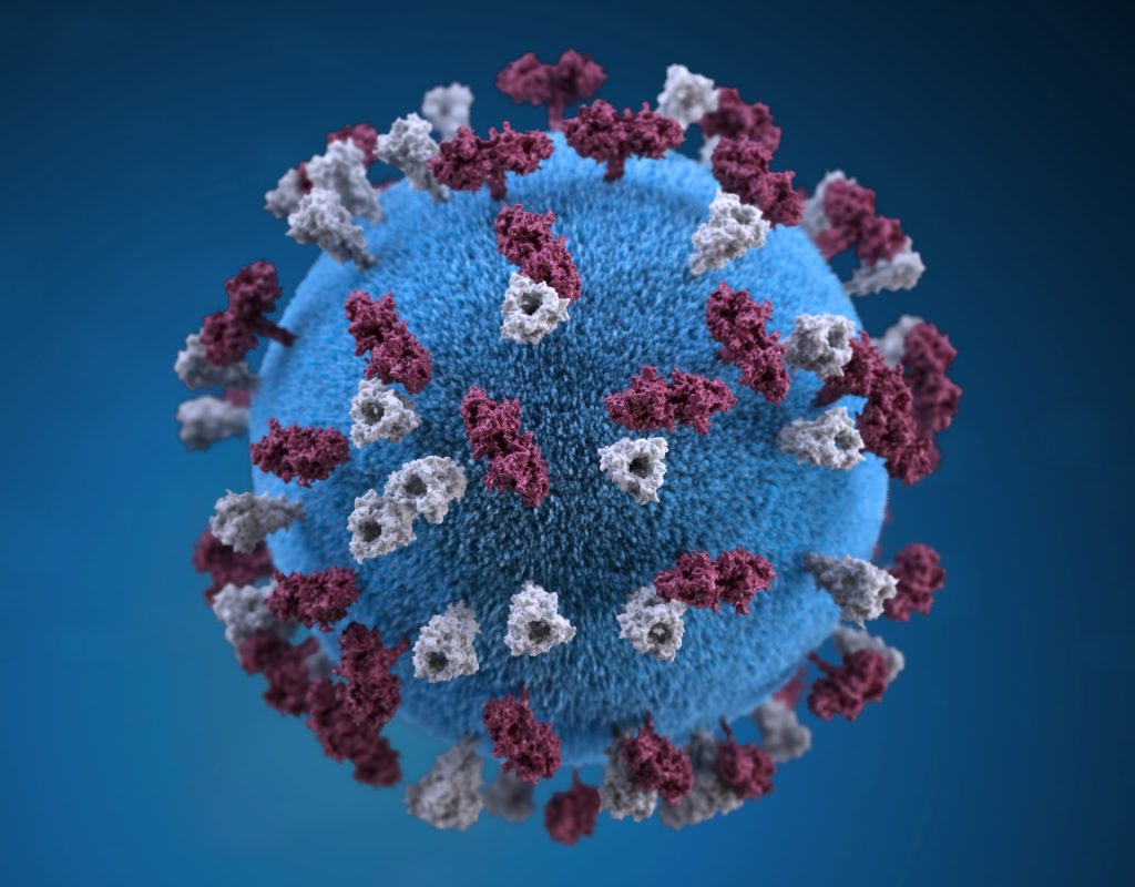
Mayo Clinic researchers mapped how the measles virus mutated and spread in the brain of a person who succumbed to a rare, lethal brain disease. New cases of this disease, which is a complication of the measles virus, may occur as measles re-emerges among the unvaccinated, say researchers.
Using the latest tools in genetic sequencing, researchers at Mayo Clinic reconstructed how a collective of viral genomes colonised a human brain.
The virus acquired distinct mutations that drove the spread of the virus from the frontal cortex outward.
The highly contagious measles virus infects the upper respiratory tract where it uses the trachea as a trampoline to launch and spread through droplets dispersed when an infected person coughs or sneezes.
Dr Cattaneo pioneered studies on how the measles virus spreads throughout the body. He first began to study the measles virus about 40 years ago and was fascinated by the rare, lethal brain disease called subacute sclerosing panencephalitis (SSPE), which occurs in about 1 in every 10 000 measles cases.
It can take about five to 10 years after the initial infection for the measles virus to mutate and spread throughout the brain.
Symptoms of this progressive neurological disease include memory loss, seizures and immobility.
Dr. Cattaneo studied SSPE for several years until the lethal disease nearly disappeared as more people were vaccinated against measles. But now, measles is resurging due to vaccine hesitancy and missed vaccinations.
During the COVID pandemic, millions of children missed receiving their measles vaccinations, which has resulted in an estimated 18% increase in measles cases and 43% increase in death from measles in 2021 compared to 2022 worldwide, according to a recent Centers for Disease Control and Prevention (CDC) report.
“We suspect SSPE cases will rise again as well. This is sad because this horrible disease can be prevented by vaccination. But now we are in the position to study SSPE with modern, genetic sequencing technology and learn more about it,” says Iris Yousaf, co-lead author of the study and a fifth-year Ph.D. candidate at Mayo Clinic Graduate School of Biomedical Sciences.
Dr Cattaneo and Yousaf had a unique research opportunity through a collaboration with the CDC. They studied the brain of a person who had contracted measles as a child and had succumbed to SSPE years later as an adult.
They investigated 15 specimens from different regions of the brain and conducted genetic sequencing on each region to piece together the puzzle of how the measles virus mutated and spread.
The researchers discovered that, after the measles virus entered the brain, its genome began to mutate in harmful ways over successive generations, creating a population of varied genomes.
“In this population, two specific genomes had a combination of characteristics that worked together to promote virus spread from the initial location of the infection – the frontal cortex of the brain – out to colonise the entire organ,” says Dr Cattaneo.
The next steps in this research are to understand how specific mutations favour virus spread in the brain. These studies will be done in cultivated brain cells brain organoids. This knowledge may help in creating effective antiviral drugs to combat virus spread in the brain. However, pharmacological intervention in advanced disease stages is challenging, and preventing SSPE through measles vaccination remains the best method.
Glioblastoma is one of the most treatment-resistant cancers, with those diagnosed surviving for less than two years. In a new study in NPJ Genomic Medicine, researchers at the University of Notre Dame have found that a largely understudied cell could offer new insight into how the aggressive, primary brain cancer is able to resist immunotherapy.
“A decade ago, we didn’t even know perivascular fibroblasts existed within the brain, and not just in the lining of the skull,” said senior author Meenal Datta, assistant professor of aerospace and mechanical engineering at Notre Dame.
“My lab’s expertise is examining tumours from an engineering and systems-based approach and looking at the novel mechanical features in rare cancers that may have been understudied or overlooked.”
Using standard bioinformatics and newer AI-based approaches, Datta’s TIME Lab began analysing different genes expressed in the tumour microenvironment related to the extracellular matrix – or the scaffolding cells create to support future cell adhesion, migration, proliferation and differentiation – and other various cell types.
What they found was a surprising, fairly new cell type: perivascular fibroblasts.
These fibroblasts are typically found in the blood vessels of a healthy brain and deposit collagen to maintain the structural integrity and functionality of brain vessels.
“It was a serendipitous discovery,” said first author Maksym Zarodniuk, graduate student in the TIME Lab and the bioengineering doctorate programme.
“We started in a completely different direction and stumbled upon this population of cells by using a combination of both bulk and single-cell RNA sequencing analyses of patient tumours.”
In their data, researchers were able to identify two groups of patients: those with a higher proportion of perivascular fibroblasts and those with significantly less.
They found that brain cancer patients with more perivascular fibroblasts in their tumours were more likely to respond poorly to immunotherapies and have poor survival outcomes.
Further study revealed that perivascular fibroblasts support the creation of an immunosuppressive tumour microenvironment, allowing the cancer to better evade the immune system.
The fibroblasts may also help the cancer resist therapies such as chemotherapy that targets cell division by promoting stem-like cancer cells that rarely divide, which are believed to be a major source of tumour relapse and metastasis.
“Moving forward, we want to do new experiments to confirm what we found in this paper and provide some good ground to start thinking about how to improve response to immunotherapy,” Zarodniuk said.
Because perivascular fibroblasts are a part of a healthy brain’s vasculature, Datta believes that these cells are breaking off and getting close to or infiltrating the glioblastoma tumour.
However, instead of supporting healthy brain function, these fibroblasts are getting reprogrammed and helping the tumour instead.
“Most people think about the brain as being very soft, with soft cells and a soft matrix. But by putting down these fibroblasts and making these very fibrous proteins, it gives us an entirely different perspective on the structure of the brain and how it can be taken advantage of by cancer cells originating in the same organ,” Datta said.
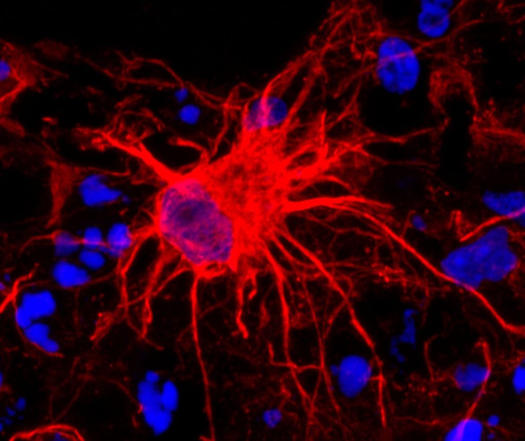
Image of an astrocyte, a subtype of glial cells. Glial cells are the most common cell in the brain.
Credit: Pasca Lab, Stanford University
NIH support from: NINDS, NIMH, NIGMS, NCATS
A new study published in PNAS Nexus provides a better understanding of how the brain responds to injuries. Researchers at the George Washington University discovered that a protein called Snail plays a key role in coordinating the response of brain cells after an injury.
The study shows that after an injury to the central nervous system (CNS), a group of localised cells start to produce Snail, a transcription factor or protein that has been implicated in the repair process. The GW researchers show that changing how much Snail is produced can significantly affect whether the injury starts to heal efficiently or whether there is additional damage.
“Our findings reveal the intricate ways the brain responds to injuries,” said senior author Robert Miller, the Vivian Gill Distinguished Research Professor and Vice Dean of the GW School of Medicine and Health Sciences.
“Snail appears to be a key player in coordinating these responses, opening up promising possibilities for treatments that can minimise damage and enhance recovery from neurological injuries.”
This study identified for the first time a special group of microglial-like cells that produce Snail. Microglial cells are found in the central nervous system. The researchers found that lowering the amount of Snail produced after an injury results in inflammation and increased cell death. During this process, the injury worsens and there are fewer connections or synapses between brain cells. In contrast, when Snail levels are increased the outcome of brain injury improves-suggesting this protein can help limit the spread of injury-induced damage.
The research raises questions about whether an experimental drug that affects Snail production could be used to limit the damage incurred after someone suffers a stroke or has been injured in an accident, Miller said.
Additional studies must be done to show that increasing Snail production could curtail injury or even promote healing of the brain.
Miller and his team also plan to study the regulation of Snail in diseases like multiple sclerosis, a disease resulting in damage to the myelin nerve sheath. If drugs targeting Snail could be used to stop that damage, many of the future symptoms of this disease could be eased, he says.
But researchers have years of work to do before new drugs targeting Snail can be tested in clinical trials. The payoff ultimately might be drugs that can lead to accelerated healing for stroke damage, head wounds and even neurodegenerative diseases like dementia.
A University of Iowa-led team of international neuroscientists have obtained the first direct recordings of the human brain in the minutes before and after a brain hub crucial for language meaning was surgically disconnected. The results reveal the importance of brain hubs in neural networks and the remarkable way in which the human brain attempts to compensate when a hub is lost, with immediacy not previously observed. The findings were reported recently in the journal Nature Communications.
Hubs are critical for connectivity
The human brain has hubs – the intersection of many neuronal pathways that help coordinate brain activity required for complex functions like understanding and responding to speech. But debate has reigned as to whether highly interconnected brain hubs are irreplaceable for certain brain functions. By some accounts the brain, as an already highly interconnected neural network, can in principle immediately compensate for the loss of a hub, in the same way that traffic can be redirected around a blocked-off city centre.
With a rare experimental opportunity, the UI neurosurgical and research teams led by Matthew Howard III, MD, professor and DEO of neurosurgery, and Christopher Petkov, PhD, professor and vice chair for research in neurosurgery, have achieved a breakthrough in understanding the necessity of a single hub. By obtaining evidence for what happens when a hub required for language meaning is lost, the researchers showed both the intrinsic importance of the hub as well as the remarkable and rapid ability of the brain to adapt and at least partially attempt to immediately compensate for its loss.
Evaluating the impact of losing a brain hub
The study was conducted during surgical treatment of two patients with epilepsy. Both patients were undergoing procedures that required surgical removal of the anterior temporal lobe – a brain hub for language meaning – to allow the neurosurgeons access to a deeper brain area causing the patients’ debilitating epileptic seizures. Before this type of surgery, neurosurgery teams often ask the patients to conduct speech and language tasks in the operating room as the team uses implanted electrodes to record activity from parts of the brain close to and distant from the planned surgery area. These recordings help the clinical team effectively treat the seizures while limiting the impact of the surgery on the patient’s speech and language abilities.
Typically, the recording electrodes are not needed after the surgical resection procedure and are removed. The innovation in this study was that the neurosurgery team was able to safely complete the procedure with the recording electrodes left in place or replaced to the same location after the procedure. This made it possible to obtain rare pre- and post-operative recordings allowing the researchers to evaluate signals from brain areas far away from the hub, including speech and language areas distant from the surgery site. Analysis of the change in responses to speech sounds before and after the loss of the hub revealed a rapid disruption of signaling and subsequent partial compensation of the broader brain network.
“The rapid impact on the speech and language processing regions well removed from the surgical treatment site was surprising, but what was even more surprising was how the brain was working to compensate, albeit incompletely within this short timeframe,” says Petkov, who also holds an appointment at Newcastle University Medical School in the UK.
The findings disprove theories challenging the necessity of specific brain hubs by showing that the hub was important to maintain normal brain processing in language.
“Neurosurgical treatment and new technologies continue to improve the treatment options provided to patients,” says Howard, who also is a member of the Iowa Neuroscience Institute.
“Research such as this underscores the importance of safely obtaining and comparing electrical recordings pre and post operatively, particularly when a brain hub might be affected.”
According to the researchers, the observation on the nature of the immediate impact on a neural network and its rapid attempt to compensate provides evidence in support of a brain theory proposed by Professor Karl Friston at University College London, which posits that any self-organising system at equilibrium works towards orderliness by minimising its free energy, a resistance of the universal tendency towards disorder.
These neurobiological results following human brain hub disconnection were consistent with several predictions of this and related neurobiological theories, showing how the brain works to try to regain order after the loss of one of its hubs.
Schizophrenia is often accompanied by extensive impairment of memory, including prospective memory, which is the ability to remember to perform future activities. In a randomised clinical trial published in Neuropsychopharmacology Reports, researchers found that repetitive transcranial magnetic stimulation (rTMS), a non-invasive method that uses alternating magnetic fields to induce an electric current in the underlying brain tissue, may help ameliorate certain aspects of prospective memory in individuals with schizophrenia.
The trial included 50 patients with schizophrenia and 18 healthy controls. Of the 50 patients, 26 completed active rTMS and 24 completed a sham rTMS. Healthy controls received no treatment.
Investigators assessed event-based prospective memory, which is remembering to perform an action when an external event occurs, such as remembering to give a message to a friend when you next see them and also time-based prospective memory, which is remembering to perform an action at a certain time, such as remembering to attend a scheduled meeting.
Both event-based prospective memory and time-based prospective memory scores at the baseline of the trial were significantly lower in patients with schizophrenia than in controls. After rTMS treatments, the scores of event-based prospective memories in patients were significantly improved and were similar to those in controls, while patients’ scores of time-based prospective memory did not improve.
“The findings of this study may provide one therapeutic option for prospective memory in patients with schizophrenia,” said co–corresponding author Su-Xia Li, MD, PhD, of Peking University, in China.
Moderate to severe traumatic brain injury carries lasting effects: trouble with focussing, recall and decision-making. Though many recover enough to live independently, their impairments prevent them from returning to school or work and from resuming their social lives. Current treatments offer little improvement, but results of a clinical trial of a new brain stimulation device, published in Nature Medicine, have shown great promise in at least partially restoring cognitive function.
“In general, there’s very little in the way of treatment for these patients,” said Jaimie Henderson, MD, professor of neurosurgery and co-senior author of the study.
But the fact that these patients had emerged from comas and recovered a fair amount of cognitive function suggested that the brain systems that support attention and arousal – the ability to stay awake, pay attention to a conversation, focus on a task – were relatively preserved.
These systems connect the thalamus, a relay station deep inside the brain, to points throughout the cortex, the brain’s outer layer, which control higher cognitive functions.
‘Dimmed lights’
“In these patients, those pathways are largely intact, but everything has been down-regulated,” said Henderson, the John and Jene Blume-Robert and Ruth Halperin Professor. “It’s as if the lights had been dimmed and there just wasn’t enough electricity to turn them back up.”
In particular, an area of the thalamus called the central lateral nucleus functions as a hub that regulates many aspects of consciousness.
“The central lateral nucleus is optimised to drive things broadly, but its vulnerability is that if you have a multifocal injury, it tends to take a greater hit because a hit can come from almost anywhere in the brain,” said Nicholas Schiff, MD, a professor at Weill Cornell Medicine and co-senior author of the study.
The researchers hoped that precise electrical stimulation of the central lateral nucleus and its connections could reactivate these pathways, turning the lights back up.
Precise placement
In the trial, the researchers recruited five participants who had lasting cognitive impairments more than two years after moderate to severe traumatic brain injury. They were aged 22 to 60, with injuries sustained three to 18 years earlier.
The challenge was placing the stimulation device in a small target in the right area, which varied across individuals. Each brain is shaped differently to begin with, and the injuries had led to further modifications.
“That’s why we developed a number of tools to better define what that area was,” Henderson said. The researchers created a virtual model of each brain that allowed them to pinpoint the location and level of stimulation that would activate the central lateral nucleus.
Guided by these models, Henderson surgically implanted the devices in the five participants.
“It’s important to target the area precisely,” he said. “If you’re even a few millimetres off target, you’re outside the effective zone.”
A pioneering moment
After a two-week titration phase to optimise the stimulation, the participants spent 90 days with the device turned on for 12 hours a day.
Their progress was measured by a standard test of mental processing speed, called the trail-making test, which involves drawing lines connecting a jumble of letters and numbers.
“It’s a very sensitive test of exactly the things that we’re looking at: the ability to focus, concentrate and plan, and to do this in a way that is sensitive to time,” Henderson said.
At the end of the 90-day treatment period, the participants had improved their speeds on the test, on average, by 32%, far exceeding the 10% the researchers had aimed for.
“The only surprising thing is it worked the way we predicted it would, which is not always a given,” Henderson said.
For the participants and their families, the improvements were apparent in their daily lives. They resumed activities that had seemed impossible – reading books, watching TV shows, playing video games or finishing a homework assignment. They felt less fatigued and could get through the day without napping.
The therapy was so effective the researchers had trouble completing the last part of their study. They had planned a blinded withdrawal phase, in which half the participants would be randomly selected to have their devices turned off. Two of the patients declined, unwilling to take that chance. Of the three who participated in the withdrawal phase, one was randomized to have their device turned off. After three weeks without stimulation, that participant performed 34% slower on the trail-making test.
The clinical trial is the first to target this region of the brain in patients with moderate to severe traumatic brain injury, and it offers hope for many who have plateaued in their recovery.
“This is a pioneering moment,” Schiff said. “Our goal now is to try to take the systematic steps to make this a therapy. This is enough of a signal for us to make every effort.”
An international study published recently in the journal Brain has reported promising results in restoring function lost in mice and rat models of stroke. Researchers were able to restore lost brain function using small molecules that in the future could potentially be developed into a stroke recovery therapy.
“Communication between nerve cells in large parts of the brain changes after a stroke and we show that it can be partially restored with the treatment,” says Tadeusz Wieloch, senior professor of neurobiology at Lund University in Sweden.
“Concomitantly, the rodents regain lost somatosensory functions, something that around 60 per cent of all stroke patients experience today. The most remarkable result is that the treatment began several days after a stroke,” Wieloch continues.
In an ischaemic stroke, lack of blood flow to affected parts of the brain lead to loss of function such as paralysis, sensorimotor impairment and vision and speech difficulties, but also to pain and depression.
There are currently no approved drugs that improve or restore the functions after a stroke, apart from clot-dissolving treatment in the acute phase (within 4.5 hours of the stroke). Some spontaneous improvements occur, but many stroke patients suffer chronic loss of function.
For example, about 60% of stroke sufferers, experience lost somatosensory functions such as touch and position sense.
The new study shows that rats that were treated with a class of substances that inhibit the metabotropic glutamate receptor (mGluR5), a receptor that regulates communication in the brain’s nerve cell network.
“Rodents treated with the GluR5 inhibitor regained their somatosensory functions,” says Tadeusz Wieloch, who led the study.
Two days after the stroke, ie when the damage had developed and function impairment was most prominent, the researchers started treating the rodents that exhibited the greatest impaired function.
“A temporary treatment effect was seen after just 30 minutes, but treatment for several weeks is needed to achieve a permanent recovery effect. Some function improvement was observed even when the treatment started 10 days after a stroke,” says Tadeusz Wieloch.
Importantly, sensorimotor functions improved, even though the extent of the brain damage was not diminished.
This, explains Tadeusz Wieloch, is due to the intricate network of nerve cells in the brain, known as the connectome – the way brain areas are inter connected and communicate form the basis for various brain functions.
“Impaired function after a stroke is due to cell loss, but also because of reduced activity in large parts of the connectome in the undamaged brain. The receptor mGluR5 is apparently an important factor in the reduced activity in the connectome, which is prevented by the inhibitor which therefore restores the lost brain function,” says Tadeusz Wieloch.
The results also showed that sensorimotor function was further improved if treatment with the mGluR5 inhibitor is combined with somatosensory training by housing several rodents in cages enriched with toys, chains, grids, and plastic tubes.
The researchers hope that in the future their results could lead to a clinical treatment that could be initiated a few days after an ischaemic stroke.
“Combined with rehabilitation training, it could eventually be a new promising treatment. However, more studies are needed. The study was conducted on mice and rats, and of course needs to be repeated in humans. This should be possible since several mGluR5 inhibitors have been studied in humans for the treatment of neurological diseases other than stroke, and shown to be tolerated by humans,” says Tadeusz Wieloch.
Researchers have identified objective evidence of how the neck muscles are involved in primary headaches. The study findings, being presented at the annual meeting of the Radiological Society of North America (RSNA), could lead to better treatments.
The distinct underlying causes of primary headaches, comprising tension-type headaches and migraines, are still not fully understood.
“Our imaging approach provides first objective evidence for the very frequent involvement of the neck muscles in primary headaches, such as neck pain in migraine or tension-type headache, using the ability to quantify subtle inflammation within muscles,” said Nico Sollmann, MD, PhD, resident at University Hospital Ulm and University Hospital Rechts der Isar in Munich, Germany.
In tension-type headaches there is often the perception of a tightening in the head and mild to moderate dull pain on both sides of the head. While these headaches are typically associated with stress and muscle tension, their exact origin is not fully understood.
Migraines are characterised by a severe throbbing pain and generally occur or are worse on one side of the head. Migraines may also cause nausea, weakness and light sensitivity.
Neck pain is commonly associated with primary headaches but there are no objective biomarkers for myofascial involvement. Myofascial pain is associated with inflammation or irritation of muscle or of the connective tissue, known as fascia, that surrounds the muscle.
For the study, Dr Sollmann and colleagues aimed to investigate the involvement of the trapezius muscles in primary headache disorders by quantitative magnetic resonance imaging (MRI) and to explore associations between muscle T2 values and headache and neck pain frequency.
The prospective study recruited 50 participants, mostly women, ranging in age from 20 to 31 years old. Of the participants, 16 had tension-type headache, and 12 had tension-type headache plus migraine episodes. The groups were matched with 22 healthy controls.
All participants underwent 3D turbo spin-echo MRI. The bilateral trapezius muscles were manually segmented, followed by muscle T2 extraction.
Associations between muscle T2 values and the presence of neck pain, number of days with headache, and number of myofascial trigger points as determined by manual palpation of the trapezius muscles were analysed (adjusting for age, sex and body mass index).
The tension-type headache plus migraine group demonstrated the highest muscle T2 values. Muscle T2 was significantly associated with the number of headache days and the presence of neck pain.
The increased muscle T2 values could be interpreted as a surrogate of inflammation arising from the nervous system and increased sensitivity of nerve fibres within myofascial tissues.
“The quantified inflammatory changes of neck muscles significantly correlate with the number of days lived with headache and the presence of subjectively perceived neck pain,” Dr Sollmann said.
“Those changes allow us to differentiate between healthy individuals and patients suffering from primary headaches.”
Muscle T2 mapping could be used to stratify patients with primary headaches and to track potential treatment effects for monitoring.
“Our findings support the role of neck muscles in the pathophysiology of primary headaches,” Dr Sollmann said. “Therefore, treatments that target the neck muscles could lead to a simultaneous relief of neck pain, as well as headache.”
Dr Sollmann pointed out that non-invasive treatment options that directly target the site of pain in the neck muscles could be highly effective and safer than systemic drugs.
“Our imaging approach with delivery of an objective biomarker could facilitate therapy monitoring and patient selection for certain treatments in the near future,” he added.
Researchers from the University of Birmingham have designed and developed a novel diagnostic device to detect traumatic brain injury (TBI) by shining a safe laser into the eye.
The technique is radically different from other diagnostic methods and is expected to be developed into a hand-held device for use in the critical ‘golden hour’ after traumatic brain injury, when life critical decisions on treatment must be made.
The device, described in Science Advances, incorporates a class 1, CE marked, eye-safe laser and a unique Raman spectroscopy system, which uses light to reveal the biochemical and structural properties of molecules by detecting how they scatter light, to detect the presence and levels of known biomarkers for brain injury.
There is an urgent need for new technologies to improve the timeliness of TBI diagnosis. TBI is caused by sudden shock or impact to the head, which can cause mild to severe injury to the brain, and rapid intervention is necessary to prevent further irreversible damage.
Diagnosis at the point of injury is difficult. Moreover, radiological investigations such as X-ray or MRI are very expensive and slow to show results.
Birmingham researchers, led by Professor Pola Goldberg Oppenheimer from the School of Chemical Engineering, designed and developed the novel diagnostic hand-held device to assess patients as soon as injury occurs.
It is fast, precise and non-invasive for the patient, causing no additional discomfort, can provide information on the severity of the trauma, and will be suitable to be used on-site to assess TBI.
Professor Pola Goldberg Oppenheimer said: “Early diagnosis of TBI is crucial, as life-critical decisions on treatment must be made with the first ‘golden hour’ after injury. However current diagnostic procedure relies on observation by ambulance crews, and MRI or CT scans at a hospital – which may be some distance away.”
The device works by scanning the retina where the optic nerve sits. Since the optic nerve is so closely linked to the brain, it carries the same biological information in the form of protein and lipid biomarkers.
These biomarkers exist in a very tightly regulated balance, meaning even the slightest change may have serious effects on the ‘brain-health’. TBI causes these biomarkers to change, indicating that something is wrong.
Previous research has demonstrated the technology can accurately detect the changes in animal brain and eye tissues with different levels of brain injuries — picking up the slightest changes.1,2,3
The device detailed in the current paper detects and analyses the composition and balance of these biomarkers to create ‘molecular fingerprints’.
The current study details the development, manufacture, and optimisation of a proof-of-concept prototype, and its use in reading biochemical fingerprints of brain injury on the optic nerve, to see whether it is a viable and effective approach for initial ‘on the scene’ diagnosis of TBI.
The researchers constructed a phantom eye to test its alignment and ability to focus on the back of the eye, used animal tissue to test whether it could discern between TBI and non-TBI states, and also developed decision support tools for the device, using AI, to rapidly classify TBIs.
The device is now ready for further evaluation including clinical feasibility and efficacy studies, and patient acceptability.
The researchers expect the diagnostic device to be developed into a portable technology which is suitable for use in point-of-care conditions capable to rapidly determine whether TBI occurs as well as classify whether it is mild, moderate or severe, and therefore, direct triage appropriately and in timely manner.
A small study published in September found that some ceramic plates and bowls bought from South African chain stores are coated in glaze that contains lead, a toxic heavy metal which can damage multiple organs when consumed. The paper comes in the wake of research that finds that due to its harmful effects on the cardiovascular system, lead exposure is linked to the deaths of somewhere between 2.3 and 8.2 million people a year worldwide (these findings are dissected in part one of this Spotlight special series on lead poisoning).
It is estimated that about 7.8 million children in South Africa (aged 0-14) have lead poisoning, which is about 53% of all young people in that age-range. This means that they have more than five micrograms of lead per 100mL of blood, the clinical threshold for lead poisoning set by the National Institute for Communicable Diseases. Lead increases the risk of health problems at any level, however if a healthcare worker finds that a patient exceeds this threshold then this indicates that the problem is severe enough that they should notify the health department.
But why are children in the country exposed to so much lead?
Scientists from the South African Medical Research Council (SAMRC) have found several sources over the last two decades. These include lead-based paints (which can chip and generate lead dust which people breathe in), certain traditional ayurvedic medicines that contain lead, fishing sinkers (which are sometimes melted down, producing toxic fumes), lead ammunition (which can generate lead dust when fired, and may contaminate hunted game meat), as well as gold mining waste facilities, which can contaminate the surrounding soil.
The recent paper on ceramics adds to a growing body of evidence that cookware and crockery also likely play a role.
Toxic pottery
Research for the new paper was conducted in 2018, when SAMRC scientists purchased 44 randomly selected plates and bowls from six large retail chain stores in Johannesburg. After testing the glaze, they found that almost 60% of the items contained more than the maximum amount of lead recommended by the United Nations – which is 0.009% of total content. Indeed, the average item contained about 47 times this amount.
Glaze is a liquid coating that is applied to ceramic to make it shinier and more durable. Once it’s coated, the ceramic is fired, leaving it with a glossy sheen. Lead is often used in these glazes to add extra colour and increase water-resistance, but if the ceramic isn’t heated at a high enough temperature then the glaze won’t completely solidify. In the case of ceramic crockery, this means that lead may run off into food or water prepared in these dishes, particularly if they are used for cooking or simply holding acidic foods.
Indeed, this is precisely what has happened throughout parts of Mexico. Research in that country finds that children have higher amounts of lead in their blood if they live in households where food is prepared in lead-glazed pottery (a result which researchers have found repeatedly). Recently, health inspectors in the US linked cases of lead poisoning to the use of ceramic cookware bought in Mexico. After the affected individuals stopped using the ceramics, their blood-lead levels went down.
In order to test whether lead is leaching off the South African ceramics, the SAMRC researchers left an acidic solution in the plates and bowls. When they returned 24 hours later, lead was found to have run off one of the 44 items.
Angela Mathee, the head of the SAMRC’s Environment and Health Research Unit and the paper’s lead author, says that while this is comforting, the results may be deceiving: “our speculative concern is that particularly for people who are poor and keep their ceramic ware for a very long time, that with knocks and cracks and wear and tear over the years, it’s possible that the product could start leaching – even if it wasn’t at the time of purchase. Though that is untested”.
A second caveat is that of the 44 bowls and plates, only one was originally made in South Africa, and it’s this item that released lead.
Additionally, even if lead-based ceramics don’t leach, the production of these items may still cause harm. For instance, a study in Brazil found that children who simply lived near artisanal pottery workshops were more likely to have high amounts of lead in their blood. Caregivers of these children did not report having any lead-glazed ceramics or being involved in pottery making. Thus, researchers suspect that children were simply breathing in lead dust generated by the nearby potters.
Lead leaching from cooking pots
Although this is the first time lead has been found in ceramic glazes in South Africa, other kinds of kitchenware products have previously been shown to contain lead. In 2020, researchers published a study in which they purchased 20 cooking pots from informal traders and artisanal manufacturers across South Africa. Each pot was made from recycled aluminium.
They found lead in every pot, and some also contained dangerous amounts of arsenic (a known carcinogenic). The researchers cut the pots up, and boiled a piece from each one in an acidic solution. They found 11 out of the 20 pieces leached more lead than the maximum permissible limit set by the EU. (The experiment was repeated twice more on the same metal pieces, with similar results).
Thus, the authors conclude that artisanal aluminium pots are a likely source of lead exposure in the country. And the issue may extend past individual households, as the SAMRC has documented the use of artisanal aluminium pots in school feeding programmes.
Not only can lead-based artisanal pots cause lead poisoning by leaching into food, but researchers note that simply manufacturing them likely generates lead dust. As demonstrated in a small follow-up study on informal metal workshops in Kwazulu-Natal and Limpopo which found that workers had a lot more lead dust on their hands by the end of the work day than at the start.
It’s also possible that production facilities like this end up contaminating nearby residential areas. A 2018 study in the Johannesburg suburb of Bertrams found that nearly a third of all garden soil samples contained dangerous amounts of lead (i.e. lead levels that exceeded South Africa’s guidelines for safe soil). The scientists hypothesised that one reason may be that various cottage industries, including scrap metal recyclers, are interspersed among suburban homes.
Are regulations on lead being ignored?
South Africa has already taken legislative steps to deal with lead coatings. In the 2000s, a number of alarming studies found lead-based paints covering homes and playground equipment in public parks across several cities. In response, a law came into effect in 2009 that made it illegal to sell household paint or glaze that is more than 0.06% lead. Draft regulations published in 2021 will further slash this limit to 0.009% in line with recommendations by the UN. These will only become enforceable once the finalised regulations are gazetted.
Though evidence is scant, these laws may have had a positive effect. A study last year found that paints produced by large companies being sold in Botswana, but manufactured in South Africa, were all below the lead-threshold set by the 2009 law (and broadly in line with the new draft regulations as well).
However, the research on ceramics suggests the regulations have not always been adhered to, at least when it comes to glazes. The only South African-made piece of crockery which was tested in the study described earlier had a coating that contained over 100 times the amount of lead legally permissible under the 2009 law (despite the tests being conducted nine years after it was passed).
If additional research finds that the problem is widespread, then Mexico’s experience may offer one path forward. There, a ban on lead glaze has long gone unenforced. NGOs in parts of the country have responded by assisting artisanal potters to switch to lead-free glazes and to develop higher-temperature kilns (which would prevent metals from leaching). This has been coupled with public awareness campaigns about the harms of lead-based pottery and a certification program for potters using lead-free coatings.
But stakeholders say the government needs to play its part as well. The South African Paint Manufacturing Association (SAPMA) has previously urged the government to do more to enforce its regulations. In 2021 they stated that “random samples taken from hardware shelves by the government regularly showed that hazardous levels of paint were still being sold. But no report of any offender being charged by the police appeared in the press”.
The National Department of Health didn’t respond to a request for comment about this at the time of publication.
Speaking to Spotlight for this article however, the executive director of SAPMA, Tara Benn, says “I believe manufacturers are adhering to the current regulation and most if not all have already adopted the new regulation of less than 90 parts per million [i.e. 0.009%], but this regulation has not been published as yet”.
Data and investment needed
Except for a few (mostly wealthy) nations like the United States, very few countries run nationally representative blood-lead surveys. In countries like South Africa, researchers have only been able to make very rough calculations about how many people have lead poisoning by pooling together different studies that have been done in particular communities.
As a result, policy makers lack good data about the extent of the problem. National blood-lead monitoring schemes would also allow health officials to work out which communities are most affected, which in turn, could help them identify the sources of lead exposure.
Bjorn Larsen, an environmental economist who consults for the World Bank, explains: “The first thing that needs to be done is we have to get in place routine blood-lead measurements that are nationally representative…This can be done by adding a [blood-lead] module to existing routine household surveys, for example UNICEF’s Multiple Indicator Cluster Survey…countries also have their own routine household surveys, [blood-lead tests] could be added to those”.
In the United States, all children who are enrolled in Medicaid (the government-run insurance scheme) receive blood-lead tests at ages one and two (these can be done via a simple finger-prick test) . This is in addition to nationally representative surveys which are done by the Centres for Disease Control and Prevention (CDC). Overall, the CDC receives about four million lead test results from across the country each year.
In addition, experts are increasingly calling for greater international health financing for the prevention of lead poisoning in low- and middle-income countries. Last month, a group of experts, including researchers from Stanford and officials from UNICEF, released a joint statement on lead poisoning in developing nations. It argues that “despite the extraordinary health, learning, and economic toll attributable to lead, we find the global lead poisoning crisis remains almost entirely absent from the global health, education, and development agendas”.
The statement argues that $350 million in international aid over the next seven years would be enough to make a significant dent in the problem. They provide a breakdown of these funds, which include international assistance with enforcing anti-lead laws, purchasing lead-testing equipment and assisting companies (such as paint manufacturers) with moving away from lead-based sources.
Note: This is the second in a two-part Spotlight special series on lead poisoning. You can read part one here.