Category: Neurology

The Power of Touch: Skin-to-skin Contact Linked to Preemie Brain Growth

Photo by Hush Naidoo on Unsplash

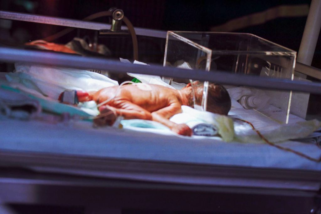
Preterm infants born before 32 weeks who received more skin-to-skin contact while in the hospital showed stronger brain development in areas tied to emotion and stress regulation than babies who received less skin-to-skin care, according to a study published in Neurology®, the medical journal of the American Academy of Neurology. The study can only show an association and cannot establish causation.

“Skin-to-skin contact in preterm infants has been shown to have many benefits, with previous studies linking it to improved bonding, sleep, heart and lung function and growth, as well as reduced pain and stress,” said study author Katherine E. Travis, PhD, of Burke Neurological Institute in White Plains, New York. “Our findings in infants born very preterm suggest skin-to-skin care may also play a role in shaping early brain development, highlighting the potential importance of caregiving experiences during the earliest weeks of a preemie’s life.”

he study included 88 preterm infants with an average gestational age of 29 weeks who weighed an average of 2.65 pounds. The average stay in the hospital was two months. The goal was to find out whether skin-to-skin holding, also called kangaroo care, was linked to brain development in areas that help regulate emotions and stress. Researchers tracked skin-to-skin care with family members throughout each infant’s hospitalisation, including how long each session lasted and the total minutes per day. Families visited an average of once per day. When they provided skin-to-skin care, the average session was around 70 minutes with 73% of sessions provided by mothers. For the entire hospital stay, the average amount of skin-to-skin care per day was 24 minutes.

Each infant received a brain scan before going home from the hospital – around the time they would have reached full-term age of around 40 weeks. The brain scans measured how water moves through brain tissue. This movement helps reveal how white matter – the brain’s communication network – is developing. Researchers then compared the markers of white matter with the amount of time the preemies received skin-to-skin care per session and per day.

For skin-to-skin duration per session, researchers found longer sessions were linked to higher mean diffusivity – how freely water moves through the brain – in two key brain regions: the cingulum, which supports attention and emotion regulation; and the anterior thalamic radiations, which connects areas involved in emotional processing and memory.

Longer sessions were also linked to lower fractional anisotropy – how water movement is influenced by developing cellular tissues – in the anterior thalamic radiations. For daily total minutes of skin-to-skin care, researchers found higher amounts were linked to higher mean diffusivity in the anterior thalamic radiations. They were also linked to lower fractional anisotropy in the anterior thalamic radiations. These associations remained significant even after researchers accounted for factors that could influence brain development, including gestational age at birth, age at time of scan, socioeconomic status and how often family visited.

“Our findings add to growing evidence that white matter development is sensitive to a preterm infant’s experience while in the hospital,” said Travis. “Skin-to-skin care not only provides preterm infants with family connections through bonding, it may also be encouraging new connections within the brain itself, improving a baby’s brain health overall.”

A limitation of the study is that it was conducted at a single hospital and researchers reviewed existing medical records. The authors note that future research should explore how early caregiving experiences – like skin-to-skin care – might shape brain development and support later behavioural outcomes as preterm infants grow.

Source: American Academy of Neurology

Could a New Way to Restore Lithium Deficiency in Alzheimer’s Really Work?

Neurons in the brain of an Alzheimer’s patient, with plaques caused by tau proteins. Credit: NIH

It has been known that brain lithium (Li) levels are depleted in individuals with mild cognitive impairment, a precursor for Alzheimer’s disease. For years, there have been attempts to restore Li levels to prevent Alzheimer’s disease by administering lithium carbonate. But now, it has been shown that the Li from this compound has been sequestered and not actually restoring the endogenous Li levels. Now, scientists have tried using lithium oxide (LiO) salts instead – and the treatment appears to be effective in prevention and even reversal of a mouse model of Alzheimer’s.

Join our QuickNews podcast as the arguments for and against this lithium-based approach are unpacked and debated.

Poor Sleep May Accelerate Brain Ageing

Poor sleep may accelerate brain ageing, a new study shows. Photo by Andrea Piacquadio

People who sleep poorly are more likely than others to have brains that appear older than they actually are. This is according to a comprehensive brain imaging study from Karolinska Institutet, published in the journal eBioMedicine. Increased inflammation in the body may partly explain the association.

Poor sleep has been linked to dementia, but it is unclear whether unhealthy sleep habits contribute to the development of dementia or whether they are rather early symptoms of the disease. In a new study, researchers at Karolinska Institutet have investigated the link between sleep characteristics and how old the brain appears in relation to its chronological age. 

The study includes 27 500 middle-aged and older people from the UK Biobank who underwent magnetic resonance imaging (MRI) of the brain. Using machine learning, the researchers estimated the biological age of the brain based on over a thousand brain MRI phenotypes. 

Low-grade inflammation 

The participants’ sleep quality was scored based on five self-reported factors: chronotype (being a morning/evening person), sleep duration, insomnia, snoring, and daytime sleepiness. They were then divided into three groups: healthy (≥ 4 points), intermediate (2-3 points), or poor (≤ 1 point) sleep. 

“The gap between brain age and chronological age widened by about six months for every 1-point decrease in healthy sleep score,” explains Abigail Dove, researcher at the Department of Neurobiology, Care Sciences and Society, Karolinska Institutet, who led the study. “People with poor sleep had brains that appeared on average one year older than their actual age.” 

To understand how poor sleep can affect the brain, the researchers also examined levels of low-grade inflammation in the body. They found that inflammation could explain just over ten per cent of the link between poor sleep and older brain age. 

“Our findings provide evidence that poor sleep may contribute to accelerated brain ageing and point to inflammation as one of the underlying mechanisms,” says Abigail Dove. “Since sleep is modifiable, it may be possible to prevent accelerated brain ageing and perhaps even cognitive decline through healthier sleep.” 

Several possible explanations 

Other possible mechanisms that could explain the association are negative effects on the brain’s waste clearance system, which is active mainly during sleep, or that poor sleep affects cardiovascular health, which in turn can have a negative impact on the brain. 

Participants in the UK Biobank are healthier than the general UK population, which could limit the generalisability of the findings. Another limitation of the study is that the results are based on self-reported sleep. 

The study was conducted in collaboration with researchers from the Swedish School of Sport and Health Sciences, and Tianjin Medical University and Sichuan University in China, among others. It was funded by the Alzheimer’s Foundation, the Dementia Foundation, the Swedish Research Council, the Loo and Hans Osterman Foundation for Medical Research, and the Knowledge Foundation. The researchers report no conflicts of interest. 

Source: Karolinska Institutet

No ‘Beneficial’ Level of Alcohol Consumption for Dementia Risk

Photo by Apolo Photographer on Unsplash

Any amount of alcohol consumption may increase risk of dementia, according to the most comprehensive study of alcohol consumption and dementia risk to date.

Led by the University of Oxford, Yale University, and the University of Cambridge, the research challenges previous suggestions that light-to-moderate drinking may have a protective effect against dementia. The study is published in BMJ Evidence-Based Medicine.

Alcohol consumption is widespread and is linked with an increased risk of many diseases. Heavy drinking has previously been linked to higher risk of dementia. The connection between moderate levels of drinking and higher risk of dementia was uncertain with some studies suggesting that moderate drinking may even reduce dementia risk. However, recent studies involving brain scans have shown that drinking alcohol even at low levels may increase the risk of dementia.

This study combined observational data from more than half a million participants in two large and diverse population studies: the US Million Veteran Program and UK Biobank to assess whether self-reported alcohol use was linked with risk of developing a broad range of types of dementia.

The researchers also investigated links between genetically-predicted likelihood of drinking alcohol and alcohol use disorder for more than 2.4 million participants in 45 individual studies. This approach helped the researchers overcome some of the difficulties in distinguishing correlation from causation.

Key findings:

  • Observational analyses seemed to support previous findings that current low and moderate drinking is associated with lower risk of dementia when compared with non-drinking and heavy drinking; however, some current non-drinkers were previously heavy drinkers, which could account for their increased dementia risk compared to consistently low drinkers;
  • Genetic analyses, however, revealed a continuously increasing trend of higher dementia risk with greater alcohol intakes, suggesting that any level of alcohol consumption increases the risk of dementia, with no evidence that drinking alcohol may have a protective effect;
  • A doubled increase in a person’s genetically-predicted risk of alcohol use disorder was associated with a 16% higher risk of dementia, while a three times higher increase in number of alcoholic drinks per week increased the risk of dementia risk by 15%;
  • The study also showed that people who later developed dementia reduced their alcohol intake before diagnosis, another explanation for prior findings of protective effects of alcohol, rather than true benefit.

Dr Anya Topiwala, Senior Clinical Researcher at Oxford Population Health, Consultant Psychiatrist, and lead author of the study, said ‘Our findings challenge the common belief that low levels of alcohol are beneficial for brain health. Genetic evidence offers no support for a protective effect – in fact, it suggests the opposite. Even light or moderate drinking may increase the risk of dementia, indicating that reducing alcohol consumption across the population could play a significant role in dementia prevention.’

Dr Stephen Burgess, Statistician at the University of Cambridge, said ‘The random nature of genetic inheritance allows us to compare groups with higher and lower levels of alcohol drinking in a way that allows us to make conclusions that untangle the confusion between correlation and causation. Our findings do not only hold for those who have a particular genetic predisposition, but for anyone who chooses to drink, our study suggests that greater alcohol consumption leads to higher risk of dementia.’

Dr Joel Gelernter, Professor at Yale University and senior author of the study, said ‘These results, which add to our understanding of the relationship between alcohol and dementia, have clinical implications – there was a time when medical knowledge seemed to support that light drinking would be beneficial to brain health, and this work adds to the evidence that this is not correct’.

This study adds to growing evidence that alcohol use, even at moderate levels, may have no safe threshold when it comes to brain health, reinforcing the case for preventive strategies that reduce alcohol consumption in the general population.

The study, ‘Alcohol use and risk of dementia in diverse populations: evidence from cohort, case–control and Mendelian randomisation approaches‘, is published in BMJ Evidence-Based Medicine.

Source: Oxford University

Psychedelics Alter Far More Neurons than Expected

The discovery challenges decades of assumptions and points to new hope for patients with depression, Alzheimer’s, and beyond

The most basic assumption about how psychedelic medicine works is at least partially flawed: Psychedelics are altering not just a few specific brain cells, but the vast majority of them, according to a new University of Michigan study.

The research, published in Molecular Psychiatry, shows that even neurons without serotonin 2A receptors – which are important for physiological processes, including mood regulation, perception and cognitive functions – can dramatically benefit from psychedelic compounds. This means that the therapeutic use of psychedelic medicine is far broader than currently appreciated, with important implications for Alzheimer’s disease and PTSD.

“We identified brain regions where most neurons are completely lacking serotonin 2A receptors. Surprisingly, psychedelic treatment was still able to strongly boost connectivity onto these neurons,” said the study’s senior author Omar Ahmed, U-M professor of psychology whose lab studies behavioural neural circuits and attempts to repair them when they go awry in specific disorders.

Psychedelic medicine is being successfully used in clinical trials to treat major depression. For decades it has been presumed that psychedelics work therapeutically by targeting the serotonin 2A receptor found on neurons in the frontal cortex and boosting connections onto those neurons. It has been assumed that frontal neurons with this serotonin 2A receptor were the only neurons benefiting from psychedelic therapy. This is why psychedelic medicine has focused on treating conditions relating to frontal dysfunction, such as major depression, Ahmed said.

When the research team studied the genes expressed in neurons of the entire cortex of the brain, they identified brain regions that did not express the serotonin 2A receptor that is supposed to be needed for psychedelic therapy to work. Ahmed’s lab, including co-first authors Tyler Ekins and Chloe Rybicki-Kler, showed that the retrosplenial cortex, a brain region important for memory, orientation and even imagining oneself in the future, was remarkably devoid of these receptors. The retrosplenial cortex is one of the first brain regions to be impaired in Alzheimer’s disease.

The team then recorded from these neurons lacking serotonin 2A receptors and found that they also show robust neuroplasticity (more synapses) after psychedelic treatment.

“This was a very unexpected finding given the current assumptions about how psychedelic medicine works,” Ahmed said.

The next step used a genetic engineering technique called CRISPR-Cas to reveal the rules that govern this surprising boost in brain connectivity, leading to a revised theory of how psychedelics control the brain’s ability to adapt and change. These new rules do not require neurons to have serotonin 2A receptors themselves to receive a synaptic boost from psychedelics, dramatically increasing the number of brain connections that can be potentially repaired by psychedelic medicine.

“The most successful medicines are those where we fully understand how they work. That is why it is so important to understand the fundamentals of how psychedelic medicine actually works,” Ahmed said.

The new findings are cause for both caution and optimism, he said. Caution, because they show that we need to be wary of psychedelics acting on unintended neurons. Optimism, because they open up the possibility of using psychedelic-like compounds to restore brain connections in Alzheimer’s disease and other disorders involving the retrosplenial cortex, such as PTSD.

“We are actively working on essential preclinical research to test this hypothesis related to Alzheimer’s disease,” Ahmed said.

Source: University of Michigan

Combination of Diet and Medication Reprograms Paediatric Neuroblastoma

Credit: National Cancer Institute

Researchers at Children’s Hospital of Philadelphia (CHOP) found that combining a specialised diet with an approved medication interrupts the growth of high-risk neuroblastoma, a deadly paediatric cancer, by reprogramming tumour behaviour. The findings were published in the journal Nature.

Neuroblastoma originates from primitive cells meant to form nerve tissues but that remain “undifferentiated,” indicating cancer cells that haven’t specialized, often suggesting a more aggressive and unfavourable prognosis. These tumours rely on a steady supply of chemicals called polyamines that are essential for rapid cell growth and tumour progression. A medicine called difluoromethylornithine (DFMO) was approved by the Food and Drug Administration (FDA) to treat children with high-risk neuroblastoma, as DFMO blocks polyamine production. However, researchers sought to improve the effectiveness of the drug by using it at high doses and combining it with a diet that is depleted of the nutrients used by the body to make polyamines (arginine). This two-step approach was anticipated to lower polyamines substantially more than low dose DFMO alone.

“Our findings show that this treatment reduced polyamines in tumours to roughly 10% of their usual levels. This reduction greatly slowed tumour growth, and in many cases, completely eliminated the tumours,” said Michael D. Hogarty, MD, a lead author and an Attending Physician in the Division of Oncology at Children’s Hospital of Philadelphia. “Notably, the treatment altered the way the tumour cells make proteins, making it harder for them to grow and easier for them to mature, or differentiate.”

Hogarty and his team used a preclinical model to mimic MYCN-driven neuroblastoma, directly addressing the strong association between extra MYCN gene copies and aggressive neuroblastoma with poor prognosis. Animal models with tumours were divided into groups: one fed a normal diet and the other lacking amino acids for polyamine production. Each group either received DFMO in their drinking water or did not. The special diet or DFMO alone partially lowered polyamines and extended survival, but the combination had the most significant impact on tumours due to the profound polyamine depletion it caused.

The researchers plan to conduct additional preclinical studies, followed hopefully by clinical trials in children to determine the safety and efficacy of targeting this specific metabolic dependency of neuroblastoma cells. By complementing existing treatments, they hope to substantially improve patient outcomes, and because the therapy targets polyamines it may be effective in many other types of cancer that have frequent MYC gene activation. 

Source: Children’s Hospital of Philadelphia

Most Epilepsy Patients Wait a Year After Starting Treatment for Seizure Relief

Source: Pixabay

Antiseizure medications help the majority of people with focal epilepsy, a common form of the neurological disorder. Yet most will still have episodes for at least a year after their treatment begins, until their doctors can find the right drug and dosage for them, a new study shows.

Accounting for about 60% of people with epilepsy, focal epilepsy occurs when nerve cells in a certain brain region send out a sudden, excessive burst of electrical signals. This uncontrolled activity, which is called a focal seizure, can cause problems such as abnormal emotions or feelings and unusual behaviours. Much attention has been paid to the minority of patients who do not respond well to available treatments, but the current study looks at another group: those who may not respond to the first medication or regimen prescribed but who might respond to another tried later.

Led by researchers at NYU Langone Health as part of the international Human Epilepsy Project, the study is among the first in a decade to focus on those whose seizures ultimately can be prevented or controlled with drugs. Results for nearly 450 men, women, and teens newly diagnosed with the disorder revealed that although more than half eventually received a medication or regimen that worked for them, major improvements were not achieved until an average of 12 months. Many needed even longer to find relief.

“Our findings suggest that those with focal epilepsy should expect a long adjustment period as their healthcare provider determines the best treatment regimen for them,” said study senior author and neurologist Jacqueline A. French, MD.

A possible explanation for this delay is that physicians are not selecting the ideal antiseizure therapy on their first try, adds Dr French, a professor in the Department of Neurology at the NYU Grossman School of Medicine and co-principal investigator of the Human Epilepsy Project.

Neurologists commonly start patients on levetiracetam, a drug that can target many types of seizures and has few interactions with other medications. Based on the new results, however, they may want to rethink this approach, says Dr French, noting that although 57% of the study participants were initially prescribed levetiracetam, only a quarter became seizure free on their first try.

A report on the findings appears in JAMA Neurology.

Thirty-four epilepsy centres in the United States, Europe, and Australia were involved in the study, which took place from 2012 to 2019. The team collected data about the patients’ medical histories, demographic factors, and the details of their epilepsy diagnoses. All were provided annual follow-ups for either three or six years.

During this time, participants tracked their seizure frequency in an electronic diary, describing each day as either “seizure-free” or “had a seizure.” The time, duration, and type of episode, along with other notes, were also recorded. The study volunteers also reported information about their antiseizure medications, noting the type, dose, and reasons for discontinuing a regimen.

Patients were considered seizure-free if they did not have an episode for at least a year (or longer if their seizures were infrequent).

The study further showed that together, 63% of all participants experienced ongoing or even worsening seizures during the first year of therapy, whether or not they would eventually find relief.

Notably, those who had seizures only a few times per year prior to treatment were more likely to respond to medication than those who had them weekly. In addition, participants with a history of psychological disorders such as anxiety and depression were almost twice as likely to resist the drugs than those without such a history.

“Our results show that the best way to a new treatment plan is sometimes through making better use of the tools we already have instead of always searching for the next breakthrough drug,” said Dr French, who is also a member of NYU Langone’s Comprehensive Epilepsy Center.

The researchers next plan to more closely examine those who did not become seizure-free during the study period, says Dr French.

Dr French cautions that the investigation did not directly assess the role of regimen choice, dose, or side effects on the way patients responded to treatment, and it did not exclude participants who failed to adhere to their prescribed regimen.

Source: NYU Langone Health

Scientists Repair Stroke Damage in Mice Using Stem Cells

This image shows a coronal section through the mouse brain after stroke and neural stem cell transplantation. The dashed circle indicates the stroke area. The neurite projections of the transplanted human cells are stained in dark brown. Neurites extend locally into the cortex (CX) but also via the corpus callosum (CC) into the other brain hemisphere. (Image: UZH)

One in four adults suffer a stroke in their lifetime, leaving around half of them with residual damage such as paralysis or speech impairment because internal bleeding or a lack of oxygen supply kill brain cells irreversibly. No therapies currently exist to repair this kind of damage. “That’s why it is essential to pursue new therapeutic approaches to potential brain regeneration after diseases or accidents,” says Christian Tackenberg, the Scientific Head of Division in the Neurodegeneration Group at the University of Zurich (UZH) Institute for Regenerative Medicine.

Neural stem cells have the potential to regenerate brain tissue, as a team led by Tackenberg and postdoctoral researcher Rebecca Weber has now compellingly shown in two studies that were conducted in collaboration with a group headed by Ruslan Rust from the University of Southern California. “Our findings show that neural stem cells not only form new neurons, but also induce other regeneration processes,” Tackenberg says.

The first study is published in Nature Communications, the second in Science Advances.

New neurons from stem cells

The studies employed human neural stem cells, from which different cell types of the nervous system can form. The stem cells were derived from induced pluripotent stem cells, which in turn can be manufactured from normal human somatic cells. For their investigation, the researchers induced a permanent stroke in mice, the characteristics of which closely resemble manifestation of stroke in humans. The animals were genetically modified so that they would not reject the human stem cells.

One week after stroke induction, the research team transplanted neural stem cells into the injured brain region and observed subsequent developments using a variety of imaging and biochemical methods. “We found that the stem cells survived for the full analysis period of five weeks and that most of them transformed into neurons, which actually even communicated with the already existing brain cells,” Tackenberg says.

Brain regenerates itself

The researchers also found other markers of regeneration: new formation of blood vessels, an attenuation of inflammatory response processes and improved blood-brain barrier integrity. “Our analysis goes far beyond the scope of other studies, which focused on the immediate effects right after transplantation,” Tackenberg explains. Fortunately, stem cell transplantation in mice also reversed motor impairments caused by stroke. Proof of that was delivered in part by an AI-assisted mouse gait analysis.

Clinical application moving closer to reality

Human neural stem cells in culture. Cell nuclei are stained in blue, the neural stem cell-specific filament protein Nestin is shown in green, and the neural stem cell transcription factor Sox1 in red. (Image: UZH)

When he was designing the studies, Tackenberg already had his sights set on clinical applications in humans. That’s why, for example, the stem cells were manufactured without the use of reagents derived from animals. The Zurich-based research team developed a defined protocol for that purpose in collaboration with the Center for iPS Cell Research and Application (CiRA) at Kyoto University. This is important for potential therapeutic applications in humans. Another new insight discovered was that stem cell transplantation works better when it is performed not immediately after a stroke but a week later, as the second study verified. In the clinical setting, that time window could greatly facilitate therapy preparation and implementation.

Despite the encouraging results of the studies, Tackenberg warns that there is still work to be done. “We need to minimize risks and simplify a potential application in humans,” he says. Tackenberg’s group, again in collaboration with Ruslan Rust, is currently working on a kind of safety switch system that prevents uncontrolled growth of stem cells in the brain. Delivery of stem cells through endovascular injection, which would be much more practicable than a brain graft, is also under development. Initial clinical trials using induced stem cells to treat Parkinson’s disease in humans are already underway in Japan, Tackenberg reports. “Stroke could be one of the next diseases for which a clinical trial becomes possible.”

Source: University of Zurich

Landmark Study Finds Perispinal Etanercept of No Benefit to Stroke Trial Participants

A treatment for stroke patients was no more effective than an inactive drug

Source: CC0

The first international trial of an unproven stroke treatment available in the US has concluded that, while harmless, perispinal etanercept is no more effective than an inactive dummy drug, or placebo.

Survivors of stroke have travelled at considerable expense to private clinics in the US to be treated with the arthritis drug etanercept.

In the clinics, the drug is injected into the cervical spinal area, and the patient is then tilted head-down in the belief that this allows the drug to enter the brain.

Stroke is a leading cause of disability throughout the world, affecting more than 7 million people a year. Despite advances, treatments for impairment after stroke remain limited. Some patients call perispinal etanercept a “miracle cure”.

Florey leading stroke researcher, neurologist Professor Vincent Thijs led the Perispinal Etanercept to improve STroke Outcomes – or “PESTO” – trial to investigate this further, supported by funding from the Australian Government.

“We understand why people living with the long-term effects of stroke seek hope and new options,” Professor Thijs said. “With support from the Stroke Foundation and the Medical Research Future Fund, we put this treatment to the test using the gold standard of clinical research – a double-blind randomised trial.”

Half of the PESTO participants were treated with the drug, and half were treated with an inactive dummy drug, with patients and doctors “blind” to who was getting which.

This type of trial eliminates biases because neither doctors nor patients knew who was getting etanercept and who was getting the placebo. Because the results for the 2 patient groups were so similar, we concluded that while the drug did not cause harm, we found no evidence that it led to improved quality of life compared to placebo.

Professor Thijs, who leads the Young Stroke Service at The Florey, said improvements could be due to the placebo effect, a well-established medical phenomenon where some patients in a trial may notice an improvement, despite only receiving a dummy treatment.

Key PESTO trial results, published in Neurology:

  • 126 people from Australia and New Zealand participated in PESTO.
  • 63 received the treatment, 63 the placebo.
  • Their stroke symptoms were measured before the trial and 28 days after.
  • There were no adverse side effects.
  • Among participants who received perispinal etanercept, 52 per cent (33 out of 63) felt better.
  • Among participants who received the placebo, 57 per cent (36 out of 63) felt better.
  • The difference in results between the 2 groups is deemed statistically insignificant.

“It’s important for doctors and the stroke survivor community in Australia and around the world to know that we found no evidence that perispinal etanercept improved quality of life,” Professor Thijs said.

Kelvin Hill, Executive Director of Stroke Programs, Research and Innovation at Stroke Foundation said: “Every Australian stroke patient should have access to the best, evidence-based treatment. Findings of the PESTO study underscore the critical importance of robust research and clinical trials in discovering if treatments work or not.

“Australians experience around 46 000 stroke events every year (one every 11 minutes), and there are now over 440 000 survivors of stroke living in Australia. Stroke Foundation will continue to advocate for more research funding to unlock new effective treatments for stroke; and ensure that advice provided in the Living Clinical Guidelines for Stroke Management enables clinicians to provide the best stroke care possible,” Mr Hill added.

Source: Florey Institute of Neuroscience and Mental Health

Oxytocin Shines a Light into Parental Attachment and Sex Differences

The developing brain of a two-week-old mouse pup under the microscope. The oxytocin system appears in green, the light-sensitive protein in red and cells that carry both show up in yellow. Cell nuclei are in blue. Credit: Weizmann Institute of Science

According to attachment theory, the attachment between an infant and a primary caregiver shapes the baby’s future social ties. Yet little is known about the biological mechanisms underlying childhood attachment, mainly because it is so difficult to study the young brain in natural conditions.

Now, scientists in Prof Ofer Yizhar’s laboratory at the Weizmann Institute of Science have developed a new, noninvasive research method that makes it possible to silence selected nerve cells deep within the brains of mouse pups without disrupting their natural behaviour. Using this method, the researchers investigated the role of oxytocin, a short protein released from nerve cells in the brain. While most oxytocin research has focused on adults, the new findings, published in Science, show that oxytocin also shapes the social behaviour of pups and may underlie emotional differences between males and females that emerge early in life.

Oxytocin, sometimes referred to as the “love hormone,” was once thought to simply promote sociability in adults. Over time, however, it became clear that its role is far more complex: In some circumstances, it intensifies behaviors and emotions far removed from love, such as anxiety or aggression. Recent research has also shown that young mammalian brains – including those of human children – are especially sensitive to oxytocin. In brain regions responsible for sensory processing, emotional regulation and social behavior, the number of oxytocin receptors peaks during early childhood: around ages two to three in humans, and two to three weeks in mice. Some studies have even linked oxytocin deficiency to childhood autism. Still, without sufficiently precise tools to examine neural activity deep within the developing brain, many aspects of the role of oxytocin in early life have remained a mystery.

“The findings may offer a clue as to why males and females diverge in their social behaviors and emotional worlds long before puberty”

To shed light on the subject, a team led by Dr Daniel Zelmanoff, a physician-scientist in Yizhar’s lab, developed a noninvasive technique to probe specific nerve cells in the young brain. The group, pioneers in the field of optogenetics – a technology that uses light to switch individual cells on or off – devised a method in which the targeted brain cells of mouse pups are infected with an engineered virus. This otherwise harmless virus introduces a foreign gene of mosquito origin that encodes a light-sensitive protein; when exposed to light, the protein “turns off” the nerve cell. In fact, the protein is so light-sensitive that the researchers could silence selected nerve cells deep inside the brain simply by shining red light on the pups’ heads.

“This new method allows us to peek inside the brain without disturbing the pups’ everyday lives, making it a powerful tool for studying nervous system development,” Yizhar explains. “It is especially useful for studying oxytocin because this hormone’s effects depend on social context – and our method lets us switch off the oxytocin system on demand, only during the exact situation we want to study.”

The researchers focused on oxytocin’s role during the temporary separation of a mouse pup from its mother and their reunion a few hours later – a situation familiar to every parent of a young child. The scientists observed increased oxytocin activity in the pup’s brain during separation, which returned to normal after reunion with the mother. Pups with an active oxytocin system during the separation gradually adapted to being alone in an unfamiliar environment, producing fewer ultrasonic vocalizations – the mouse equivalent of a baby’s cry. In contrast, pups whose oxytocin system was silenced did not adapt; they continued emitting distress calls at the same rate until reunited with their mothers. These findings show that the so-called “love hormone” also plays a critical role in coping with loneliness.

Attachment theory holds that children who are securely attached to their parents show distress when separated from them but are able to calm down over time, feeling free to explore their surroundings. “We discovered that mouse pups need an active oxytocin system in order to adapt to separation from their mothers,” says Yizhar. “This suggests that the oxytocin system plays a role not only in the brain of the parent, which was already known, but also in that of the infant. In addition, since oxytocin receptors are present in the sensory processing centers of the young brain, we hypothesize that this hormone also helps sharpen a pup’s senses when it is alone.”

Children do not quickly forget the experience of being separated from their parents, and this separation shapes how they behave when reunited. For example, a securely attached child separated from a parent for a few hours will seek contact upon reunion, and is quickly calmed. The researchers found that activation of the oxytocin system in mouse pups during separation not only strengthened them in the moment but also determined how they behaved when their mothers returned. These pups emitted more ultrasonic calls than usual, and the frequency of the calls grew as they got closer to their mothers. Using artificial intelligence, the team identified a distinct vocal pattern: Before attaching to the mother’s nipple, the pups made high-pitched, frequent calls; afterwards, their calls dropped in pitch and slowed in tempo.

“Activating the oxytocin system during separation increases the pup’s motivation to regain closeness to the mother when reunited,” Yizhar explains. “This is reflected in the heightened rate and unique pattern of their calls. We now understand that these ultrasonic vocalizations are much more than just crying: The high-pitched, rapid calls appear to signal a request for closeness, while the lower-pitched, slower-paced calls likely express a quick return to calm and a wish to remain attached. Of course, more research is needed to pin down the exact meaning of each vocalization type.”

In the next stage, the researchers explored whether oxytocin’s role in pups differs between the sexes, as it does in older animals. They found that female pups with an active oxytocin system emitted many more ultrasonic calls when reunited with their mothers than females with silenced oxytocin systems, whereas the calls of male pups were unaffected by the status of their oxytocin systems. “This is the first sex difference observed in oxytocin system activity at such an early stage of development,” Yizhar notes. “It may offer a clue as to why males and females diverge in their social behaviours and emotional worlds long before puberty.”

“Most known functions of oxytocin are shared by all mammals,” Yizhar concludes. “Still, future studies must check whether the hormone affects the development of social behaviour, emotional maturity and maternal attachment in the brains of children. If so, this could help us better understand what can go wrong in emotional and social development – as in autism spectrum disorder, for example – and how to intervene at an early stage.”

Source: Weizmann Institute of Science