Category: Neurology

Students Lead Breakthrough Study on Diabetes Drugs and Dementia Risk

Source: Pixabay CC0

Two undergraduate medicine students at University of Galway have led a major study examining how cardioprotective glucose-lowering therapies affect the risk of developing dementia.

The research has been published in JAMA Neurology.

The new study involved a systematic review and meta-analysis of 26 clinical trials involving more than 160 000 participants.

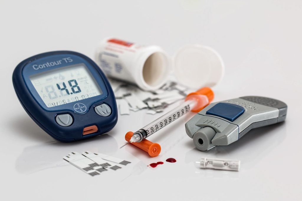
The researchers found that while most glucose-lowering therapies were not significantly associated with a reduction in dementia risk, one class of drugs – known as GLP-1 receptor agonists (GLP-1Ras) was linked to a significant reduction.

The study was conducted by medical students Allie Seminer and Alfredi Mulihano, alongside researchers from University of Galway, the HRB Clinical Research Facility Galway and University Hospital Galway.

Key Findings:

  • The research analysed data from 26 randomised controlled trials with a total of 164 531 participants.
  • While glucose-lowering therapies as a whole did not significantly reduce dementia risk, GLP-1 receptor agonists (GLP-1Ras) were linked to a 45% lower risk of dementia.
  • The findings provide crucial insights into the potential for diabetes medications to influence long-term brain health.

Dr Catriona Reddin, senior author, researcher at the University of Galway and Registrar in Geriatric Medicine at HSE West North West, said: “This research represents a significant contribution to our understanding of how some diabetes medications may impact brain health. Diabetes is a known risk factor for dementia, but whether glucose-lowering therapies can help prevent cognitive decline has remained unclear. Our findings suggest that GLP-1 receptor agonists, in particular, may have a protective effect on brain health.”

Professor Martin O’Donnell, Dean of the College of Medicine, Nursing and Health Sciences at University of Galway and Consultant Stroke Physician with HSE West North-West said: “Given the increasing prevalence of both diabetes and dementia, findings from this study have important public health implications for prevention of dementia.

“What makes this study particularly exciting for the College of Medicine, Nursing and Health Sciences at University of Galway, is that it was led by two of our undergraduate medicine students. We place a strong emphasis on research as a core component of our undergraduate medicine programme, ensuring that students have opportunities to engage in high-impact studies that shape global healthcare.”

Allie Seminer, a third year student from New York and co-lead author, said: “Being involved in a study of this scale as an undergraduate has been an incredible experience. What stood out for me was the sense of responsibility – knowing that our work could help shape understanding of a global health issue. It was incredibly motivating to be part of a team working at this level, and it has shown me how research is an essential part of becoming a well-rounded doctor. It highlights how research is not just an add-on to our degree but an essential part of how we learn to advance medical knowledge.”

Alfredi Mulihano, a third year student from Dundalk and co-lead author, said:  “Being part of this study has completely changed how I see my role as a future doctor. It brought together clinical insight, data analysis, and critical thinking in a way that lectures alone cannot. The experience opened my eyes to the impact we can have beyond the bedside – contributing to knowledge that could change how diseases like dementia are prevented.”

Source: University of Galway

Researchers Discover Natural Compound may Slow ALS and Dementia

Researchers from the University of Missouri have discovered that kaempferol, a natural antioxidant found in certain fruits and vegetables, such as kale, berries and endives, may support nerve cell health and holds promise as a potential treatment for ALS. Photo: Pixabay CC0

A natural compound found in everyday fruits and vegetables may hold the key to protecting nerve cells — and it’s showing promise as a potential treatment for ALS and dementia, according to new research from the University of Missouri.

“It’s exciting to discover a naturally occurring compound that may help people suffering from ALS or dementia,” Smita Saxena, a professor of physical medicine and rehabilitation at the School of Medicine and lead author of the study, said. “We found this compound had a strong impact in terms of maintaining motor and muscle function and reducing muscle atrophy.”

The study, which appears in Acta Neurologica, discovered that kaempferol, a natural antioxidant found in certain fruits and vegetables, such as kale, berries and endives, may support nerve cell health and holds promise as a potential treatment for ALS.

In lab-grown nerve cells from ALS patients, the compound helped the cells produce more energy and eased stress in the protein-processing center of the cell called the endoplasmic reticulum. Additionally, the compound improved overall cell function and slowed nerve cell damage. Researchers found that kaempferol worked by targeting a crucial pathway that helps control energy production and protein management — two functions that are disrupted in individuals with ALS.

“I believe this is one of the first compounds capable of targeting both the endoplasmic reticulum and mitochondria simultaneously,” Saxena said. “By interacting with both of these components within nerve cells, it has the potential to elicit a powerful neuroprotective effect.”

The challenge

The catch? The body doesn’t absorb kaempferol easily, and it could take a large amount to see real benefits in humans. For instance, an individual with ALS would need to consume at least 4.5kg of kale in a day to obtain a beneficial dose.

“Our bodies don’t absorb kaempferol very well from the vegetables we eat,” Saxena said. “Because of this, only a small amount reaches our tissues, limiting how effective it can be. We need to find ways to increase the dose of kaempferol or modify it so it’s absorbed into the bloodstream more easily.”

Another hurdle is getting the compound into the brain. The blood-brain barrier — a tightly locked layer of cells that blocks harmful substances — also makes it harder for larger molecules like kaempferol to pass through.

What’s next?

Despite its challenges, kaempferol remains a promising candidate for treating ALS, especially since it works even after symptoms start. It also shows potential for other neurodegenerative diseases including Alzheimer’s and Parkinson’s.

To make the compound easier for the body to absorb, Saxena’s team at the Roy Blunt NextGen Precision Health building is exploring ways to boost its uptake by neurons. One promising approach involves packaging lipid-based nanoparticles — tiny spherical particles made of fats that are commonly used in drug delivery.

“The idea is to encapsulate kaempferol within lipid-based nanoparticles that are easily absorbed by the neurons,” Saxena said.  “This would target kaempferol to neurons to greatly increase its beneficial effect.”

The team is currently generating the nanoparticles with hopes of testing them by the end of the year.

Source: University of Missouri-Columbia

Childhood Experiences Shape White Matter with Cognitive Effects Seen Years Later

Photo by Caleb Woods on Unsplash

Mass General Brigham investigators have linked difficult early life experiences with reduced quality and quantity of the white matter communication highways throughout the adolescent brain. This reduced connectivity is also associated with lower performance on cognitive tasks. However, certain social resiliency factors like neighbourhood cohesion and positive parenting may have a protective effect. Results are published in Proceedings of the National Academy of Sciences (PNAS).

White matter are the communication highways that allow the brain networks to carry out the necessary functions for cognition and behaviour. They develop over the course of childhood, and childhood experiences may drive individual differences in how white matter matures. Lead author Sofia Carozza, PhD, and senior author Amar Dhand, MD, PhD, of the Department of Neurology at Brigham and Women’s Hospital, a founding member of the Mass General Brigham healthcare system, wanted to understand what role this process plays in cognition once children reach adolescence.

“The aspects of white matter that show a relationship with our early life environment are much more pervasive throughout the brain than we’d thought. Instead of being just one or two tracts that are important for cognition, the whole brain is related to the adversities that someone might experience early in life,” said Carozza.

The team studied data from 9082 children (about half of them girls, with an average age of 9.5) collected in the Adolescent Brain Cognitive Development (ABCD) study. This study, funded by the National Institutes of Health and conducted at 21 centres across the U.S., gathered information on brain activity and structure, cognitive abilities, environment, mood and mental health. The researchers looked at several categories of early environmental factors, including prenatal risk factors, interpersonal adversity, household economic deprivation, neighbourhood adversity, and social resiliency factors.

Carozza and Dhand used diffusion imaging scanning of the brain to measure fractional anisotropy (FA)—a way of estimating the integrity of the white matter connections—and streamline count, an estimate of their strength. They then used a computational model to compare how these features of white matter were related to both childhood environmental factors and current cognitive abilities such as language skills and mental arithmetic.

Their analysis revealed widespread differences in white matter connections throughout the brain depending on the children’s early-life environments. In particular, the researchers found lower quality of white matter connections in parts of the brain tied to mental arithmetic and receptive language. These white matter differences accounted for some of the relationship between adverse life experiences in early childhood and lower cognitive performance in adolescence.

“We are all embedded in an environment, and features of that environment such as our relationships, home life, neighbourhood, or material circumstances can shape how our brains and bodies grow, which in turn affects what we can do with them,” said Carozza. “We should work to make sure that more people can have those stable, healthy home lives that the brain expects, especially in childhood.”

The researchers note that their study is based on observational data, which means they cannot draw strong causal conclusions. Brain imaging was also only available at a single timepoint, offering a snapshot but not allowing researchers to track changes over time. Prospective studies—following children over time and collecting brain imaging information at multiple time points—would be needed to more definitively connect adversity and cognitive performance.

Source: Mass General Brigham

Does the Brain Produce Oestrogen to Control Appetite?

Photo by Fakurian Design on Unsplash

Although a woman’s ovaries produce the most oestrogen, various types of oestrogen are also synthesised throughout different tissues in the body, including the brain’s neurons. New research in The FEBS Journal indicates that such neuro-oestrogens help suppress appetite.

Knowing that the enzyme aromatase is important for the production of oestrogens, investigators depleted or knocked out the gene encoding aromatase in mice, so that the animals were unable to synthesise oestrogens in a systemic or body-wide manner. These mice demonstrated increased food intake and body weight compared with their aromatase-expressing counterparts. Restoring aromatase expression specifically in the brain reduced food intake and increased sensitivity to leptin, the “fullness” hormone, confirming that neuro-oestrogens can influence appetite.

To further investigate the role of neuro-oestrogens independently of ovarian oestrogen involvement, the researchers removed the ovaries in female mice. The brain’s hypothalamus (the central hub for appetite signals) in ovariectomised mice showed increased expression of the gene encoding aromatase, and these mice decreased their food intake.

“Our results imply that neuro-oestrogens likely contribute to appetite regulation and may be relevant for body weight reduction” the authors wrote.

Source: Wiley

An Arthritis Drug Might Unlock Lasting Relief from Epilepsy and Seizures

Source: Pixabay

A drug typically prescribed for arthritis halts brain-damaging seizures in mice that have a condition like epilepsy, according to researchers at the University of Wisconsin–Madison. The drug, called tofacitinib, also restores short-term and working memory lost to epilepsy in the mice and reduces inflammation in the brain caused by the disease.

If the drug proves viable for human patients, it would be the first to provide lasting relief from seizures even after they stopped taking it.

“It ticks all the boxes of everything we’ve been looking for,” says Avtar Roopra, a neuroscience professor in the UW–Madison School of Medicine and Public Health and senior author of the study, which appears in Science Translational Medicine.

Epilepsy is one of the most common neurological diseases, afflicting more than 50 million people around the world. While there are many known causes, the disease often appears after an injury to the brain, like a physical impact or a stroke.

Some days, months or even years after the injury, the brain loses the ability to calm its own activity. Normally balanced electrical activity through the brain goes haywire.

“The system revs up until all the neurons are firing all the time, synchronously,” says Roopra. “That’s a seizure that can cause massive cell death.”

And the seizures repeat, often at random intervals, forever. Some drugs have been useful in addressing seizure symptoms, protecting patients from some of the rampant inflammation and memory loss, but one-third of epilepsy patients do not respond to any known drugs, according to Olivia Hoffman, lead author of the study and a postdoctoral researcher in Roopra’s lab. The only way to stop the most damaging seizures has been to remove a piece of the brain where disruptive activity starts.

On their way to identifying tofacitinib’s potential in epilepsy, Hoffman and co-authors used relatively new data science methods to sift through the way thousands of genes were expressed in millions of cells in the brains of mice with and without epilepsy. They found a protein called STAT3, key to a cell signaling pathway called JAK, at the centre of activity in the seizure-affected mouse brains.

“When we did a similar analysis of data from brain tissue removed from humans with epilepsy, we found that was also driven by STAT3,” Hoffman says.

Meanwhile, Hoffman had unearthed a study of tens of thousands of arthritis patients in Taiwan aimed at describing other diseases associated with arthritis. It turns out, epilepsy was much more common among those arthritis patients than people without arthritis — but surprisingly less common than normal for the arthritis patients who had been taking anti-inflammatory drugs for more than five-and-a-half years.

“If you’ve had rheumatoid arthritis for that long, your doctor has probably put you on what’s called a JAK-inhibitor, a drug that’s targeting this signaling pathway we’re thinking is really important in epilepsy,” Hoffman says.

The UW researchers ran a trial with their mice, dosing them with the JAK-inhibitor tofacitinib following the administration of a brain-damaging drug that puts them on the road to repeated seizures. Nothing happened. The mice still developed epilepsy like human patients.

Remember, though, that epilepsy doesn’t often present right after a brain-damaging event. It can take years. In the lab mice, there’s usually a lull of weeks of relatively normal time between the brain damage and what the researchers call “reignition” of seizures. If it’s not really epilepsy until reignition, what if they tried the drug then? They devised a 10-day course of tofacitinib to start when the mouse brains fell out of their lull and back into the chaos of seizures.

“Honestly, I didn’t think it was going to work,” Hoffman says. “But we believe that initial event sort of primes this pathway in the brain for trouble. And when we stepped in at that reignition point, the animals responded.”

The drug worked better than they could have imagined. After treatment, the mice stayed seizure-free for two months, according to the paper. Collaborators at Tufts University and Emory University tried the drug with their own mouse models of slightly different versions of epilepsy and got the same, seizure-free results.

Roopra’s lab has since followed mice that were seizure-free for four and five months. And their working memory returned.

“These animals are having many seizures a day. They cannot navigate mazes. Behaviourally, they are bereft. They can’t behave like normal mice, just like humans who have chronic epilepsy have deficits in learning and memory and problems with everyday tasks,” Roopra says. “We gave them that drug, and the seizures disappear. But their cognition also comes back online, which is astounding. The drug appears to be working on multiple brain systems simultaneously to bring everything under control, as compared to other drugs, which only try to force one component back into control.”

Because tofacitinib is already FDA-approved as safe for human use for arthritis, the path from animal studies to human trials may be shorter than it would be for a brand-new drug. The next steps toward human patients largely await NIH review of new studies, which have been paused indefinitely amid changes at the agency.

For now, the researchers are focused on trying to identify which types of brain cells are shifted back to healthy behavior by tofacitinib and on animal studies of even more of the many types of epilepsy. Hoffman and Roopra have also filed for a patent on the use of the drug in epilepsy.

Source: University of Wisconsin-Madison

Do Seizures in Newborns Increase Children’s Risk of Developing Epilepsy?

Photo by Lucy Wolski on Unsplash

Seizures in newborns are one of the most frequent acute neurological conditions among infants admitted to neonatal care units. A study published in Developmental Medicine & Child Neurology indicates that newborns experiencing such neonatal seizures face an elevated risk of developing epilepsy.

For the study, investigators analysed data on all children born in Denmark between 1997 and 2018, with the goal of comparing the risk of epilepsy in children with and without neonatal seizures.

Among 1,294,377 children, the researchers identified 1,998 who experienced neonatal seizures. The cumulative risk of epilepsy was 20.4% among children with neonatal seizures compared with 1.15% among children without. This indicates that 1 in 5 newborns with neonatal seizures will develop epilepsy.

Epilepsy was diagnosed before 1 year of age in 11.4% of children with neonatal seizures, in an additional 4.5% between 1 and 5 years, 3.1% between 5 and 10 years, and 1.4% between 10 and 22 years. Stroke, hemorrhage, or structural brain malformations in newborns, as well as low Apgar scores, were associated with the highest risks of developing epilepsy.

“Our study highlights that there are risk factors that may be used to identify infants for tailored follow-up and preventive measures,” said corresponding author Jeanette Tinggaard, MD, PhD, of Copenhagen University Hospital – Rigshospitalet. “Importantly, four out of five neonatal survivors with a history of neonatal seizures did not develop epilepsy, and we suggest future studies to explore a potential genetic predisposition.”

Source: Wiley

Bacteria Invade Brain after Implanting Medical Devices

Deep brain stimulation illustration. Credit: NIH

Brain implants hold immense promise for restoring function in patients with paralysis, epilepsy and other neurological disorders. But a team of researchers at Case Western Reserve University has discovered that bacteria can invade the brain after a medical device is implanted, contributing to inflammation and reducing the device’s long-term effectiveness. 

The groundbreaking research, recently published in Nature Communications, could improve the long-term success of brain implants now that a target has been identified to address.

“Understanding the role of bacteria in implant performance and brain health could revolutionize how these devices are designed and maintained,” said Jeff Capadona, Case Western Reserve’s vice provost for innovation, the Donnell Institute Professor of Biomedical Engineering and senior research career scientist at the Louis Stokes Cleveland VA Medical Center.

Capadona’s lab led the study, which examined the presence of bacterial DNA in the brains of mouse models implanted with microelectrodes.

To their surprise, researchers found bacteria linked to the gut inside the brain. The discovery suggests that a breach in what is known as “the blood-brain barrier,” caused by implanting the device, could allow microbes to enter.

“This is a paradigm-shifting finding,” said George Hoeferlin, the study’s lead author, who was a biomedical engineering graduate student at Case Western Reserve in Capadona’s lab. “For decades, the field has focused on the body’s immune response to these implants, but our research now shows that bacteria—some originating from the gut—are also playing a role in the inflammation surrounding these devices.”

In the study, mouse models treated with antibiotics had reduced bacterial contamination and the performance of the implanted devices improved—although prolonged antibiotic use proved detrimental.

The discovery’s implications go beyond device failure. Some of the bacteria found in the brain have been linked to neurological diseases, including Alzheimer’s, Parkinson’s and stroke.

“If we’re not identifying or addressing this consequence of implantation, we could be causing more harm than we’re fixing,” Capadona said. “This finding highlights the urgent need to develop a permanent strategy for preventing bacterial invasion from implanted devices, rather than just managing inflammation after the fact. The more we understand about this process, the better we can design implants that work safely and effectively.”

Capadona said his lab is now expanding the research to examine bacteria in other types of brain implants, such as ventricular shunts used to treat hydrocephalus, an abnormal buildup of fluid in the brain.

The team also examined the faecal matter of a human subject implanted with a brain device and found similar results.

“This finding stresses the importance of understanding how bacterial invasion may not just be a laboratory phenomenon, but a clinically relevant issue,” said Bolu Ajiboye, professor in biomedical engineering at the Case School of Engineering and School of Medicine and scientist at the Cleveland VA Medical Center. “Through our strong translational pipeline between CWRU and the VA, we are now investigating how this discovery can directly contribute to safer, more effective neural implant strategies for patients.”

Source: Case Western Reserve University

The Pupil as a Window into the Sleeping Brain

The eye of the sleeping subject was kept open with a special fixation device to record the pupil movements for several hours.  (Image: Neural Control of Movement Lab / ETH Zurich)

For the first time, researchers have been able to observe how the pupils react during sleep over a period of several hours. A look under the eyelids showed them that more happens in the brain during sleep than was previously assumed.

While eyes are typically closed in sleep, there is a flurry of activity taking place beneath the eyelids: a team of researchers, led by principal investigators Caroline Lustenberger, Sarah Meissner and Nicole Wenderoth from the Neural Control of Movement Lab at ETH Zurich, have observed that the size of the pupil fluctuates constantly during sleep. As they report in Nature Communications, sometimes it increases in size, sometimes it decreases; sometimes these changes occur within seconds, other times over the course of several minutes.

“These dynamics reflect the state of arousal, or the level of brain activation in regions that are responsible for sleep-wake regulation,” says Lustenberger. “These observations contradict the previous assumption that, essentially, the level of arousal during sleep is low.”

Instead, these fluctuations in pupil size show that even during sleep, the brain is constantly switching between a higher and lower level of activation. These new findings also confirm for humans what other research groups have recently discovered in studies on rodents, who also exhibit slow fluctuations in the activation level (known in the field as arousal).

New method for an old mystery

The regions of the brain which control the activation level are situated deep within the brainstem, making it previously difficult to directly measure these processes in humans during sleep. Existing methods are technically demanding and have not yet been established in this context. The ETH researchers’ study therefore relies on pupil measurements. Pupils are known to indicate the activation level when a person is awake. They can therefore be used as markers for the activity in regions situated deeper within the brain.

The ETH researchers developed a new method for examining the changes in people’s pupils while asleep: using a special adhesive technique and a transparent plaster, they were able to keep the eyes of the test subjects open for several hours.

“Our main concern was that the test subjects would be unable to sleep with their eyes open. But in a dark room, most people forget that their eyes are still open and they are able to sleep,” explains the study’s lead author, Manuel Carro Domínguez, who developed the technique.

Analysis of the data showed that pupil dynamics is related not just to the different stages of sleep, but also to specific patterns of brain activity, such as sleep spindles and pronounced deep sleep waves – brain waves that are important for memory consolidation and sleep stability. The researchers also discovered that the brain reacts to sounds with varying degrees of intensity, depending on the level of activation, which is reflected in the size of the pupil.

A central regulator of the activation level is a small region in the brainstem, known as the locus coeruleus. In animals, scientists have been able to show that this is important for the regulation of sleep stages and waking. The ETH researchers were unable to prove in this study whether the locus coeruleus is indeed directly responsible for pupil changes. “We are simply observing pupil changes that are related to the level of brain activation and heart activity,” Lustenberger explains.

In a follow-up study, the researchers will attempt to influence the activity of the locus coeruleus using medication, so that they can investigate how this affects pupil dynamics. They hope to discover whether this region of the brain is in fact responsible for controlling the pupils during sleep, and how changes in the level of activation affect sleep and its functions.

Using pupillary dynamics to diagnose illnesses

Understanding pupil dynamics during sleep could also provide important insights for the diagnosis and treatment of sleep disorders and other illnesses. The researchers therefore want to investigate whether pupil changes during sleep can provide indications of dysfunctions of the arousal system. These include disorders such as insomnia, post-traumatic stress disorder and possibly Alzheimer’s. “These are just hypotheses that we want to investigate in the future,” says Lustenberger.

Another goal is to make the technology usable outside of sleep laboratories, such as in hospitals where it could help to monitor waking in coma patients or to diagnose sleep disorders more accurately. The pupil as a window onto the brain could thus pave the way for new opportunities in sleep medicine and neuroscience.

Source: ETH Zurich

New Intervention Boosts Diagnosis and Care of Brain Infection

Source: CC0

University of Liverpool researchers have worked with global partners to identify and successfully implement an intervention package that has significantly improved the diagnosis and management of brain infections in hospitals across Brazil, India, and Malawi.

The study, published in The Lancet, was coordinated by researchers at the University of Liverpool in collaboration with international partners and implemented across 13 hospitals.

The intervention included:

• A clinical algorithm which offered a flowchart of guidance for clinicians on how to manage the first crucial hours and days of suspected brain infections, including which tests (blood tests, lumbar puncture, brain scans) and treatments to administer.
• A lumbar puncture pack, providing clinicians with sample containers, equipment, and guidance to ensure proper cerebrospinal fluid collection and testing, addressing challenges like knowing how much fluid to take and which tests to request.
• A panel of laboratory tests to enable correct and timely testing for a wide range of pathogens, addressing gaps in availability and sequencing of tests, with the main goal of identifying the cause of infection.
• Training for clinicians and lab staff to enhance their knowledge and skills in diagnosing and managing brain infections, including proper use of the new intervention tools.

These measures led to significant improvements in diagnosing patients with suspected acute brain infections, such as encephalitis and meningitis. Both conditions cause significant mortality and morbidity, especially in low- and middle-income countries (LMICs), where diagnosis and management are hindered by delayed lumbar punctures, limited testing, and resource constraints. Improved diagnosis and optimal management are a focus for the World Health Organization (WHO) in tackling meningitis and reducing the burden of encephalitis.

As a result of the intervention package, the proportion of patients receiving a syndromic diagnosis (confirming they had a brain infection) increased from 77% to 86%, while the microbiological diagnosis rate (identifying the exact pathogen) rose from 22% to 30%. In addition to improving diagnosis, the intervention enhanced the performance of lumbar punctures, optimised initial treatment, and improved patients’ functional recovery after illness.

Lead author Dr Bhagteshwar Singh, Clinical Research Fellow, Clinical Infection, Microbiology & Immunology said: “Following patients and their cerebrospinal fluid (CSF) samples through the hospital system, we tailored our intervention to address key gaps in care. The results speak for themselves: better diagnosis, better management, and ultimately, better outcomes for patients. Unlike most studies, we embedded improvements into routine care, so the impact continues well beyond the study.”

Corresponding author Professor Tom Solomon, Chair of Neurological Science at the University of Liverpool and Director of The Pandemic Institute, added: “We increased microbiological diagnoses by one-third across very diverse countries, which has profound implications for treatment and public health globally. As we scale this up in more hospitals and feed it into national and international policy, including WHO’s work on defeating meningitis and controlling encephalitis, the potential impact is enormous.”

The intervention was co-designed by clinicians, lab specialists, hospital administrators, researchers, and policymakers in each country, ensuring it was feasible and sustainable. Professor Priscilla Rupali, lead researcher from Christian Medical College, Vellore, India, also commented: “The co-design process ensured that the intervention would work within local healthcare settings and could be sustained beyond the study. We are already incorporating the findings into India’s national Brain Infection Guidelines, ensuring long-term benefits for patient care.”

The intervention package is freely available as a toolkit for adaptation in different settings: https://braininfectionsglobal.tghn.org/resources/brain-infections-global-tools/.

Source: University of Liverpool

Innovative In Vivo Imaging Offers New Treatment and Hope for Chronic TMJ Pain

Photo by engin akyurt on Unsplash

Facial pain and discomfort related to the temporomandibular joint (TMJ) is the second-leading musculoskeletal disorder, after chronic back pain, affecting 8% to 12% of Americans. Current treatments for TMJ disorders are not always sufficient, leading researchers to further explore the vast nerve and vessel network connected to this joint – the second largest in the human body.

In a study published in December 2024 in the journal Paina research team led by Yu Shin Kim, PhD, associate professor at the The University of Health Science Center at San Antonio (UT Health San Antonio), observed for the first time the simultaneous activity of more than 3000 trigeminal ganglion (TG) neurons, which are cells clustered at the base of the brain that transmit information about sensations to the face, mouth and head.

“With our novel imaging technique and tools, we can see each individual neuron’s activity, pattern and dynamics as well as 3000 neuronal populational ensemble, network pattern and activities in real time while we are giving different stimuli,” said Kim.

When the TMJ is injured or misaligned, it sends out signals to increase inflammation to protect the joint. However, this signaling can lead to long-term inflammation of the joint and other parts of the highly connected nerve network, leading to chronic pain and discomfort. About 80% to 90% of TMJ disorders occur in women, and most cases develop between the ages of 15–50.

Activation at the cellular level

Previous animal studies observed behavioural changes related to pain, but this study was the first to record reactions at the cellular level and their activities. To see which portions of the nerve pathway respond to various types of pain, Kim’s team created different models of pain and observed the neuronal activity with high-resolution confocal imaging, which uses a high-resolution camera and scanning system to observe neurons in action.

The team discovered that during TMJ activation, more than 100 neurons spontaneously fire at the same time. Activation was observed in localised areas of the TMJ innervated to TG neurons. The localisation of this activation highlights the specific neural pathways involved in TMJ pain, offering deeper insight into how pain develops and spreads to nearby areas. The study is also the first to quantify the degree of TG neuronal sensitivity and network activities.

Potential link to migraine, headaches

Chronic TMJ pain in humans is often linked to other pain comorbidity such as migraines and other headaches. Kim’s team observed this crossover in the in vivo model as inflammation of TG neurons spread to the nearby orofacial areas. Kim’s previous research demonstrated how stress-related migraine pain originates from a certain molecule, begins in the dura and innervates throughout the dura and TG neurons. This current study and novel imaging technique further reveals potential connections between the TMJ, migraines and other headaches.

Potential of CGRP treatment

Calcitonin gene-related peptides (CGRP), molecules involved in transmitting pain signals and regulating inflammation, are often found in higher amounts in synovial fluid of TMJ disorder patients. Synovial fluid surrounds joints in the body, helping to reduce friction between bones and cartilage. Higher amounts of CGRP are often associated with increased pain and inflammation. Kim hypothesised in this study that a reduction in CGRP may reduce TMJ disorder symptoms. He found that CGRP antagonist added to the synovial fluid relieved both TMJ pain and hypersensitivity of TG neurons.

Currently, there are no Federal Drug Administration-approved medications for TMJ disorders other than non-steroidal anti-inflammatory drugs (NSAIDS). While some CGRP antagonist medications are FDA-approved for treating migraines, this study suggests these drugs may also provide relief for TMJ disorders. Confirmation of the positive effect of the drug on TMJ pain is a major leap forward in understanding how CGRP affect TMJ pain, said Kim.

“This imaging technique and tool allows us to see pain at its source – down to the activity of individual neurons – offering unprecedented insights into how pain develops and spreads. Our hope is that this approach will not only advance treatments for TMJ disorders but also pave the way for understanding and managing various chronic pain conditions more effectively,” said Kim.

Source: University of Texas Health Science Center at San Antonio