Category: Neurodegenerative Diseases

Did the Ancient Greeks and Romans Suffer from Dementia?

Bust of Hippocrates. By ESM – Own work, CC BY-SA 4.0, https://commons.wikimedia.org/w/index.php?curid=90175388

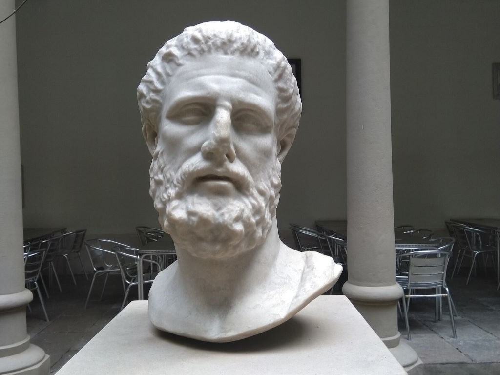
Age-related dementia is often assumed to having been with us all along, stretching back to the ancient world. But a new analysis of classical Greek and Roman medical texts suggests that it was extremely rare 2000 to 2500 years ago, in the time of Aristotle, Galen and Pliny the Elder.

The USC-led research, published in the Journal of Alzheimer’s Disease, bolsters the idea that Alzheimer’s disease and related dementias are diseases of modern environments and lifestyles, with sedentary behaviour and exposure to air pollution largely to blame.

“The ancient Greeks had very, very few – but we found them – mentions of something that would be like mild cognitive impairment,” said first author Caleb Finch, a University Professor at the USC Leonard Davis School of Gerontology.

“When we got to the Romans, and we uncovered at least four statements that suggest rare cases of advanced dementia – we can’t tell if it’s Alzheimer’s. So, there was a progression going from the ancient Greeks to the Romans.”

Ancient Greeks recognised that aging commonly brought memory issues that we would recognise as mild cognitive impairment, but nothing approaching a major loss of memory, speech and reasoning as caused by Alzheimer’s and other types of dementia.

Finch and co-author Stanley Burstein, a historian at California State University, Los Angeles, pored over a major body of ancient medical writing by Hippocrates and his followers.

The text catalogues ailments of the elderly such as deafness, dizziness and digestive disorders – but makes no mention of memory loss.

Centuries later in ancient Rome, a few mentions crop up. Galen remarks that at the age of 80, some elderly begin to have difficulty learning new things.

Pliny the Elder notes that the senator and famous orator Valerius Messalla Corvinus forgot his own name.

Cicero prudently observed that “elderly silliness … is characteristic of irresponsible old men, but not of all old men.”

Finch speculates that as Roman cities grew denser, pollution increased, driving up cases of cognitive decline.

In addition, Roman aristocrats used lead cooking vessels, lead water pipes and even added lead acetate into their wine to sweeten it – unwittingly poisoning themselves with the powerful neurotoxin.

(A few ancient writers recognised the toxicity of lead-containing material, but little progress was made in dealing with the problem until well into the 20th century. Some scholars blame lead poisoning for the fall of the Roman Empire.)

For this paper, Finch did not just think about the Roman Empire or the Greeks.

In the absence of demographic data for ancient Greece and Rome, Finch turned to a surprising model for ancient aging: today’s Tsimane Amerindians, an Indigenous people of the Bolivian Amazon.

The Tsimane, like the ancient Greeks and Romans, have a preindustrial lifestyle that is very physically active, and they have extremely low rates of dementia.

An international team of cognitive researchers led by Margaret Gatz, a professor of psychology, gerontology and preventive medicine at the USC Leonard Davis School, found among older Tsimane people, only about 1% suffer from dementia.

In contrast, 11% of people aged 65 and older living in the United States have dementia, according to the Alzheimer’s Association.

“The Tsimane data, which is quite deep, is very valuable,” Finch said.

“This is the best-documented large population of older people that have minimal dementia, all of which indicates that the environment is a huge determinant on dementia risk. They give us a template for asking these questions.”

Source: University of Southern California

Alzheimer’s Disease Cases Caused by Growth Hormone Treatment

Neurons in the brain of an Alzheimer’s patient, with plaques caused by tau proteins. Credit: NIH

Five cases of Alzheimer’s are believed to have arisen as a result of medical treatments decades earlier, according to a new paper published in Nature Medicine. Alzheimer’s disease is caused by the amyloid-beta protein, and is usually a sporadic condition of late adult life, or more rarely as an inherited condition from a faulty gene.

The study, by a team of UCL and UCLH researchers, provides the first evidence of Alzheimer’s disease in living people that appears to have been medically acquired and due to transmission of the amyloid-beta protein.

The people described in the paper had all been treated as children with a type of human growth hormone extracted from pituitary glands from deceased individuals (cadaver-derived human growth hormone or c-hGH). This was used to treat at least 1848 people in the UK between 1959 and 1985, and used for various causes of short stature.

It was withdrawn in 1985 after it was recognised that some c-hGH batches were contaminated with prions (infectious proteins) which had caused Creutzfeldt-Jakob disease (CJD) in some people.

c-hGH was then replaced with synthetic growth hormone that did not carry the risk of transmitting CJD.

These researchers previously reported that some patients with CJD due to c-hGH treatment (called iatrogenic CJD) also had prematurely developed deposits of the amyloid-beta protein in their brains.* The scientists went on to show in a 2018 paper that archived samples of c-hGH were contaminated with amyloid-beta protein and, despite having been stored for decades, transmitted amyloid-beta pathology to laboratory mice when it was injected.

They suggested that individuals exposed to contaminated c-hGH, who did not succumb to CJD and lived longer, might eventually develop Alzheimer’s disease.

This latest paper reports on eight people referred to UCLH’s National Prion Clinic at the National Hospital for Neurology and Neurosurgery in London, who had all been treated with c-hGH in childhood, often over several years.

Five of these people had symptoms of dementia, and either had already been diagnosed with Alzheimer’s disease or would otherwise meet the diagnostic criteria for this condition; another person met criteria for mild cognitive impairment. These people were between 38 and 55 years old when neurological symptoms started. Biomarker analyses supported the diagnoses of Alzheimer’s disease in two patients with the diagnosis, and was suggestive of Alzheimer’s in one other person; an autopsy analysis showed Alzheimer’s pathology in another patient.

The unusually young age at which these patients developed symptoms suggests they did not have the usual sporadic Alzheimer’s which is associated with old age. In the five patients in whom samples were available for genetic testing, the team ruled out inherited Alzheimer’s disease.

As c-hGH treatment is no longer used, there is no risk of any new transmission via this route. There have been no reported cases of Alzheimer’s acquired from any other medical or surgical procedures. There is no suggestion that amyloid-beta can be passed on in day-to-day life or during routine medical or social care.

However, the researchers caution that their findings highlight the importance of reviewing measures to ensure there is no risk of accidental transmission of amyloid-beta via other medical or surgical procedures which have been implicated in accidental transmission of CJD.

The lead author of the research, Professor John Collinge, Director of the UCL Institute of Prion Diseases and a consultant neurologist at UCLH, said: “There is no suggestion whatsoever that Alzheimer’s disease can be transmitted between individuals during activities of daily life or routine medical care. The patients we have described were given a specific and long-discontinued medical treatment which involved injecting patients with material now known to have been contaminated with disease-related proteins.

“However, the recognition of transmission of amyloid-beta pathology in these rare situations should lead us to review measures to prevent accidental transmission via other medical or surgical procedures, in order to prevent such cases occurring in future.

“Importantly, our findings also suggest that Alzheimer’s and some other neurological conditions share similar disease processes to CJD, and this may have important implications for understanding and treating Alzheimer’s disease in the future.”

Source: University College London

Crafting a ‘Key’ to Cross the Blood-brain Boundary

Source: Pixabay CC0

Researchers led by Michael Mitchell of the University of Pennsylvania are close to gaining access through the blood-brain barrier, a long-standing boundary in biology, by granting molecules a special ‘key’ to gain access.

Their findings, published in the journal Nano Letters, present a model that uses lipid nanoparticles (LNPs) to deliver mRNA, offering new hope for treating conditions like Alzheimer’s disease and seizures.

“Our model performed better at crossing the blood-brain barrier than others and helped us identify organ-specific particles that we later validated in future models,” says Mitchell, associate professor of bioengineering at Penn’s School of Engineering and Applied Science, and senior author on the study.

“It’s an exciting proof of concept that will no doubt inform novel approaches to treating conditions like traumatic brain injury, stroke, and Alzheimer’s.”

Search for the key

To develop the model, Emily Han, a PhD candidate and NSF Graduate Research Fellow in the Mitchell Lab and first author of the paper, explains that it started with a search for the right in vitro screening platform, saying, “I was combing through the literature, most of the platforms I found were limited to a regular 96-well plate, a two-dimensional array that can’t represent both the upper and lower parts of the blood-brain barrier, which correspond to the blood and brain, respectively.”

Han then explored high-throughput transwell systems with both compartments but found they didn’t account for mRNA transfection of the cells, revealing a gap in the development process.

This led her to create a platform capable of measuring mRNA transport from the blood compartment to the brain, as well as transfection of various brain cell types including endothelial cells and neurons.

“I spent months figuring out the optimal conditions for this new in vitro system, including which cell growth conditions and fluorescent reporters to use,” Han explains.

“Once robust, we screened our library of LNPs and tested them on animal models. Seeing the brains express protein as a result of the mRNA we delivered was thrilling and confirmed we were on the right track.”

The team’s platform is poised to significantly advance treatments for neurological disorders.

It’s currently tailored for testing a range of LNPs with brain-targeted peptides, antibodies, and various lipid compositions.

However, it could also deliver other therapeutic agents like siRNA, DNA, proteins, or small molecule drugs directly to the brain after intravenous administration.

What’s more, this approach isn’t limited to the blood-brain barrier as it shows promise for exploring treatments for pregnancy-related diseases by targeting the blood-placental barrier, and for retinal diseases focusing on the blood-retinal barrier.

Next Steps

The team is eager to use this platform to screen new designs and test their effectiveness in different animal models.

They are particularly interested in working with collaborators with advanced animal models of neurological disorders.

“We’re collaborating with researchers at Penn to establish brain disease models,” Han says.

“We’re examining how these LNPs impact mice with various brain conditions, ranging from glioblastoma to traumatic brain injuries. We hope to make inroads towards repairing the blood-brain barrier or target neurons damaged post-injury.”

Source: University of Pennsylvania

Could Bizarre Visual Symptoms Be a Telltale Sign of Alzheimer’s?

Photo by Mari Lezhava on Unsplash

A team of international researchers, led by UC San Francisco, has completed the first large-scale study of posterior cortical atrophy, a baffling constellation of visuospatial symptoms that present as the first signs of Alzheimer’s disease. These symptoms occur in up to 10% of cases of Alzheimer’s disease.

The study, which appears in The Lancet Neurology,  includes data from more than 1000 patients at 36 sites in 16 countries.

Posterior cortical atrophy (PCA) overwhelmingly predicts Alzheimer’s, the researchers found. Some 94% of the PCA patients had Alzheimer’s pathology and the remaining 6% had conditions like Lewy body disease and frontotemporal lobar degeneration. In contrast, other studies show that 70% of patients with memory loss have Alzheimer’s pathology.

Unlike memory issues, patients with PCA struggle with judging distances, distinguishing between moving and stationary objects and completing tasks like writing and retrieving a dropped item despite a normal eye exam, said co-first author Marianne Chapleau, PhD, of the UCSF Department of Neurology, the Memory and Aging Center and the Weill Institute for Neurosciences.

Most patients with PCA have normal cognition early on, but by the time of their first diagnostic visit, an average 3.8 years after symptom onset, mild or moderate dementia was apparent with deficits identified in memory, executive function, behaviour, and speech and language, according to the researchers’ findings.

At the time of diagnosis, 61% demonstrated “constructional dyspraxia,” an inability to copy or construct basic diagrams or figures; 49% had a “space perception deficit,” difficulties identifying the location of something they saw; and 48% had “simultanagnosia,” an inability to visually perceive more than one object at a time. Additionally, 47% faced new challenges with basic math calculations and 43% with reading.

We need better tools and training to identify patients

“We need more awareness of PCA so that it can be flagged by clinicians,” said Chapleau. “Most patients see their optometrist when they start experiencing visual symptoms and may be referred to an ophthalmologist who may also fail to recognise PCA,” she said. “We need better tools in clinical settings to identify these patients early on and get them treatment.”

The average age of symptom onset of PCA is 59, several years younger than the typical memory symptoms of Alzheimer’s. This is another reason why patients with PCA are less likely to be diagnosed, Chapleau added.

Early identification of PCA may have important implications for Alzheimer’s treatment, said co-first author Renaud La Joie, PhD, also of the UCSF Department of Neurology and the Memory and Aging Center. In the study, levels of amyloid and tau, identified in cerebrospinal fluid and imaging, as well as autopsy data, matched those found in typical Alzheimer’s cases. As a result, patients with PCA may be candidates for anti-amyloid therapies, like lecanemab (Leqembi), approved by the U.S Food and Drug Administration in January 2023, and anti-tau therapies, currently in clinical trials, both of which are believed to be more effective in the earliest phases of the disease, he said.

“Patients with PCA have more tau pathology in the posterior parts of the brain, involved in the processing of visuospatial information, compared to those with other presentations of Alzheimer’s. This might make them better suited to anti-tau therapies,” he said.

Patients have mostly been excluded from trials, since they are “usually aimed at patients with amnestic Alzheimer’s with low scores on memory tests,” La Joie added. “However, at UCSF we are considering treatments for patients with PCA and other non-amnestic variants.”

Better understanding of PCA is “crucial for advancing both patient care and for understanding the processes that drive Alzheimer’s disease,” said senior author Gil Rabinovici, MD, director of the UCSF Alzheimer’s Disease Research Center. “It’s critical that doctors learn to recognise the syndrome so patients can receive the correct diagnosis, counseling and care.

“From a scientific point of view, we really need to understand why Alzheimer’s is specifically targeting visual rather than memory areas of the brain. Our study found that 60% of patients with PCA were women – better understanding of why they appear to be more susceptible is one important area of future research.”

Source: University of California San Francisco

Researchers Shine a Light on the Mechanism Behind Guillain-​Barré Syndrome

Source: CC0

Patients with Guillain-​Barré syndrome (GBS) face a rare and heterogeneous disorder of the peripheral nervous system that is often triggered by preceding infections and causes severe muscle weakness. In Europe and the USA, around 1 to 2 cases per 100 000 people occur every year.

Although GBS is considered an autoimmune disease, the underlying mechanisms remain largely unknown, making an accurate diagnosis and effective treatment a challenge.

A recent study published in the journal Nature, has revealed a pivotal aspect of GBS pathophysiology.

The work, led by Daniela Latorre, an SNSF PRIMA group leader at the Institute of Microbiology at ETH Zurich, investigated autoimmune factors that are potentially responsible for this illness in close collaboration with clinical scientists at the University Hospital Zurich and the Neurocenter of Southern Switzerland (EOC) in Lugano.

GBS usually begins with weakness and tingling in the legs, which can then spread to the arms and upper body, making it difficult to walk or move. In severe cases, paralysis can affect respiration.

Autoreactive T cells target peripheral nerves

By employing sensitive experimental approaches, Latorre’ s group was able to reveal that in GBS patients, specific cells of the immune system known as T lymphocytes invade the nerve tissue and target the insulating covering of nerve fibres called myelin.

Normally, T lymphocytes play a vital role in our immune system by identifying and eliminating threats like infections and abnormal cells.

However, in rare cases, they can mistakenly attack the body’s own tissues, leading to autoimmune diseases.

“We found that these autoreactive T lymphocytes were exclusive to patients with a type of GBS characterised by nerve demyelination and showed a specific disease-associated signature, distinguishing them from healthy individuals,” Latorre explains.

These findings mark the first evidence of the contribution of autoreactive T lymphocytes to the disease in humans.

Furthermore, the researchers identified T lymphocytes reactive to both self-antigens of peripheral nerves (myelin) and viral antigens in a subset of post-viral GBS patients, supporting a direct link between disease development and triggers of a preceding infection.

Current treatments are effective for many GBS patients, but they lack specificity, and around 20% of patients remain severely disabled or die. Overall, the work of the research team offers novel insights into our understanding of GBS, opening avenues for further investigations on larger patient groups to decipher immune mechanisms in different GBS variants. This new knowledge could lead to targeted therapies for specific GBS subtypes, potentially improving patient care.

Source: ETH Zurich

Soft Robotic Garments Help Parkinson’s Patients to Walk More Freely

Photo by Kampus Production on Pexels

Freezing is one of the most common and debilitating symptoms of Parkinson’s disease, when they suddenly lose the ability to move their feet, often mid-stride, resulting in a series of staccato stutter steps that get shorter until the person stops altogether. These episodes are one of the biggest contributors to falls among people living with Parkinson’s disease. 

Today, freezing is treated with a range of pharmacological, surgical or behavioural therapies, none of which are particularly effective. What if there was a way to stop freezing altogether?

In a Nature Medicine report, researchers used a soft, wearable robot to help a person living with Parkinson’s walk without freezing. The robotic garment, worn around the hips and thighs, gives a gentle push to the hips as the leg swings, helping the patient achieve a longer stride. The device completely eliminated the participant’s freezing while walking indoors, allowing them to walk faster and further. 

The soft robotic apparel was developed by researchers from the Harvard John A. Paulson School of Engineering and Applied Sciences (SEAS) and the Boston University Sargent College of Health & Rehabilitation Sciences.

“We found that just a small amount of mechanical assistance from our soft robotic apparel delivered instantaneous effects and consistently improved walking across a range of conditions for the individual in our study,” said Conor Walsh, professor at SEAS and co-corresponding author of the study. 

For over a decade, Walsh’s Biodesign Lab at SEAS has been developing assistive and rehabilitative robotic technologies to improve mobility for individuals’ post-stroke and those living with ALS or other diseases that impact mobility. Some of that technology, specifically an exosuit for post-stroke gait retraining, received support to develop and commercialise the technology.

“Leveraging soft wearable robots to prevent freezing of gait in patients with Parkinson’s required a collaboration between engineers, rehabilitation scientists, physical therapists, biomechanists and apparel designers,” said Walsh, whose team collaborated closely with that of Terry Ellis,  Professor and Physical Therapy Department Chair and Director of the Center for Neurorehabilitation at Boston University.

The team spent six months working with a 73-year-old man with Parkinson’s disease, who, despite using both surgical and pharmacologic treatments, endured substantial and incapacitating freezing episodes more than 10 times a day, causing him to fall frequently. These episodes prevented him from walking around his community and forced him to rely on a scooter to get around outside.

In previous research, Walsh and his team leveraged human-in-the-loop optimization to demonstrate that a soft, wearable device could be used to augment hip flexion and assist in swinging the leg forward to provide an efficient approach to reduce energy expenditure during walking in healthy individuals.

Here, the researchers used the same approach but to address freezing. The wearable device uses cable-driven actuators and sensors worn around the waist and thighs. Using motion data collected by the sensors, algorithms estimate the phase of the gait and generate assistive forces in tandem with muscle movement.

The effect was instantaneous. Without any special training, the patient was able to walk without any freezing indoors and with only occasional episodes outdoors. He was also able to walk and talk without freezing, a rarity without the device.

“Our team was really excited to see the impact of the technology on the participant’s walking,” said Jinsoo Kim, former PhD student at SEAS and co-lead author on the study.

During the study visits, the participant told researchers: “The suit helps me take longer steps and when it is not active, I notice I drag my feet much more. It has really helped me, and I feel it is a positive step forward. It could help me to walk longer and maintain the quality of my life.”

“Our study participants who volunteer their time are real partners,” said Walsh. “Because mobility is difficult, it was a real challenge for this individual to even come into the lab, but we benefited so much from his perspective and feedback.”

The device could also be used to better understand the mechanisms of gait freezing, which is poorly understood.

“Because we don’t really understand freezing, we don’t really know why this approach works so well,” said Ellis. “But this work suggests the potential benefits of a ‘bottom-up’ rather than ‘top-down’ solution to treating gait freezing. We see that restoring almost-normal biomechanics alters the peripheral dynamics of gait and may influence the central processing of gait control.”

Source: Harvard John A. Paulson School of Engineering and Applied Sciences

Could Stimulating Gamma Brain Waves Help Treat Alzheimer’s?

Photo by JD Mason on Unsplash

A review in the Journal of Internal Medicine explores the potential of non-invasive interventions such as light, sound, and magnets to stimulate gamma brain waves for the treatment of Alzheimer’s disease. Such strategies may be beneficial because Alzheimer’s disease is characterised by reduced fast brain oscillations in the gamma range (30–100Hz).

The authors note that recent studies reveal that it is feasible and safe to induce 40Hz brain activity in patients with Alzheimer’s disease through a range of methods. Also, preliminary evidence suggests that such treatment can yield beneficial effects on brain function, disease pathology, and cognitive function in patients.

Various cells in the brain beyond neurons, including microglial cells, astrocytes and vascular cells, seem to be involved in mediating these effects.

“We found that increased gamma activity elicited by the non-invasive 40Hz sensory stimulation profoundly alters the cellular state of various glial cell types,” said corresponding author Li-Huei Tsai, PhD, of MIT. “We are actively investigating the mechanism by which the 40Hz brain activity recruits diverse cell types in the brain to provide neuroprotective effects.”

Source: Wiley

Explaining the Parallels between Vitamin B12 Deficiency and Multiple Sclerosis

This is a pseudo-colored image of high-resolution gradient-echo MRI scan of a fixed cerebral hemisphere from a person with multiple sclerosis. Credit: Govind Bhagavatheeshwaran, Daniel Reich, National Institute of Neurological Disorders and Stroke, National Institutes of Health

For decades, scientists have noted an intriguing similarity between a deficiency in vitamin B12 – an essential nutrient that supports healthy development and functioning of the central nervous system (CNS) – and multiple sclerosis (MS), a chronic disease in which the body’s immune system attacks the CNS and which can produce neurodegeneration.

Both vitamin B12 deficiency and MS produce similar neurological symptoms, including numbness or tingling in hands and feet, vision loss, difficulty walking or speaking normally and cognitive dysfunction, such as problems with memory.

In a new study, published in Cell Reports, researchers at Sanford Burnham Prebys, with collaborators elsewhere, describe a novel molecular link between vitamin B12 and MS that takes place in astrocytes – important non-neuronal glial cells in the brain.

The findings by senior study author Jerold Chun, MD, PhD, professor and senior vice president of neuroscience drug discovery, and Yasuyuki Kihara, PhD, research associate professor and co-corresponding author, and colleagues suggest new ways to improve the treatment of MS through CNS-B12 supplementation.

“The shared molecular binding of the brain’s vitamin B12 carrier protein, known as transcobalamin 2 or TCN2, with the FDA-approved MS drug fingolimod provides a mechanistic link between B12 signaling and MS, towards reducing neuroinflammation and possibly neurodegeneration,” said Chun.   

“Augmenting brain B12 with fingolimod or potentially related molecules could enhance both current and future MS therapies.”

In their paper, the team at Sanford Burnham Prebys, with collaborators at University of Southern California, Juntendo University in Japan, Tokyo University of Pharmacy and Life Sciences and State University of New York, focused on the molecular functioning of FTY720 or fingolimod (Gilenya®), a sphingosine 1-phosphate (S1P) receptor modulator that suppresses distribution of T and B immune cells errantly attacking the brains of MS patients.

Working with an animal model of MS as well as human post-mortem brains, the researchers found that fingolimod suppresses neuroinflammation by functionally and physically regulating B12 communication pathways, specifically elevating a B12 receptor called CD320 needed to take up and use needed B12 when it is bound to TCN2, which distributes B12 throughout the body, including the CNS.  This known process was newly identified for its interactions with fingolimod within astrocytes. Importantly, the relationship was also observed in human MS brains.

Of particular note, the researchers reported that lower levels of CD320 or dietary B12 restriction worsened the disease course in an animal model of MS and reduced the therapeutic efficacy of fingolimod, which occurred through a mechanism in which fingolimod hitchhikes by binding to the TCN2-B12 complex, allowing delivery of all to the astrocytes via interactions with CD320, with component losses disrupting the process and worsening disease.

These new findings further support to the use of B12 supplementation – especially in terms of delivering the vitamin to astrocytes within the brain – while revealing that fingolimod can correct the impaired astrocyte-B12 pathway in people with MS. 

The scientists said it is possible that other S1P receptor modulators on the market, such as Mayzent®, Zeposia® and Ponvory®, may access at least parts of this CNS mechanism.  The study supports B12 supplementation with S1P receptor modulators with the goal of improving drug efficacy for this class of medicines.

The study also opens new avenues on how the B12-TCN2-CD320 pathway is regulated by sphingolipids, specifically sphingosine, a naturally occurring and endogenous structural analogue of fingolimod, toward improving future MS therapies, Chun said. 

“It supports creating brain-targeted B12 formulations. In the future, this mechanism might also extend to novel treatments of other neuroinflammatory and neurodegenerative conditions.”

Source: Sanford-Burnham Prebys

New Drug with a Different Approach Holds Promise as a Treatment for Multiple Sclerosis

This is a pseudo-colored image of high-resolution gradient-echo MRI scan of a fixed cerebral hemisphere from a person with multiple sclerosis. Credit: Govind Bhagavatheeshwaran, Daniel Reich, National Institute of Neurological Disorders and Stroke, National Institutes of Health

Researchers have found in pre-clinical studies of a small molecule drug that it has promise as a potential new treatment for multiple sclerosis (MS). The results from the Centre for Addiction and Mental Health-led study have been published in the journal Science Advances.

Expanding on Dr Fang Liu’s earlier work that identified a novel drug target for the treatment of MS, she and her team have now created a small molecule compound that is effective in two different animal models of MS. This represents a key advancement that brings this MS research closer to the clinic to impact patient care.

MS is a progressive neurological disease that currently has no cure.

It is associated with a wide-range of debilitating symptoms, including problems with coordination, cognition, muscle weakness and depression. For unknown reasons, it is more common in northern latitudes and more than twice as common in women.

It is known that MS damages myelin, a protective sheath that forms around nerves in the brain and spinal cord. As the myelin damage is triggered by inflammation in the immune system, up until now all current drug treatments for MS target the immune system.

In this study, CAMH Senior Scientist Dr Fang Liu and her team treated MS in a completely different way – targeting the glutamate system. Study results showed that the newly synthesised lead compound not only reduced MS-like symptoms, it also may repair the damaged myelin in two different pre-clinical models of MS.

“Our compound had a stunning effect on rescuing myelin and motor function in the lab models, and I hope these effects will translate to the clinic to add to current treatments and bring new hope to patients with MS,” said Dr Liu.

“As with cancer chemotherapy drug cocktails, simultaneous targeting of the MS disease pathway at multiple points can have synergistic effects and result in better outcomes.”

Dr Iain Greig, Reader in Medicinal Chemistry at the University of Aberdeen, alongside his team, are working to turn the molecules identified by Dr Liu into advanced “drug-like” molecules suitable for continued development towards clinical use in patients.

He added: “In all my years as a medicinal chemist, I have never seen a more promising starting point for a drug development project. It has been a huge pleasure to be involved in this program and I am looking forward to continuing to drive it towards to the clinic.”

Much of the funding for this novel treatment for MS, which Dr. Fang and her team have been investigating for over a decade, has come from the Multiple Sclerosis Society of Canada and the National Multiple Sclerosis Society USA’s Fast Forward commercial research program.

“We are pleased to have helped enable the early development of a novel neuroprotective strategy for MS, and look forward to seeing it progress through the critical next stages needed to determine its potential benefits for people living with MS,” said Walt Kostich, PhD, head of the National MS Society (USA)’s Fast Forward commercial research programme.

Dr. Liu believes that the evidence of efficacy and tolerability generated in this study for the small molecule drug makes it a good candidate to be developed for human trials. The next steps in drug development will involve some further pre-clinical research, including investigating safety and stability of the compound. CAMH and the University of Aberdeen have already filed patent applications to protect this research and are actively seeking industry partners to further advance this work towards clinical trials over the next few years.

Source: Centre for Addiction and Mental Health

Light Therapy may Improve Symptoms of Alzheimer’s Disease

New meta-analysis included 15 randomised controlled trials involving 598 patients with Alzheimer’s disease and found improvements in sleep and psycho-behavioural symptoms.

Photo by Matteo Vistocco on Unsplash

Light therapy leads to significant improvements in sleep and psycho-behavioural symptoms for patients with Alzheimer’s disease, according to a new study published this week in the open-access journal PLOS ONE by Qinghui Meng of Weifang Medical University, China, and colleagues.

The cognitive decline associated with Alzheimer’s disease is often accompanied by sleep disturbances and psycho-behavioural symptoms including apathetic and depressive behaviour, agitation and aggression. Photobiomodulation is a non-pharmacological therapy that uses light energy to stimulate the suprachiasmic nucleus (SCN), a sleep modulator in the brain. Despite light therapy receiving increased attention as a potential intervention for Alzheimer’s, a systematic evaluation of its efficacy and safety has been unavailable.

In the new study, researchers searched multiple research databases to identify all randomised controlled trials related to light therapy intervention for Alzheimer’s disease or dementia. Fifteen high-quality trials with available methods and relevant outcomes were selected for further analysis. The included trials were written in English, published between 2005 and 2022, and performed in seven countries. They included a combined 598 patients.

The meta-analysis of all fifteen trials found that light therapy significantly improved sleep efficiency, increased interdaily stability (a measure of the strength of circadian rhythms), and reduced intradaily variability (a measure of how frequently someone transitions between rest and activity during the day). In patients with Alzheimer’s disease, light therapy also alleviated depression and reduced patient agitation and caregiver burden.

Given the limited sample sizes in studies included in this meta-analysis, the authors advocate for larger future studies, which could also explore if bright light exposure could cause any adverse behaviour in patients. They conclude that light therapy is a promising treatment option for some symptoms of Alzheimer’s disease.  

The authors add: “Light therapy improves sleep and psycho-behavioral symptoms in patients with Alzheimer’s disease and has relatively few side effects, suggesting that it may be a promising treatment option for patients with Alzheimer’s disease.”

Provided by PLOS One