Category: Lab Tests and Imaging

Blood Test for Alzheimer’s Proves Highly Accurate
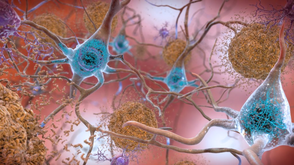
Plaques and neurons. Source: NIAH

A study in the journal Neurology has shown that a less expensive blood test to detect Alzheimer’s is highly accurate at early detection, providing further evidence that the test should be considered for routine screening and diagnosis. 

“Our study shows that the blood test provides a robust measure for detecting amyloid plaques associated with Alzheimer’s disease, even among patients not yet experiencing cognitive declines,” said senior author Professor Randall J. Bateman, MD.

“A blood test for Alzheimer’s provides a huge boost for Alzheimer’s research and diagnosis, drastically cutting the time and cost of identifying patients for clinical trials and spurring the development of new treatment options,” Prof Bateman said. “As new drugs become available, a blood test could determine who might benefit from treatment, including those at very early stages of the disease.”

Developed by Prof Bateman and colleagues, the blood test assesses whether amyloid plaques have begun accumulating in the brain based on the ratio of the levels of the amyloid beta proteins Aβ42 and Aβ40 in the blood.

The gold standard PET scan evaluation requires a radioactive brain scan, at an average cost of $5000–$8000 (R75 000–R120 000) per scan. Another common test, which analyses levels of amyloid-beta and tau protein in cerebrospinal fluid, costs about $1000 (R15 000) but requires a spinal tap process.

This study estimates that prescreening with a $500 (R7500) blood test could halve both the cost and the time it takes to enrol patients in clinical trials that use PET scans. Using only blood testing for screening could be done in under six months, a tenth or less of the cost. The test is currently only available in the US and Europe.

The current study shows that the blood test remains highly accurate, even when performed in different labs following different protocols, and in different cohorts across three continents.

Scientists didn’t know if small differences in sampling methods (such as anticoagulant use) could have a big impact on test accuracy because results are based on subtle shifts in amyloid beta protein levels in the blood. Subtle interfernece in these amyloid protein ratios could have triggered a false negative or positive result.

To confirm the test’s accuracy, researchers tested blood samples from current Alzheimer’s studies in the United States, Australia and Sweden, each of which uses different protocols for the processing of blood samples and related brain imaging.

Findings from this study confirmed that the Aβ42/Aβ40 blood test using a high-precision immunoprecipitation mass spectrometry technique developed at Washington University provides highly accurate and consistent results for both cognitively impaired and unimpaired individuals across all three studies.

When blood amyloid levels were combined with another major Alzheimer’s risk factor – the presence of the genetic variant APOE4 – the blood test accuracy was 88% compared to brain imaging and 93% when compared to spinal tap.

“These results suggest the test can be useful in identifying nonimpaired patients who may be at risk for future dementia, offering them the opportunity to get enrolled in clinical trials when early intervention has the potential to do the most good,” Prof Bateman said. “A negative test result also could help doctors rule out Alzheimer’s in patients whose impairments may be related to some other health issue, disease or medication.”
Source: Washington University School of Medicine

A New Test to Diagnose Dizziness without Deafening

Source: Miika Luotio on Unsplash

Swedish researchers have developed a new way to diagnose dizziness problems in a simpler and less painful way than the old method. A bone conduction speaker, easily attached behind the ear, can make the diagnosis more efficient and safer – especially for patients with pre-existing hearing problems.

For patients with dizziness, the relationship of dizziness and hearing is used for diagnosis. Typically, a ‘VEMP’ test (Vestibular Evoked Myogenic Potentials) is performed. With loud sounds, the test evokes a muscle reflex contraction in the neck and eye muscles, triggered by the vestibular system. In their new approach, reported in Communications Medicine, researchers at Chalmers University instead made use of bone-conducted sounds to achieve better results.

“We have developed a new type of vibrating device called B250 that is placed behind the ear of the patient during the test,” said Bo Håkansson, a professor at Chalmers University. “The vibrating device is small and compact in size and optimised to provide an adequate sound level for triggering the reflex at frequencies as low as 250 Hz, which we have found to be optimal for VEMP stimulation. Previously, no vibrating device has been available that was directly adapted for this type of test of the balance system.”

In bone conduction transmission, sound waves are transformed into vibrations through the skull, stimulating the cochlea within the ear, in the same way as when sound waves normally go through the ear canal, the eardrum and the middle ear. This can be used in various technologies such as in hearing aids.

Half of over-65s suffer from dizziness, but the causes can be difficult to diagnose for several reasons. Dizziness in 50% of those cases results from vestibular system problems. But current VEMP methods have major shortcomings and can cause hearing loss and discomfort for patients. The VEMP test uses very high sound levels which can cause permanent hearing damage. Additionally, if certain types of hearing loss are already present, the test can be inconclusive.

“The previous test was like a machine gun going off next to the ear – with this bone-conduction method it will be much more comfortable. The sound levels to which patients are exposed can be minimised. The test can be performed at 40 decibels lower than today’s method, which uses air-conducted sounds through headphones. This eliminates the risk that the test itself could cause hearing damage,” said researcher Karl-Johan Fredén Jansson, who made all the measurements in the project.

“The benefits also include safer testing for children, and that patients with impaired hearing function due to chronic ear infections or congenital malformations in the ear canal and middle ear can still be diagnosed for the origin of their dizziness,” said Prof Håkansson.

The device has now been tested and developed in several patient studies that have been published internationally, both with healthy individuals to obtain normal data, and in patients suffering from various types of dizziness. The device is compatible with standardised equipment for balance diagnostics in healthcare, which makes it easy to use. In addition to the benefits for patients, the cost of the new technology is also judged to be lower than the corresponding equipment used today.

Source: News-Medical.Net

Lab Results are Influenced by Ambient Daily Temperatures

Photo by Louis Reed on Unsplash

Ambient temperature influences many common lab tests, and these distortions likely affect medical decision making, such as whether to prescribe medications, according to new research published in the journal Med. 

To account for this, the researchers suggest that laboratories could statistically adjust for ambient temperature on test days when reporting lab results.

“When a doctor orders a laboratory test, she uses it to shed light on what’s going on inside your body, but we wondered if the results of those tests could also reflect something that’s going on outside of your body” said study co-author Ziad Obermeyer of the University of California, Berkeley. “This is exactly the kind of pattern that doctors might miss. We’re not looking for it, and lab tests are noisy.”

Delving into this problem, Obermeyer and Devin Pope of the University of Chicago analysed a large dataset of test results from different climates. In a sample of more than four million patients, they modelled more than two million test results based on temperature. They measured how day-to-day temperature fluctuations influenced results, over and above the patients’ average values, and seasonal variation.

Temperature was found to affect more than 90% of individual tests and 51 of 75 assays, including measures of kidney function, cellular blood components, and lipids such as cholesterol and triglycerides. “It’s important to note that these changes were small: less than one percent differences in most tests under normal temperature conditions,” Obermeyer said.

These small fluctuations did not likely reflect long-term physiological trends. For example, lipid panels checked on cooler days appeared to suggest a lower cardiovascular risk, resulting in almost 10% fewer prescriptions for cholesterol-lowering drugs called statins to patients tested on the coolest days compared to the warmest days, despite the results likely not reflecting stable changes in cardiovascular risk.

Since the study wasn’t an experiment, the exact mechanisms underlying the fluctuations in lab results could not be pinpointed. However, blood volume, specific assay performance, specimen transport, or changes in lab equipment might explain them. “Whatever their cause, temperature produces undesirable variability in at least some tests, which in turn leads to distortions in important medical decisions,” Pope said.

Laboratories could get around this by statistically adjusting for ambient temperature on the test day when reporting lab results. This could be a way to reduce weather-related variability without expensive temperature control equipment. 

In practice, decisions on adjustment would need to be at the discretion of the laboratory staff and the treating physician, potentially on a case-by-case basis.

According to the authors, the study may also have broader clinical implications. “The textbook way of thinking about medical research is bench to bedside. First, we come up with a hypothesis, based on theory, then we test it with data,” Obermeyer said. “As more and more big data comes online, like the massive dataset of lab tests we used, we can flip that process on its head: discover fascinating new patterns and then use bench science to get to the bottom of it. I think this bedside-to-bench model is just as important as its better-known cousin because it can open up totally new questions in human physiology.”

Source: Science Daily

A New Alternative to Skin Biopsies

Source: Pixabay

A new ‘virtual histology’ technology being developed by researchers could replace many skin tissue biopsies. The technology is detailed in Light: Science & Applications. 

Histology is the microscopic study of tissues and organs through sectioning, staining, and examining those sections under a microscope. Often called microscopic anatomy and histochemistry, histology allows for the visualisation of tissue structure and characteristic changes the tissue may have undergone.

“This process bypasses several standard steps typically used for diagnosis – including skin biopsy, tissue fixation, processing, sectioning and histochemical staining. Images appear like biopsied, histochemically stained skin sections imaged on microscope slides,” said the study’s senior author, Aydogan Ozcan, Chancellor’s Professor and Volgenau Chair for Engineering Innovation of the Electrical and Computer Engineering Department at UCLA Samueli.

The technology may enable rapid diagnosis of malignant skin tumours, reducing unnecessary invasive skin biopsies and allowing skin cancer diagnosis at an earlier stage. This technology had only previously been applied to microscopy slides of unstained tissue, acquired through a biopsy. This is the first time virtual histology has been applied to intact, unbiopsied tissue.

“The current standard for diagnosing skin diseases, including skin cancer, relies on invasive biopsy and histopathological evaluation. For patients, this often leads to unnecessary biopsies and scars as well as multiple visits to doctors. It also can be costly for patients and the health care system,” said Dr Philip Scumpia, assistant professor of dermatology and dermatopathology at the David Geffen School of Medicine at UCLA and the West Los Angeles Veterans Affairs Hospital and a member of the UCLA Jonsson Comprehensive Cancer Center. “Our approach potentially offers a biopsy-free solution, providing images of skin structure with cellular-level resolution.”

The research team, led by Ozcan, Scumpia and Dr. Gennady Rubinstein, a dermatologist at the Dermatology & Laser Centre in Los Angeles, created a deep-learning framework to transform images of intact skin acquired by an emerging noninvasive optical technology, reflectance confocal microscopy (RCM), into a format that is user-friendly for dermatologists and pathologists. Analysing RCM images requires special training because they are in black and white, and unlike standard histology, they lack nuclear features of cells.

“I was surprised to see how easy it is for this virtual staining technology to transform the images into ones that I typically see of skin biopsies that are processed using traditional chemical fixation and tissue staining under a microscope,” Dr Scumpia said.

The researchers trained a neural network to rapidly transform RCM images of unstained skin into virtually stained 3D images like the H&E (haematoxylin and eosin) images familiar to dermatologists and dermatopathologists.

“This framework can perform virtual histology on a variety of skin conditions, including basal cell carcinoma. It also provides detailed 3D images of several skin layers,” said Ozcan. “In our studies, the virtually stained images showed similar color contrast and spatial features found in traditionally stained microscopic images of biopsied tissue. This approach may allow diagnosticians to see the overall histological features of intact skin without invasive skin biopsies or the time-consuming work of chemical processing and labeling of tissue.”

“The only tool currently used in clinics to help a dermatologist are dermatoscopes, which magnify skin and polarise light. At best, they can help a dermatologist pick up patterns,” said Rubinstein, who also uses reflectance confocal microscopes in clinic.

The authors said there is a way to go before clinical use, but they aim to introduce the technology at various scales alongside other optical-imaging systems. Once the neural network is “trained,” with many tissue samples and the use of powerful graphics processing units (GPUs), it will be able to run on a computer or network, enabling rapid transformation from a standard image to a virtual histology image.

This technology could usher in a new age of “digital dermatology” and change how dermatology is practised. Additionally, the research team will see if this artificial intelligence platform can work with other AI technologies to look for patterns and further aid in clinical diagnosis.

Source: UCLA

Discrepancies in Radiology Interpretation

Source: National Cancer Institute

Researchers who conducted an analysis of nearly six million acute examinations suggest that leaders in imaging practice consider efforts to match interpretation of subspecialty examinations with radiologists’ fellowship training in the acute community setting.

Pointing out that major and minor discrepancy rates were not higher for acute community setting examinations outside of interpreting radiologists’ fellowship training, “discrepancy rates increased for advanced examinations,” acknowledged lead investigators Suzanne Chong from Indiana University in Indianapolis and Tarek Hanna of Emory University. The study was published in the American Journal of Roentgenology.

Using the databank of a large US teleradiology company, Chong, Hanna, and colleagues performed an analysis of 5 883 980 acute examinations that were preliminarily interpreted by 269 teleradiologists with a fellowship of neuroradiology, abdominal radiology, or musculoskeletal radiology. When providing final interpretations, client on-site radiologists voluntarily submitted quality assurance (QA) requests if preliminary and final interpretations were discrepant; the teleradiology company’s own QA committee categorised discrepancies as major (n=8444) or minor (n=17 208).

Among initial teleradiology interpretations of acute community setting examinations, common examinations’ major and minor discrepancies rates were not significantly different when concordant versus discordant with radiologists’ fellowship training. However, advanced examinations’ discrepancy rates were higher when concordant with radiologists’ fellowship (relative risk = 1.45 and 1.17, respectively).

Noting that their findings support multispecialty radiologist practice in acute community settings, “efforts to match examination and interpreting radiologist subspecialty may not reduce diagnostic discrepancies,” the article authors cautioned.

A supplement to the published article is available here [PDF].

Source: American Roentgen Ray Society

New HPV Test Enables Precision Treatment

Source: NCI on Unsplash

Researchers have made advances in improving detection of the human papillomavirus (HPV) in the bloodstream, which could further hone precision treatment of the illness.

The team sequenced circulating tumour DNA, which can lead to the detection of HPV in a person’s blood. Previous science in the field has proven that the virus, which causes cancers in the throat, mouth, and genital areas, can be found in the bloodstream but tests have had limited sensitivity. The new study enables ‘ultrasensitive’ detection, which could pave the way toward greater use of precision medicine for patients with cancers affecting these vulnerable areas of the body.

In a cohort of patients with advanced cervix cancer, the new sequencing method detected 20-fold lower levels of HPV circulating tumour DNA, making it a promising new method to monitor the disease.

The results come from the laboratory of Senior Scientist Dr Scott Bratman at Princess Margaret Cancer Center and are published in Clinical Cancer Research. “Increasingly, as clinicians we’re focused on precision medicine and making sure we’re not over-treating people while still curing them, that’s a very difficult balance to strike,” Dr Bratman said.

One way is to use liquid biopsy approaches or blood-based biomarkers, such as circulating tumour DNA, in order to monitor how the treatment is progressing, he added.

“We’re really at the cusp of a revolution from a technology, clinical implementation and standard of care standpoint, where five to 10 years from now we will not be treating everybody with the same dose of radiation and chemotherapy, and then waiting months to see if the treatment was effective,” he said. “I’m confident we will be giving much more tailored doses.”

When physicians scale back on these treatments, there is a risk of the cancer reoccurring. With more sensitive tests, reoccurrences can be detected early and patients returned to treatment.

“Patients who need more treatment will then be able to continue on, or different treatments can be added,” Dr Bratman said. “We can spare the vast majority of patients who will not need those interventions and provided them with a greater quality of life once they’re cured of the cancer.”

The work will enable further study in the field, refining the approach using larger study groups, and eventually, practice-changing clinical trials. This technique could also be applied to other cancer-causing viruses such as certain types of stomach cancer and lymphomas.

Source: Princess Margaret Cancer Center

Joint Statement Says Prior Radiation Should not Affect Decisions to Image

Photo by National Cancer Institute on Unsplash

Previous radiation exposure should not be considered when assessing the clinical benefit of radiological exams, according to a statement by three scientific groups representing medical physicists, radiologists, and health physicists.

Medical radiation exposure is a hot topic. People receive average annual background radiation levels of around 3 mSv; exposure from a chest X-ray is about 0.1 mSv, and exposure from a whole-body CT scan is about 10 mSv. The annual radiation limit for nuclear workers is 20mSv.

The American Association of Physicists in Medicine, along with the American College of Radiology and the Health Physics Society, issued a joint statement opposing cumulative radiation dose limits for patient imaging, saying that there could be negative impacts on patient care. The statement opposes the position taken by several organisations and recently published papers.

“It is the position of the American Association of Physicists in Medicine (AAPM), the American College of Radiology (ACR), and the Health Physics Society (HPS) that the decision to perform a medical imaging exam should be based on clinical grounds, including the information available from prior imaging results, and not on the dose from prior imaging-related radiation exposures,” the statement reads.

“AAPM has long advised, as recommended by the International Commission on Radiological Protection (ICRP), that justification of potential patient benefit and subsequent optimization of medical imaging exposures are the most appropriate actions to take to protect patients from unnecessary medical exposures. This is consistent with the foundational principles of radiation protection in medicine, namely that patient radiation dose limits are inappropriate for medical imaging exposures.

“Therefore, the AAPM recommends against using dose values, including effective dose, from a patient’s prior imaging exams for the purposes of medical decision-making. Using quantities such as cumulative effective dose may, unintentionally or by institutional or regulatory policy, negatively impact medical decisions and patient care.

“This position statement applies to the use of metrics to longitudinally track a patient’s dose from medical radiation exposures and infer potential stochastic risk from them. It does not apply to the use of organ-specific doses for purposes of evaluating the onset of deterministic effects (e.g., absorbed dose to the eye lens or skin) or performing epidemiological research.”

The Radiological Society of North America also endorses the position.

The AAPM emphasises the importance of patient safety in their position. Radiation usage must be both justified and optimised and benefits should outweigh the risks.

“This statement is an important reminder that patients may receive substantial clinical benefit from imaging exams,” said James Dobbins, AAPM President. “While we want to see prudent use of radiation in medical imaging, and many of our scientific members are working on means of reducing overall patient radiation dose, we believe it is an important matter of patient safety and clinical care that decisions on the use of imaging exams be made solely on the presenting clinical need and not on prior radiation dose.

“AAPM is pleased to partner with our fellow societies—the American College of Radiology and the Health Physics Society—to bring a broadly shared perspective on the important issue of whether previous patient radiation exposure should play a role in future medical decision making.”

The AAPM cites the International Commission on Radiological Protection, which stresses that setting radiation exposure limits to patients is not appropriate. This is partly due to a lack of standardised dose estimates.

The position only addresses stochastic risks from radiation exposure, which are chance effects whose risk for a given imaging exam, like cancer,is unrelated to the amount of prior radiation. Deterministic effects, incremental, direct exposure responses, such as skin damage, result from different biological mechanisms and are not included.

The AAPM compiled a list of answers to frequently asked questions on the topic of medical radiation safety along with references to research papers which support the organisation’s position.

Source: News-Medical.Net

Study Uncovers Assortment of New Biomarkers for Dementia

Source: National Cancer Institute on Unsplash

An international study identified 15 novel biomarkers that are linked to late-onset dementias. These protein biomarkers predict cognitive decline and subsequent increased risk of dementia 20 years before the disease onset. 

The proteins identified by the study are involved with immune system dysfunction, blood-brain-barrier dysfunction, vascular pathologies, and central insulin resistance. Six of these proteins can be modified with currently available medications.  

“These findings provide novel avenues for further studies to examine whether drugs targeting these proteins could prevent or delay the development of dementia,” explained lead author Joni Lindbohm MD, PhD from the University College London and University of Helsinki.

The study findings have been published in Alzheimer’s & Dementia: The Journal of the Alzheimer’s Association.

Pathophysiological research on dementia aetiology has focused on amyloid beta and tau proteins, but thus far prevention and treatment trials targeting these biomarkers have been unsuccessful. This has spurred the search for other potential mechanisms that could predispose to dementia. Recent development of scalable platforms has made it possible to analyse a wide range of circulating proteins, which may reveal novel dementia-linked biological processes.

In this study, the researchers analysed proteins with a novel large-scale protein panel from stored blood samples of the British Whitehall II and US Atherosclerosis Risk in Communities (ARIC) study collected 20 years ago. Using a panel of 5000 proteins, the researchers identified proteins in plasma that predicted cognitive decline in 5-yearly screenings and subsequent onset of clinical dementia. The 15 proteins that were identified were predictive of dementia in both the British and US cohorts.

“This new study is the first step in our 5-year Wellcome Trust funded research programme. We will next examine whether the identified proteins have a causal association with dementia, and whether they are likely to be modifiable, and druggable”, said study author Professor Mika Kivimäki, Director of the Whitehall II study at University College London.

The research programme ultimately aims to identify novel drug targets for dementia prevention.

Source: EurekAlert!

T-Cells Could Identify ‘The Bends’ in Divers

Photo by USGS on Unsplash

A new study investigated genetic changes that occur in a serious condition affecting scuba divers — ‘the bends’ — and found that inflammatory genes and white blood cell activity are upregulated. The findings could lead to biomarkers that will help doctors to diagnose the condition more precisely.

The bends, more formally known as decompression sickness, is a potentially lethal condition that can affect divers. Symptoms include joint pain, a skin rash, and visual disturbances. In some patients, the condition can be severe, potentially leading to paralysis and death. The bends can also affect people working in submarines, flying in unpressurised aircraft or in spacewalks.

It has been studied for a long time: a 1908 paper correctly hypothesised that it involves bubbles of gas forming in the blood and tissue due to pressure decrease. Yet even after a century the precise mechanisms underlying the condition are not well understood. Animal studies have suggested that inflammatory processes may have a role in decompression sickness, but no-one had studied this in humans.

Nowadays, getting ‘the bends’ is rare as divers have well-established methods to mitigate risk, such as controlled ascents from the depths. Nevertheless, doctors have no means to test for the condition, if they do encounter it, and instead rely on observing symptoms and seeing whether patients respond to hyperbaric oxygen therapy.

To investigate decompression sickness, the researchers sampled the blood of divers who had been diagnosed with decompression sickness and also divers who had completed a dive without it. The blood samples were drawn at two times: within 8 hours of the divers emerging from the water, and 48 hours afterwards, when those divers with decompression sickness had undergone hyperbaric oxygen treatment. RNA sequencing analysis was done to measure gene expression changes in white blood cells.

“We showed that decompression sickness activates genes involved in white blood cell activity, inflammation and the generation of inflammatory proteins called cytokines,” explained Dr Nikolai Pace of the University of Malta, a researcher involved in the study. “Basically, decompression sickness activates some of the most primitive body defense mechanisms that are carried out by certain white blood cells.”

These genetic changes had diminished in samples from 48 hours after the dive, after the patients had been treated with hyperbaric oxygen therapy — an interesting finding. The results provide a first step towards a diagnostic test for decompression sickness, and may also reveal new treatment targets.

“We hope that our findings can aid the development of a blood-based biomarker test for human decompression sickness that can facilitate diagnosis or monitoring of treatment response,” said Prof Ingrid Eftedal of the Norwegian University of Science and Technology, who was also involved in the project. “This will require further evaluation and replication in larger groups of patients.”

Source: EurekaAlert!

Journal information: “Acute effects on the human peripheral blood transcriptome of decompression sickness secondary to scuba diving” Frontiers in Physiology, DOI: 10.3389/fphys.2021.660402

Training Humans to Spot Abnormalities in 50 Milliseconds

Photo by Kony Xyzx from Pexels

After looking for just one-twentieth of a second, experts in camouflage breaking can accurately detect not only that something is hidden in a scene, but precisely identify the camouflaged target, with great potential in medical diagnostic settings as well for the military.

Medical College of Georgia neuroscientist Dr Jay Hegdé and his colleagues developed a relatively rapid method for training civilian novices to become expert camouflage breakers, a skill that even allowed them to sense that something was amiss even when there was no specific target to identify.

Experienced radiologists also have this intuitive sense, detecting subtle changes in mammograms, sometimes years before there is a detectable lesion. One of the main goals of radiology education is training novices to develop advanced or ‘expert’ search methods to improve their recognition of abnormalities, While artificial intelligence may significantly improve diagnosis, there is also the potential to improve the skills of humans. 

The researchers behind the camouflage breaking technique wanted to know if trainees could detect the actual camouflaged target or just sense that something is out of place, an issue that is highly significant in real world circumstances.

They already knew that they could train most nonmilitary individuals with good vision to break camouflage in as little as an hour daily for two weeks, which could benefit the military.

“The potential for rapid training of novices in the camouflage-breaking paradigm is very promising as it highlights the potential for application to a wide variety of detection and localisation tasks,” said Dr Frederick Gregory, programme manager, US Army Combat Capabilities Development Command Army Research Laboratory. “Results in experts highlight an opportunity to extend the training to real world visual search and visualisation problems that would be of prime importance for the Army to solve.“

This sort of enhanced ability to spot something amiss could have great applications in medical diagnosis and in search and rescue situations, to name a few.

For this study, six adult volunteers with normal or corrected-to-normal vision were trained to break camouflage using Hegdé’s deep-learning method, but received no specific  training on how to pinpoint the target. Participants viewed digitally synthesised camouflage scenes such as foliage or fruit and each scene had a 50-50 chance of containing no target versus a camouflaged target like a human head or a novel, 3D digital image. Similar to how computer scientists ‘trained’ self-driving cars, the idea is to get viewers to get to know the lay of the land that is their focus. “If it turns out there is something that doesn’t belong there, you can tell,” he said.

Trainees could then either look at the image for 50 milliseconds or as long as they wanted, then proceed to the next step where they quickly viewed a random field of pixels, that work like a visual palate cleanser, before acknowledging whether the camouflage image contained a target then using a mouse to show where the target was. “You have to work from memory to say where it was,” he notes.

When the participants could look at the image for as long as they wanted, the reported target location was not much different from when they only had 50 milliseconds — which is not a lot of time for their eyes to move around, Dr Hegdé said.

Again, participants had no subsequent training on identifying precisely where the target was. Yet even without that specific training, they could do both equally well. “This was not a given,” Dr Hegdé noted.

In a second experiment with seven different individuals they used a much-abbreviated training process, which basically ensured participants knew which buttons to push when, using a clearly more pronounced ‘pop-out’ target with scenarios like a black O-shaped target among a crowd of black C shapes. Both the longer and shorter viewing times yielded similar results to the more extensively trained camouflage-breakers both in accuracy and reaction time.

Camouflage is used extensively by the military, from deserts to jungles, with the visual texture changing to blend with the natural environment. “You often are recognised by your outline, and you use these patterns to break up your outline, so the person trying to break your camouflage doesn’t know where you leave off and the background begins,” he said. Animals have also used camouflage for millions of years to evade predators, or to sneak up on prey.

Context is another important factor for recognition, he pointed out, giving the example of not recognising a person whose face you have seen several times when you see them in a different setting. His current Army-funded studies aim to further explore the importance of context, and the ramifications of ‘camouflage breaking’ in identifying medical problems.

He noted that even with his training, some people are inherently better at breaking camouflage than others (he is really bad at it, he admitted) and the reason why is a goal for future research.

Source: Augusta University