In a study of 210 community-dwelling older adults who had surgery following hip fracture, participants who reported feeling high levels of psychological resilience were later able to walk faster and longer than those feeling less resilient. The results, published in the Journal of the American Geriatrics Society, will help the development of interventions such as targeted exercise programmes.
Walking capacity is a critically important outcome following hip fracture, in that it serves both as a surrogate measure of functional ability and physical health more globally, as well as a predictor of future survival, institutionalisation, social interaction and community engagement. Moreover, poor recovery after hip fracture causes considerable suffering for patients and imposes a financial burden on the social and health care sector. In 29%–50% of cases, older adults with hip fracture do not reach their pre-fracture levels of functioning a year after the fracture. Poorer pre-fracture function, greater cognitive impairment, greater co-morbidity burden, poorer social support, and poorer nutrition, have been identified as predictors for diminished post-fracture recovery.
Psychological resilience was measured through a questionnaire provided at the start of the study, and they assessed walking capacity at the start as well as 16 weeks later.
“We believe these results support opportunities to improve walking capacity following hip fracture in older adults by devising multicomponent interventions combining targeted exercise with psychological resilience-enhancing programmes,” said corresponding author Richard H. Fortinsky, PhD, of the University of Connecticut School of Medicine and the UConn Center of Aging.
By studying damage involving the connection between the brain’s hemispheres, researchers are finding new ways to leverage neural plasticity to promote functional recovery after a spinal cord injury.
In a study published in JCI Insight, the team of researchers used models in the lab to investigate a unilateral spinal cord injury similar to Brown-Sequard Syndrome, a rare neurological condition where damage to the spinal cord in a person results in weakness or paralysis on one side of the body and a loss of sensation on the opposite side.
Assistant Professor Wei Wu at Indiana University School of Medicine, said that the spinal cord injury model damaged the connection between the left hemisphere of the brain and the right side of the body, leading to significant loss of function in the right forelimb.
“The skilled function of upper limbs is very important for the quality of life in the patients with cervical spinal cord injury, but such functional recovery is very difficult to achieve in the severe injury,” said Asst Prof Wu, first author of the paper. “We found that the intact corticospinal system in the opposite side of the brain and spinal cord can be modulated to at least partially take over the control of the forelimb that is damaged by the spinal cord injury, resulting in a forelimb functional improvement.”
Since each hemisphere controls the opposite side of the body, researchers discovered a spontaneous shift of the neural circuits after injury from the left hemisphere to the right. Although there are connections between the right hemisphere of the brain and the right side of the body through some relayed pathways after injury, Asst Prof Wu said that’s not sufficient to support the motor recovery.
Using optogenetics to stimulate the right hemisphere of the brain, the researchers modulated the motor cortex. Additional neural circuits were shifted from the left side to the right side of the brain to dramatically increase and improve forelimb function.
“New circuits in the whisker, jaw forelimb and neck areas in the right hemisphere of the brain are recruited to control the right forelimb,” Asst Prof Wu said. “Interestingly, the beneficial neural plastic changes emerge both in the brain and the distal spinal cord after the optogenetic neuromodulation was applied on the motor cortex.”
Asst Prof Wu said results of the study showed significant improvement to the forelimb; however, there are still many challenges ahead, since complete digital recovery was not achieved.
The research team will continue explore this transhemispheric neural reorganisation to further improve the functional recovery after the spinal cord injury, Asst Prof Wu said. He hopes that these findings will be applied to treatments for spinal cord injuries.
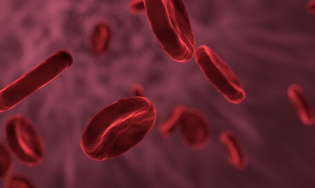
A study published in JAMA Internal Medicine shows that, after taking haemoglobin levels into count, sex and previous pregnancy of blood donors do not affect the survival of patients receiving red blood cell transfusions. Differences in recipient survival depend rather on the haemoglobin quantity in the transfusion, the researchers found.
Female donor sex and previous pregnancy are established risk factors for transfusion-related acute lung injury following plasma and platelet transfusions, which is a leading cause of transfusion-related mortality.
Previous studies have produced conflicting results as to how donor sex affects the recipient’s survivability in the recipient following red blood cell transfusion. Some studies have indicated higher mortality in patients who have received red blood cells from women, in men who have received red blood cells from women who have been pregnant, and in sex-mismatched transfusions. Other studies, however, have not reported such correlations.
This question has now been further explored by researchers from Karolinska Institutet in a register study of almost 370 000 patients in Sweden who received a red blood cell transfusion for the first time between 2010 and 2018.
The aim of the study was to see how the sex and previous pregnancy status of the donor affects survival in the recipient within two years from transfusion. It also looked at how the risk of needing more transfusions differed between patients who received red blood cells from female and male donors. Blood from women on average contains less haemoglobin than blood from men, meaning that more transfusions might be required to obtain the desired level of haemoglobin in a recipient.
The study demonstrates that the median value for haemoglobin was lower in female blood donors (135g/L than male (149g/L) and that patients who received blood from a woman had a 12% higher risk of needing another transfusion within 24 hours than blood from a man. However, this sex difference was eliminated when adjusting for the donors’ haemoglobin levels, which the researchers say was an expected effect that had not been factored into previous studies.
“When we take into account the lower haemoglobin levels in blood from women, we see no difference in survival among patients who received a blood transfusion from women compared with from men, regardless of how many times the female donors had been pregnant and of the patients’ sex and age,” said the study’s first author Jingcheng Zhao, adjunct researcher at Karolinska Institutet. “Differences in haemoglobin levels are a source of error that previous studies have not taken into consideration and that might explain the conflicting results that has been seen previously.”
Data for the study was drawn from national population, health and blood donor registries. The study also shows that donor sex is naturally randomly distributed in the patient material since no regard is paid to the sex and previous pregnancies of the donors by the blood donor centres when supplying blood. According to the researchers, this means that more far-reaching conclusions be drawn.
Dr Zhao said this allows them to determine causality. “We’ll now continue developing methods for studying causal relationships in transfusion epidemiology using observational data, on things like donor characteristics and how blood is handled. There’s still much we don’t know about blood transfusion and its effects.”
One limitation is that it was not possible to separately study transfusions where the red blood cells had not undergone leukoreduction (the filtering out of white blood cells), since this procedure has been standard in Sweden since the 1990s. The researchers therefore add a caveat about generalizing the conclusions to erythrocyte concentrates that have not undergone leukoreduction, which, however, is relatively uncommon now.
Concussion may cause different types of brain damage which lead to similar symptoms in children, according to research published in eLife. A new way of studying concussions could help inform the development of future treatments.
While most children fully recover after a concussion, some will have lasting symptoms. The findings help explain the complex relationships that exist between symptoms and the damage caused by the injury.
The researchers found that certain combinations of brain damage were associated with specific symptoms such as attention difficulties. Other symptoms, such as sleep problems, occurred in children with multiple types of injuries. For example, damage to areas of the brain that are essential for controlling sleep and wakefulness could cause challenges with sleeping, as could damage to brain regions that control mood.
The brain’s white matter holds clues
To do this, they examined how damage to the brain resulting from concussion affected its structural connection network, known as white matter. They then used statistical modelling techniques to see how these changes related to 19 different symptoms reported by the children or their caregivers.
Analysing symptoms may advance treatment
“Despite decades of research, no new treatment targets and therapies for concussions have been identified in recent years,” said lead author Guido Guberman, a Vanier Scholar and MDCM Candidate at McGill University. “This is likely because damage to the brain caused by concussions, and the symptoms that result from it, can vary widely across individuals. In our study, we wanted to explore the relationships that exist between the symptoms of concussion and the nature of the injury in more detail.”
Guberman and his colleagues analysed data collected from 306 children, aged nine to 10 years old, who had previously had a concussion. The children were all participants in the Adolescent Brain Cognitive Development (ABCD) Study.
“The methods used in our study provide a novel way of conceptualising and studying concussions,” says senior author Maxime Descoteaux, a Professor of Computer Science at Université de Sherbrooke. “Once our results are validated and better understood, they could be used to explore potential new treatment targets for individual patients. More broadly, it would be interesting to see if our methods could also be used to gather new insights on neurological diseases that likewise cause varied symptoms among patients.”
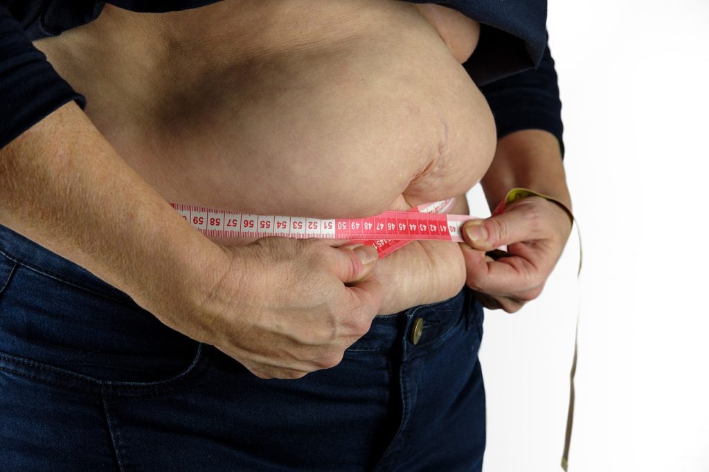
Women with obesity and overweight, particularly women with high waist circumference, are more susceptible to fractures than those with normal weight, according to new research presented at the European Congress on Obesity (ECO). In men, however, underweight, not overweight, is associated with a greater risk of broken bones.
Obesity has long been thought to help protect against fractures. This is because mechanical loading on bones, which increases with body weight, helps increase bone mineral density, an important determinant of bone strength.
However, recent studies have suggested that the relationship between obesity and fracture risk varies depending on sex, the skeletal site studied and definition of obesity used (body mass index [BMI] vs waist circumference).
To find out more, Dr Anne-Frederique Turcotte, Endocrinology and Nephrology Unit, CHU de Quebec Research Centre, Quebec City, Canada, and colleagues, analysed data from CARTaGENE, a prospective population-based cohort of almost 20 000 individuals aged 40-70 years from Quebec, Canada.
In women, greater waist circumference (WC) was linearly associated with an increased risk of fracture. For each 5cm (two inch) increase in WC, the risk of fracture at any site was 3% higher and the risk of a distal lower limb fracture was 7% higher. The association between WC and ankle fractures was particularly strong.
In women, greater BMI was associated with a greater risk of distal lower limb fractures. Compared with women with a BMI of 25 kg/m², those with a BMI of 27.5-40 kg/m² showed a greater risk of distal lower limb fractures. The increase in risk rose linearly from 5% in those with a BMI of 27.5 kg/m², to 40% in those with a BMI of 40 kg/m².
Women with a BMI of 22.5 kg/m² had a 5% lower risk of distal lower limb fractures than those with a BMI of 25 kg/m².
It isn’t known why obesity is associated with a higher risk of fractures in women. However, most fractures are a result of a fall and falls are more common in people with obesity. The ankle, unlike the hip and thighbone, is not protected by soft tissue, which could make it more prone to breaking during a fall.
Dr Turcotte added: “Waist circumference was more strongly associated with fractures in women than BMI. This may be due to visceral fat – fat that is very metabolically active and stored deep within the abdomen, wrapped around the organs – secreting compounds that adversely affect bone strength.
“We also know that people with obesity take longer to stabilise their body, when they trip, for example. This is particularly pronounced when weight is concentrated at the front of the body, suggesting that individuals with distribution of body fat in the abdominal area may be at higher risk of falling.”
In men, increases in BMI and WC were not significantly associated with fractures. However, men with underweight were at higher risk of distal upper limb fractures than those with normal weight. Men with a BMI ≤17.5 kg/m² were twice as likely to have distal upper limb fracture as men with a BMI of 25 kg/m².
The researchers say a larger number of fractures in men is needed to determine whether this is a true result or whether the pattern for men follows that for women.
The analyses were adjusted for a number of potential confounders: age, menopausal status, ethnicity, marital status, education, income, area of residence, smoking status, alcohol consumption, physical activity level, supplemental calcium and vitamin D intake, history of fracture and comorbidities and medications known to influence fracture risk.
The study authors said: : “Our findings show that the relationship between obesity and fractures is complex and varies by sex. In women, there was a linear relationship between waist circumference and the incidence of fracture at any site and at the distal lower limb, particularly at the ankle.
“Similar results were observed for women with a BMI between 27 and 40 kg/m². In men however, there was no relationship between obesity and the risk of fracture, although a BMI in the underweight range was associated with a higher risk of some fractures.”
Patients with lower levels of sex hormones are more likely to need to undergo surgery for rotator cuff tears, suggests a study in TheJournal of Bone & Joint Surgery.
Sex hormone deficiencies “was associated with a significantly increased incidence of RCR within [two] independent databases,” according to the new research by Peter N. Chalmers, MD, and colleagues at University of Utah. These findings add to previous evidence that hormone levels may be a systemic factor contributing to the development of rotator cuff tears, a common condition that is a major cause of shoulder pain.
The study used health insurance data for nearly 230 000 adults under age 65 who underwent surgery to repair a torn rotator cuff from 2008 through 2017. Patients were matched for age, sex, and type of insurance to patients who did not undergo rotator cuff surgery.
Patients undergoing rotator cuff repair had an average age of 54 years, and 58% were men. Most patient characteristics were similar between those who underwent rotator cuff repair and those who did not, except tobacco use, which was more common in the surgical cohort.
Dr Chalmers and colleagues found that 27% of women and 7% of men undergoing rotator cuff surgery had diagnosed sex hormone deficiency, compared with 20% and 4% respectively in the control group. Controlling for other factors, rotator cuff repair likelihood was 48% higher in women with oestrogen deficiency and 89% higher in men with testosterone deficiency.
To confirm their findings, the researchers then accessed the Veterans Administration Genealogy database which has data on millions of individuals. Here, they found that rotator cuff repair was about 2.5 times more likely for women with oestrogen deficiency and three times more likely for men with testosterone deficiency.
This study builds on a prior study by the same research group, which demonstrated that women with mutations in an oestrogen receptor gene were more likely to develop rotator cuff disease, with higher rates of failed rotator cuff surgery.
Despite limitations such as not accounting for hormone replacement therapy, the observed association between sex hormone deficiency and rotator cuff repair strongly supports the theory that low oestrogen and testosterone levels may contribute to the development of rotator cuff tears. The researchers concluded that “Future prospective studies will be necessary to understand the relationship of sex hormones to the pathophysiology of rotator cuff disease.”
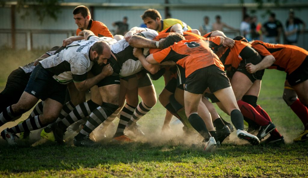
There is more risk of suffering concussions or being misdiagnosed with head trauma is greater among fighters who rapidly cut weight before bouts, usually by dehydrating, researchers have revealed for the first time.
More than 60% of athletes in combat sports such as boxing and mixed martial arts (MMA) reported that their symptoms worsened after they dehydrated to make strict weight classes.
These controversial weight cutting techniques involve stop drinking water and endure long periods in saunas.
MMA athletes reported concussion severity to be 40% higher compared to other sports, particularly boxing – believed to be due to the mix of striking and contact with the ground.
Published in the Clinical Journal of Sports Medicine, the study comes after a series of tragedies associated with weight cuts, with some athletes even dying in the attempt to achieve a perceived competitive edge, by competing in a smaller weight class.
They found that as competitors dramatically dehydrate themselves to meet stringent weight classes, the associated symptoms may ‘muddy the waters’ of baseline concussion testing, due to similar symptoms.
This is because the symptoms of hypohydration – where competitors enter a water deficit – match concussive signs, such as dizziness, headaches, and lethargy.
This study has spurred further probes into the neurological implications of rapid weight loss. The researchers have called on governing bodies to check fighters’ hydration levels before fights.
Researcher Nasir Uddin, from St Mary’s University, said: “This study shows that current concussion testing does not account for the crossover of symptoms from being dehydrated, and is potentially putting fighters at risk.
“Not only is cutting weight through dehydration in and of itself dangerous, but it might actually exacerbate concussion symptoms and, even more concerningly, means medical professionals may actually misdiagnose it.
“Going forward, governing bodies should ensure hydration and baseline concussion symptoms are taken into account before and after bouts.”
The study surveyed more than 130 anonymous athletes representing six combat sports, all aged 18 or above and who had previously cut weight.
It also found that 65% of the fighters had an experience of a weight cut “not going to plan”, suffering a lack of energy, strength, power, coordination or increased susceptibility to being ‘rocked’ during a bout.
This means the dangerous practice may not offer a competitive advantage after all.
Dr Jamie Tallent, from the University of Essex, said: “This is perhaps the most surprising finding that not only are weight cuts dangerous – they leave fighters at a disadvantage more often than not and may exacerbate the risks of being further injured.”
A clinical study from Lund University in Sweden has shown that long-term problems such as dizziness and difficulty focusing after a concussion originate in an injury to the vestibular nerve. The researchers published their findings in the Journal of Neurotrauma.
Concussion resulting from a blow to the head is a hazard in many sports. In American football, where players who have suffered from repeated concussions have developed dementia, severe depression and cognitive impairment.
Concussion usually has only temporary symptoms, but an increasing number of athletes experience long-term problems that make it difficult to work, go to school or play sports. The symptoms are aggravated by activity or impressions and include headaches, depression, anxiety, nausea, difficulty focusing and problems with balance.
“It has been unclear what causes the symptoms, and it is difficult for healthcare professionals to help these athletes. We wanted to investigate this further to find out what really causes the symptoms,” said Professor Niklas Marklund, one of the study’s researchers.
The study included 21 healthy athletes without previous trauma to the head, and 21 athletes who all suffered from sports-related concussions and who had experienced persisting symptoms for more than six months. The researchers used a 7-Tesla MRI, to study the athletes’ brains to understand more about what caused the symptoms. They discovered impaired function of the balance organs in the inner ear of 13 athletes in the group with long-term problems. In the group of healthy athletes three people had similar findings.
“The test results show that the injury is located to the vestibular nerve, which is connected to the semicircular canals in a cavity inside the skull, and which is directly adjacent to the cochlea in the ear. These injuries lead to the inward nerve impulses not working properly, and the brain therefore does not receive important information about body movements and sensory impressions required to maintain a good balance,” said Anna Gard, doctoral student at Lund University and first author of the study.
Concussion often results from the head rotating too fast.
“We have not examined athletes with short-term problems after blows to the head, so we cannot say anything about them. This study applies to athletes with prolonged symptoms after concussion. The rotation of the head that occurs in connection with a concussion could lead to a stretch of the vestibular nerve, which then leads to impaired function. Now that we have more knowledge about where the problems are located, it is easier to find possible therapies that could help these athletes,” concluded Prof Marklund.
In a discovery that could greatly benefit the treatment of traumatic injuries, scientists have identified a cluster of cells in the brainstem that control the body’s response to severe blood loss
The collection of neurons that the researchers discovered drive a response that maintains blood pressure during blood loss. However, severe blood loss eventually results in cardiovascular collapse, a condition called ‘decompensated haemorrhage’, marked by an abrupt and dangerous loss of blood pressure that presages haemorrhagic shock, where the body’s organs begin to shut down.
“During blood loss, the brain coordinates a cardiovascular response that supports blood flow to critical organs, like the heart and brain,” said researcher George Souza, PhD. “Our study shows that the cardiovascular response to blood loss depends on changes in the activity of a few hundred neurons in the brainstem.”
The new results, published in Cell Reports, shed light on an important process the body uses to maintain its blood pressure. Neurons, termed adrenergic C1 neurons, monitor blood pressure and activate during blood loss, increasing vasodilatory nerve activity that maintains proper blood pressure.
The scientists utilised advanced imaging and a technique called optogenetics controls neurons using light. Their research revealed that the C1 neurons are hyperactive during blood loss, and this keeps blood pressure study. But these neurons become inactive with severe blood loss, resulting in cardiovascular collapse.
The scientists found that re-activating the C1 neurons in lab rats restored both blood pressure and heart rate before cardiovascular collapse could lead to haemorrhagic shock.
“Our study indicates that reactivating the brain pathways controlling blood pressure during decompensated haemorrhage effectively reverses cardiovascular collapse. We think this indicates that neuromodulation of the pathways described by our study could be a beneficial adjunct therapy for low blood pressure following blood loss,” explained lead researcher Stephen Abbott, PhD.
The researchers noted that more research is needed as several factors could also cause the C1 neurons’ drop in activity during the onset of decompensated haemorrhage.
“These findings illuminate the importance of the brain-body interactions during blood loss and provide a new perspective for the underlying cause of cardiovascular collapse,” Dr Abbott said.
A newly published study has identified a key regulatory mechanism in inflammation that may lead to new targets for resolving that inflammation –and the inflammation of patients with sepsis, cancer and COVID.
In the journal PNAS, scientists reported their discovery of a regulatory pathway for immune response after infection or injury, such as burns. Dysregulation of this pathway could differentiate those who are at risk of fatal sepsis or help identify targets to resolve this unregulated inflammation.
“We are very excited about the findings in this paper and the far-reaching impacts it could have on understanding a key regulatory step in the immune response,” said co-lead author Cindy McReynolds, who holds a doctorate in pharmacology and toxicology.
In a rodent model, the research team found that the metabolites of linoleic acid formed by the enzyme, soluble epoxide hydrolase (sEH), drive damaging inflammation after injury. These metabolites, known as lipid mediators, regulate inflammation, blood pressure and pain. Drugs that inhibit the sEH enzyme and reduce inflammation could lead to better outcomes.
“Our previous work identified that these same lipid mediators were up-regulated in severe COVID infections, and we are now finding that these compounds play a role in modulating the immune response so that the body is unable to fight infection or respond properly to trauma without leading to a potentially fatal overreaction,” said Dr McReynolds.
“This dysregulation has fatal consequences in serious diseases such as COVID, cancer, sepsis, burn, where fatality rates can be as high as 40 percent in severe cases,” she said. “An understanding of these pathways can help identify patients at risk of developing serious disease or identify new therapeutic targets for treatment.”
“The immunological disbalance we see in many cases of severe burn injury, trauma and sepsis pose a huge clinical challenge as we lack the understanding of how to diagnose and treat it,” explained co-lead author Dr Christian Bergmann. “With this work, we reveal an important mechanism how immune cells are functionally disabled by sEH-derived metabolites of linoleic acid.”
“The natural compounds we are studying in this paper are metabolites of linoleic acid (LA), an essential fatty acid the body needs in very small amounts to survive and is only available through the diet,” Dr McReynolds elaborated. “At lower concentrations, these metabolites are necessary for regulating thermogenesis and heart health but promote inflammation at higher concentrations. LA is more stable and much cheaper than longer chain polyunsaturated fatty acids, so heavily processed foods have higher LA content to increase shelf-life. Additionally, agricultural practices, such as feeding animals corn-based diets, have increased LA in meats and dairy products.”
“As a result, we are consuming the highest amount of linoleic acid and have the highest recorded concentration of LA in our fatty tissue in human history,” McReynolds said. “As our bodies respond to stress or disease, we metabolise LA into the regulatory metabolites that were monitored in this paper. At higher concentrations, the immune system was unable to properly respond to infection, thereby promoting a sustained immune response. These observations are important in inflammatory-driven diseases, such as sepsis and COVID, but could also be important in understanding many of the increased chronic diseases we are seeing in our population.”