Category: Genetics

Pig-to-human Kidney Xenotransplant is the Longest Successful One Yet

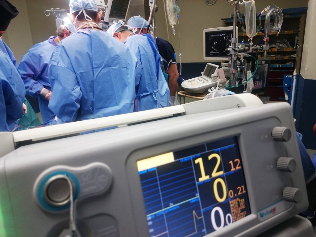
Photo by Natanael Melchor on Unsplash

Surgeons at NYU Langone Health have transplanted a genetically engineered pig kidney that continues to function well after 32 days in a man declared dead by neurologic criteria and maintained with a beating heart on ventilator support. This represents the longest period that a gene-edited pig kidney has functioned in a human, and the latest step toward the advent of an alternate, sustainable supply of organs for transplant.

Led by Robert Montgomery, MD, DPhil, the procedure was performed on July 24, 2023 and marks the fifth xenotransplant performed at NYU Langone. Observation is ongoing and the study will continue through mid-September 2023.

“This work demonstrates a pig kidney – with only one genetic modification and without experimental medications or devices – can replace the function of a human kidney for at least 32 days without being rejected,” said Dr Montgomery, who had previously performed the world’s first genetically modified pig kidney transplant into a human decedent in 2021.

Removing single troublesome gene

The first hurdle to overcome in xenotransplants is preventing so-called hyperacute rejection, which typically occurs just minutes after an animal organ is connected to the human circulatory system. By “knocking out” the gene that encodes the biomolecule known as alpha-gal, responsible for a rapid antibody-mediated rejection of pig organs by humans, immediate rejection has been avoided in all five xenotransplants at NYU Langone. Additionally, the pig’s thymus gland, which is responsible for educating the immune system, was embedded underneath the outer layer of the kidney to stave off novel, delayed immune responses. The combination of modifications has been shown to prevent rejection of the organ while preserving kidney function.

To ensure the body’s kidney function was sustained solely by the pig kidney, both of the transplant recipient’s native kidneys were surgically removed. One pig kidney was then transplanted and started producing urine immediately without any signs of hyperacute rejection. During the observation phase, intensive care clinical staff maintained the decedent on support while the pig kidney’s performance was monitored and sampled with weekly biopsies. Levels of creatinine, a bodily waste product found in the blood and an indicator of kidney function, were in the optimal range during the length of the study, and there was no evidence on biopsy of rejection.

The surgery was the latest in a larger study approved by a specific research ethics oversight board at NYU Langone and was performed after consultation with the New York State Department of Health. This important research, which study leaders say could save many lives in the future, was made possible by the family of a 57-year-old male who elected to donate his body after a brain death declaration and a circumstance in which his organs or tissues were not suitable for transplant.

A big leap toward a new organ source

“There are simply not enough organs available for everyone who needs one,” said Dr Montgomery, who received a hepatitis C-positive heart transplant himself in 2018. “Too many people are dying because of the lack of available organs, and I strongly believe xenotransplantation is a viable way to change that.”

The kidney and thymus gland used in this procedure were procured from a GalSafeTM pig, an animal engineered by Revivicor, Inc., a subsidiary of United Therapeutics Corporation. In December 2020, the U.S. Food and Drug Administration (FDA) approved the GalSafe pig as a potential source for human therapeutics as well as a food source for people with alpha-gal syndrome, a meat allergy caused by a tick bite.

Less may be more

While previous genetically engineered pig organ transplants have incorporated up to 10 genetic modifications, this latest study shows that a single-gene knockout pig kidney can still perform optimally for at least 32 days without rejection.

“We’ve now gathered more evidence to show that, at least in kidneys, just eliminating the gene that triggers a hyperacute rejection may be enough along with clinically approved immunosuppressive drugs to successfully manage the transplant in a human for optimal performance – potentially in the long-term,” said Dr Montgomery.

The NYU Langone team used standard transplant immunosuppression medications combined with enhanced screening of porcine cytomegalovirus (pCMV) in the donor pig to ensure safety. Recent studies have shown pCMV may affect organ performance and potentially trigger organ failure. No pCMV was detected after 32 days, and close surveillance of porcine endogenous retrovirus (PERV), along with six other viruses of interest, was performed.

Next steps

Monitoring of the pig kidney recipient will continue for another month with permission from the family, ethics committee approval and continued support from United Therapeutics. The additional data from the next several weeks will be analyzed further to develop a deeper understanding of this unique medical advance.

“We think using a pig already deemed safe by the FDA in combination with what we have found in our xenotransplantation research so far, gets us closer to the clinical trial phase,” said Dr Montgomery. “We know this has the potential to save thousands of lives, but we want to ensure the utmost safety and care as we move forward.”

Source: NYU Langone Health

Genetic Mechanism Increases Resistance to the Antibiotic Albicidin by 1000-fold

Photo by Sangharsh Lohakare on Unsplash

A new analysis shows that infectious bacteria exposed to the promising antibiotic albicidin rapidly develop up to a 1000-fold increase in resistance via a gene amplification mechanism. Mareike Saathoff of Freie Universität Berlin, Germany, and colleagues presented these findings in the open access journal PLOS Biology.

Bacterial resistance to antibiotics is a growing problem associated with millions of deaths around the world every year. Understanding how bacteria evolve resistance is key to developing more effective antibiotics and strategies for using them.

In recent years, albicidin has emerged as a promising antibiotic capable of killing a wide range of bacterial species by disrupting their DNA replication. Researchers are working to develop new albicidin-based medications; yet, despite its promise, some bacteria are able to develop resistance to albicidin.

To further investigate albicidin resistance mechanisms, Saathoff and colleagues conducted a suite of experiments employing a broad set of tools, including RNA sequencing, protein analysis, X-ray crystallography, and molecular modeling. They found that two bacteria often associated with human infection, Salmonella typhimurium and Escherichia coli, develop resistance to albicidin when exposed to increasingly higher concentrations of the compound. Their analysis narrowed down the source of this resistance to an increase in the number of copies of a gene known as STM3175 (YgiV) in the bacterial cells, which is amplified in each new generation of cells as they multiply. STM3175 encodes a protein that interacts with albicidin in such a way that protects the bacteria from it.

Further experiments showed that the same albicidin-resistance mechanism is widespread among both pathogenic and harmless bacteria, including the microbes Vibrio vulnificus, which can infect wounds, and Pseudomonas aeruginosa, which can cause pneumonia and other infections. These findings could help inform the ongoing development of albicidin-based antibiotic strategies.

The authors add, “Our study reveals a gene duplication and amplification-based mechanism of a transcriptional regulator in Gram-negative bacteria, that mediates resistance to the peptide antibiotic albicidin.”

Source: Science Daily

Study of African-ancestry Genomes Reveals Defensive Gene Against HIV

Photo by Ayo Ogunseinde on Unsplash

A study on almost 4000 people of African descent has identified a gene that acts as natural defence against HIV by limiting its replication in certain white blood cells. The findings were published in Nature. An international effort co-led by EPFL, Canada’s National Microbiology Laboratory, and Imperial College London, it paves the way for new treatment strategies.

“We searched for human genetic variation that associates with spontaneous control of HIV and identified a novel region in the genome that is only variable in populations of African ancestries,” says Professor Jacques Fellay at EPFL’s School of Life Sciences. “We used a combination of computational and experimental approaches to explore the biological mechanism behind the genetic association and provide evidence that the gene CHD1L acts to limit HIV replication in a subset of white blood cells.”

HIV is still a problem

Despite significant advances in treatment and access to therapy, the human immunodeficiency virus remains a global health challenge with almost 40 million affected individuals, no vaccine and no cure.

Although annual HIV infections have been declining because of widespread antiretroviral therapies, the trend has slowed substantially since 2005, and there are now alarming increases in the number of newly infected adults in some regions.

Genome-Wide Association Studies, or GWAS, analyse the entire genome of a large number of individuals to identify genetic variants associated with a clinical outcome, such as the ability to naturally control viral replication.

Measuring HIV replication control: not enough in African populations

The degree of viral infection is measured by the virus’ “setpoint viral load” (spVL), which refers to the relatively stable level of HIV replication in the body after the initial, acute phase of infection in untreated individuals.

A critical determinant of HIV infection progression and transmissibility, spVL is expressed as the number of viral copies/mL of plasma. The spVL of HIV varies widely in the infected population, depending on the ability of every individual’s immune system to control viral replication without antiretroviral drugs.

Although there have been large studies of spVL control in populations of European descent, much less has been done in populations of African ancestries, which have both a high genomic diversity and the greater burden of HIV.

A key gene for resistance to HIV replication in people of African ancestries

To address this disparity, a large international collaboration of scientists and clinicians has now performed large-scale GWAS using data from diverse populations of African ancestries. In total, the scientists analyzed the genomes from 3,879 individuals living with HIV-1. Using computational analysis and fine-mapping techniques, they identified a novel region in the genome that shows a strong association with spVL control.

This region corresponds to a gene known as CHD1L (for “Chromodomain Helicase DNA Binding Protein 1 Like”), which encodes a protein that helps DNA unwind after it has been damaged, allowing it to be repaired. But in this study, the CHD1L gene showed genetic variation specific to populations of African ancestries, and that was linked to the spontaneous control of the most common and virulent type of HIV, called HIV-1.

Having identified CHD1L as a potential modulator of HIV-1 infection, the researchers explored the biological mechanism behind the genetic association and determined that CHD1L plays a role in limiting HIV replication in a subset of white blood cells.

The study was co-led by Jacques Fellay at EPFL, Paul McLaren at the Public Health Agency of Canada’s National Microbiology Laboratory, and Manjinder Sandhu at Imperial College London.

The discovery of CHD1L’s role in limiting HIV replication could lead to improved treatment options for infected individuals. “Our findings provide insights into potential therapeutic targets, which are needed to continue the fight against HIV-1,” says Fellay. “In addition, our results underscore the importance of performing genomic studies in diverse ancestral populations to better address their specific medical needs and global health inequities.”

Source: Ecole Polytechnique Fédérale de Lausanne

Researchers Identify Sex-specific Genes for Obesity

Source: CC0

Researchers have added several genes, which appear to affect obesity risk in certain sexes and ages, to the list of genes which influence weight gain. The study, published in the journal Cell Genomics, may shed light on new biological pathways that underlie obesity and highlight how sex and age contribute to health and disease.

“There are a million and one reasons why we should be thinking about sex, age, and other specific mechanisms rather than just lumping everyone together and assuming that disease mechanism works the same way for everyone,” says senior author John Perry, a geneticist and professor at the University of Cambridge. “We’re not expecting people to have completely different biology, but you can imagine things like hormones and physiology can contribute to specific risks.”

To untangle sex’s role in obesity risk, the research team sequenced the exome (the protein-coding part of the genome) of 414 032 adults from the UK Biobank study. They looked at variants, or mutations, within genes associated with body mass index (BMI) in men and women, respectively. Five genes influencing BMI in women and two in men were identified.

Among them, faulty variants of three genes – DIDO1, PTPRG, and SLC12A5 – are linked to higher BMI in women, up to nearly 8 kg/m² more, while having no effect on men. Over 80% of the women with DIDO1 and SLC12A5 variants had BMI-indicated obesity. Those carrying DIDO1 variants had stronger associations with higher testosterone levels and increased waist-to-hip ratio, both risk indicators for obesity-related complications like diabetes and heart disease. Others with SLC12A5 variants had higher odds of having type 2 diabetes compared with non-carriers. These findings highlight previously unexplored genes that are implicated in the development of obesity in women but not men.

Perry and his colleague then repeated their method to look for age-specific factors by searching for gene variants associated with childhood body size based on participants’ recollections. They identified two genes, OBSCN and MADD, that were not previously linked to childhood body size and fat. While carriers of OBSCN variants had higher odds of having higher weight as a child, MADD variant carriers were associated with smaller body sizes. In addition, the genetic variants acting on MADD had no association with adult obesity risk, highlighting age-specific effects on body size.

“What’s quite surprising is that if you look at the function of some of these genes that we identified, several are clearly involved in DNA damage response and cell death,” says Perry. Obesity is a brain-related disorder, whereas biological and environmental factors act to influence appetite. “There’s currently no well-understood biological paradigm for how DNA damage response would influence body size. These findings have given us a signpost to suggest variation in this important biological process may play a role in the aetiology of obesity.”

Next, the research team hopes to replicate the study in a larger and more diverse population. They also plan to study the genes in animals to peer into their function and relationship with obesity.

“We’re at the very earliest stages of identifying interesting biology,” says Perry. “We hope the study can reveal new biological pathways that may one day pave the way to new drug discovery for obesity.”

Source: Science Daily

Abdominal Fat Accumulation may not be as Great a Diabetes Risk as Previously Thought

Photo by Andres Ayrton on Pexels

Conventional wisdom holds that abdominal fat accumulation increases the risk for type 2 diabetes. But surprising new findings from the University of Virginia School of Medicine suggest that naturally occurring genetic variations in our genes can lead some people to store fat at the waist but also protect them from diabetes.

The unexpected discovery, which is published in eLife, provides a more nuanced view of the role of obesity in diabetes and related health conditions. It also could pave the way for more personalised medicine, such as prioritising weight loss for patients whose genes put them at increased risk but place less emphasis on it for patients with protective gene variants, the researchers say.

“There is a growing body of evidence for metabolically healthy obesity. In this condition, people who would normally be at risk for cardiovascular diseases and diabetes because they are obese are actually protected from adverse effects of their obesity. In our study, we found a genetic link that may explain how this occurs in certain individuals,” said researcher Mete Civelek, PhD, of UVA’s Center for Public Health Genomics. “Understanding various forms of obesity is important to tailor treatments for individuals who are at high risk for adverse effects of obesity.”

As medicine grows more sophisticated, understanding the role of naturally occurring gene variations will play an important role in ensuring patients get the best, most tailored treatments. The new work by Civelek and his team, for example, indicates that variants can simultaneously predispose some people to store fat at the abdomen, thought to put them at increased risk for metabolic syndrome, while also protecting them from type 2 diabetes. (Metabolic syndrome raises the risk for diabetes, stroke and other serious health issues.)

One of the metrics doctors use to determine if a patient has metabolic syndrome is abdominal obesity. This is often calculated by comparing the patient’s waist and hip measurements. But Civelek’s research suggest that, for at least some patients, it may not be that simple, with doctors using genetic testing to guide patients to good health.

“We found that among the hundreds of regions in our genomes which increase our propensity to accumulate excess fat in our abdomens, there are five which have an unexpected role,” said Yonathan Aberra, the lead author of the study and a PhD candidate at UVA’s Department of Biomedical Engineering, a joint program of the School of Medicine and School of Engineering. “To our surprise, these five regions decrease an individual’s risk for type 2 diabetes.”

In addition to producing surprising findings, Civelek’s research provides important new tools for his fellow researchers seeking to understand the complexities of gene variations. The sophisticated approach Civelek and his collaborators developed to identify the relevant variants and their potential effects will be useful for future research into metabolic syndrome and other conditions.

The tools could also prove invaluable in the development of new and better treatments for metabolic syndrome, the scientists say.

“We now need to expand our studies in more women and people from different genetic ancestries to identify even more genes that underlie the metabolically health obesity phenomenon,” Civelek said. “We plan to build on our findings to perform more experiments to potentially identify a therapeutic target.”

Source: University of Virginia Health System

New CRISPR Discovery Targets Infected Cells

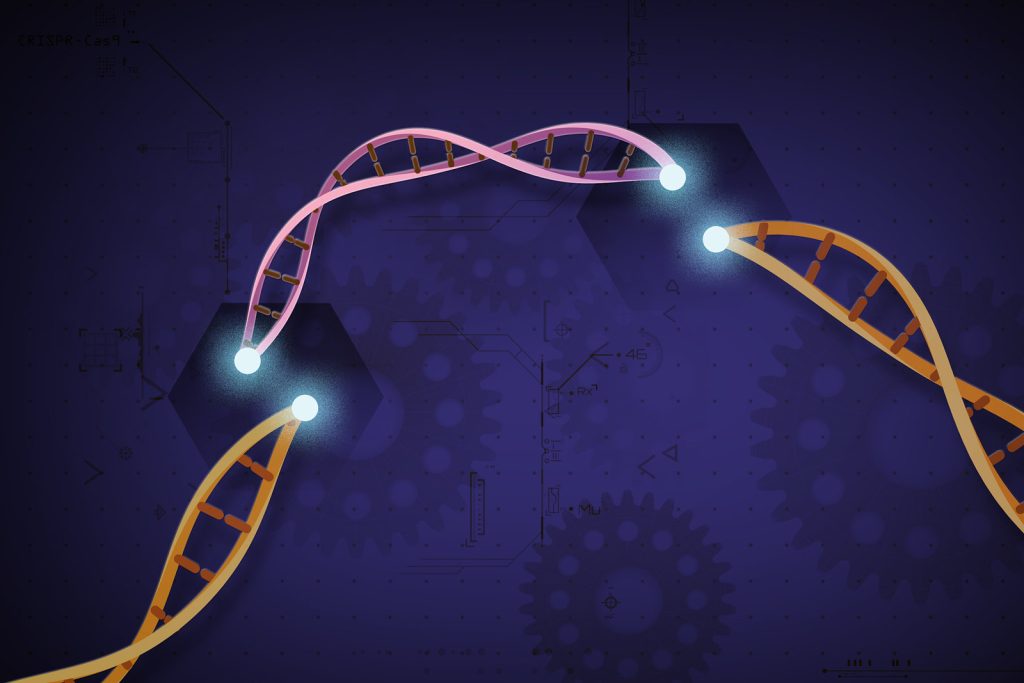
CRISPR-Cas9 is a customisable tool that lets scientists cut and insert small pieces of DNA at precise areas along a DNA strand. This lets scientists study our genes in a specific, targeted way. Credit: Ernesto del Aguila III, National Human Genome Research Institute, NIH

German and US scientists have discovered a CRISPR system in cells that shuts them down entirely to protect against viral replication, instead of merely chopping out foreign DNA that it comes across. It does this by shredding any DNA or RNA it comes across, causing the cell to become senescent and not become a virus factory. The newly identified CRISPR system is described in two papers published in Nature.

“With this new system, known as Cas12a2, we’re seeing a structure and function unlike anything that’s been observed in CRISPR systems to date,” says Jackson, assistant professor in Utah State’s Department of Chemistry and Biochemistry.

CRISPR, (Clustered Regularly Interspaced Short Palindromic Repeats) has taken science by storm with its gene-editing potential. Study of CRISPR DNA sequences and CRISPR-associated (Cas) proteins, which are actually bacterial immune systems, is still a young field.

Identified as a distinct immune system within the last five years, the Class 2, type V Cas12a2 is somewhat similar to the better-known ‘molecular scissors’ of CRISPR-Cas9, which binds to target DNA and cuts it, effectively shutting off a targeted gene. But CRISPR-Cas12a2 binds a different target than Cas9, and that binding has a very different effect.

Using cryo-electron microscopy, the team captured the CRISPR-Cas12a2 in a naturally occurring defensive strategy called abortive infection, a natural resistance strategy used by bacteria and archaea to limit the spread of viruses and other pathogens by preventing replication in the cell.

The team observed Cas12a2 in the act of cutting double-stranded DNA, bending it 90° to expose the backbone of the helix to cut it, a phenomenon that a phenomenon that elicits audible gasps from fellow scientists,” Jackson says.

Since the difference between a healthy cell and a malignant cell or infected cell is genetic, if Cas12a2 could be harnessed, “the potential therapeutic applications are significant.”

“If Cas12a2 could be harnessed to identify, target and destroy cells at the genetic level, the potential therapeutic applications are significant,” he says.

Source: Utah State University

Obesity Genetic Risk Could be Curbed by Practising Restraint

Photo by Jonathan Borba

Obesity risk genes make people feel hungrier and lose control over their eating, but practisng dietary restraint could counteract this, according to new research from University of Exeter. Published in the International Journal of Epidemiology, the study found that those with higher genetic risk of obesity can reduce the effects that are transmitted via hunger and uncontrolled eating by up to half through dietary restraint.

Lead author psychology PhD student, Shahina Begum said: “At a time when high calorie foods are aggressively marketed to us, it’s more important than ever to understand how genes influence BMI. We already know that these genes impact traits and behaviours such as hunger and emotional eating, but what makes this study different is that we tested the influence of two types of dietary restraint – rigid and flexible – on the effect of these behaviours. What we discovered for the first time was that increasing both types of restraint could potentially improve BMI in people genetically at risk; meaning that restraint-based interventions could be useful to target the problem.”

Genes linked to obesity increase BMI, with up to a quarter of this effect explained by increases in hunger and uncontrolled (including emotional) eating. There are over 900 genes that have so far been identified by researchers as being associated with BMI and several studies suggest these risk genes influence feelings of hunger and loss of control towards food.

This study examined 3780 adults aged between 22 and 92 years old from two UK cohorts: the Genetics of Appetite Study, and Avon Longitudinal Study of Parents and Children. Their weight and height were measured, and they provided a DNA sample via their blood to calculate an overall score for their genetic risk of obesity. They then completed questionnaires to measure 13 different eating behaviours, including disinhibition (a tendency to engage in binge or emotional eating) and over-eating due to hunger.

As expected, researchers found that a higher genetic risk score was associated with a higher BMI, partly due to increased disinhibition and hunger. However, results also found that those who had high levels of dietary restraint reduced those effects by almost half for disinhibition and a third for hunger, suggesting that restraint may counteract some of the effects of genetic risk.

There are different types of dietary restraint, including flexible strategies to rigid strategies, like calorie counting. The study tested the influence of both types of restraint for the first time and found both could potentially improve BMI in people genetically at risk.

Interventions to facilitate dietary restraint could include changing the food environment (by reducing the calorie content or portion size of food) or supporting individuals. To this end, members of the research team have developed a Food Trainer app (https://www.exeter.ac.uk/research/foodt/) to help achieve that. The app works as a game that trains people to repeatedly stop to high calorie food and research suggests this training may be particularly beneficial for those with a higher BMI.

Source: University of Exeter

Schizophrenia Might Stem From Genetic Mutations In Utero

Photo by Alex Green on Pexels

As an adult-onset psychiatric disorder, schizophrenia is thought to be triggered by some combination of environmental factors and genetics, although the exact cause remains unclear. In a study published in the journal Cell Genomics, researchers find a correlation between schizophrenia and somatic copy-number variants, a type of mutation that occurs early in development but after genetic material is inherited. This study is one of the first to rigorously describe the relationship between somatic genetic mutations and schizophrenia risk.

“We originally thought of genetics as the study of inheritance. But now we know that genetic mechanisms go way beyond that,” says senior author Chris Walsh, an investigator at the Howard Hughes Medical Institute and chief of genetics and genomics at Boston Children’s Hospital. “We’re looking at mutations that are not inherited from the parents.”

The researchers analysed genotype-marker data from over 20,000 blood samples of people with or without schizophrenia from the Psychiatric Genomics Consortium. They ultimately identified two genes, NRXN1 and ABCB11, that correlated with schizophrenia cases when disrupted in uteroNRXN1, a gene that helps transmit signals throughout the brain, has been associated with schizophrenia before. However, this is the first study to associate somatic, not inherited, NRXN1 mutations with schizophrenia.

Unlike inherited mutations, which are present in all the cells of the body, somatic mutations are only present in a fraction of cells based on when and where a mutation occurred. If a mutation occurs early in development, it is expected that the variant is present throughout the body in a mosaic pattern. On the basis of this principle, researchers can identify somatic mutations that occurred early in development and are present not only in the brain but also in a fraction of cells in the blood.

“If a mutation occurs after fertilisation when there are only two cells, the mutation will be present in half of the cells of the body,” says Walsh. “If it occurs in one of the first four cells, it will be present in about a quarter of the cells of the body, and so on.”

The second gene the researchers identified, ABCB11, is most known to encode a liver protein. “That one came out of nowhere for us,” says Eduardo Maury, a student in Harvard-MIT’s MD-PhD program. “There have been some studies associating mutations in this gene with treatment-resistant schizophrenia, but it hasn’t been strongly implicated in schizophrenia per se.”

When the team investigated further, they found that ABCB11 is also expressed in very specific subsets of neurons that carry dopamine from the brainstem to the cerebral cortex. Most schizophrenia drugs are thought to act on these cells to decrease an individual’s dopamine levels, so this might explain why the gene is associated with treatment resistance.

Next, the team is working towards identifying other acquired mutations that might be associated with schizophrenia. Given that the study analysed blood samples, it will be important to look at more brain-specific mutations that might have been too subtle or recent in a patient’s life for this analysis to detect. In addition, somatic deletions or duplications might be an under-investigated risk factor associated with other disorders.

“With this study, we show that it is possible to find somatic variants in a psychiatric disorder that develops in adulthood,” says Maury. “This opens up questions about what other disorders might be regulated by these kinds of mutations.”

Source: Cell Press via ScienceDaily

Backdoor to the Inner Ear Allows Delivery of Gene Therapy

Photo by Dylann Hendricks on Unsplash

An international team of researchers has developed a new method to deliver drugs into the inner ear, according to a new study in Science Translational Medicine. The discovery was possible by harnessing the natural flow of fluids in the brain and employing a little-understood backdoor into the cochlea. When combined to deliver a gene therapy that repairs inner ear hair cells, the researchers were able to restore hearing in deaf mice.

“These findings demonstrate that cerebrospinal fluid transport comprises an accessible route for gene delivery to the adult inner ear and may represent an important step towards using gene therapy to restore hearing in humans,” says lead author Barbara Canlon, professor at Karolinska Institutet.

The number of people worldwide predicted to have mild to complete hearing loss is expected to grow to around 2.5 billion by mid-century.  The primary cause is the death or loss of function of hair cells found in the cochlea – which relay sounds to the brain – due to mutations of critical genes, aging, noise exposure, and other factors. 

While hair cells do not naturally regenerate in humans and other mammals, gene therapies have shown promise and in separate studies have successfully repaired the function of hair cells in neo-natal and very young mice.

“However, as both mice and humans age, the cochlea, already a delicate structure, becomes enclosed in the temporal bone. At this point, any effort to reach the cochlea and deliver gene therapy via surgery risks damaging this sensitive area and altering hearing,” says Barbara Canlon.

In the new study, the researchers describe a little-understood passage into the cochlea called the cochlear aqueduct. The cochlear aqueduct is a thin boney channel no larger than several strands of hair. 

Channel for spinal fluid

A new study shows that the cochlear aqueduct acts as a conduit between the cerebrospinal fluid found in the inner ear and the rest of the brain. 

Scientists are developing a clearer picture of the mechanics of the glymphatic system, the brain’s unique process of removing waste. Because the glymphatic system pumps cerebrospinal fluid deep into brain tissue to wash away toxic proteins, researchers have been eyeing it as a potential new way to deliver drugs into the brain, a major challenge in developing drugs for neurological disorders. 

The new study represented an opportunity to put the drug delivery potential of the glymphatic system to the test, while at the same time targeting a previously unreachable part of the auditory system.   

Employing several imagining and modeling technologies, the researchers were able to develop a detailed portrait of how fluid from other parts of the brain flows through the cochlear aqueduct and into the inner ear.

The team then injected an adeno-associated virus into the cisterna magna, a large reservoir of cerebrospinal fluid found at the base of the skull. 

The virus found its way into the inner ear via the cochlear aqueduct and delivered a gene therapy that expresses a protein called vesicular glutamate transporter-3, which enables the hair cells to transmit signals and rescue hearing in adult deaf mice. 

“This new delivery route into the ear may not only serve the advancement of auditory research but also prove useful when translated to humans with progressive genetic-mediated hearing loss,” says Barbara Canlon.

Source: Karolinska Institutet

Food Poisoning Bacteria Species Swap Pathogenic Genes

Escherichia coli. Image by CDC on Unsplash

Researchers have identified how pathogenic genes in some Providencia spp., which have gained attention as causes of food poisoning as well as enterohaemorrhagic Escherichia coli. O157 and Salmonella, are transferred within bacterial cells. Their findings are expected to provide new insights into the identification of infection routes of Providencia spp. and the establishment of preventive methods for food poisoning.

Recently, Providencia spp. which have been detected in patients with gastroenteritis, and similar to enterohemorrhagic Escherichia coli. O157 and Salmonella spp., have been attracting attention as causative agents of food poisoning. For children with low immunity, food poisoning can be lethal as it causes severe symptoms such as diarrhoea and dehydration, so clarifying the source of infection and pathogenic factors of Providencia spp., and establishing preventive methods are urgent issues worldwide.

A joint research group led by Professor Shinji Yamasaki, Dr Sharda Prasad Awasthi, a Specially Appointed Lecturer, and graduate student Jayedul Hassan from the Graduate School of Veterinary Science, Osaka Metropolitan University, determined how the pathogenic genes in some Providencia spp. such as Providencia alcalifaciens and Providencia rustigianii are transferred within bacterial cells of genus Providencia. The group has also elucidated that the pathogenic genes of Providencia rustigianii are also transferred to other bacterial cells belonging to Enterobacteriaceae.

Professor Yamasaki concluded, “This achievement is expected to provide new insights into the identification of infection routes of Providencia spp. and the establishment of preventive methods for food poisoning.”

Source: Osaka Metropolitan University