The frequency of misattributed paternity, where the assumed father is not the biological father, is low and decreasing in Sweden, according to an analysis of nearly 2 million family units with children born mainly between 1950 and 1990.
The rates of misattributed paternity are estimated to range from 0.8% to 30% across different countries and studies. Taking information from genetic and behavioural studies, the article characterised that individuals at higher risk being those who conceive younger, live in deprived circumstances, are in long term relationships (rather than marriages), or in certain cultural groups.
In the study published in the Journal of Internal Medicine, the overall rate of misattributed paternity was 1.7%, with rates closer to 1% in more recent decades.
The researchers used nationwide ABO blood group data and a nationwide register of familial relationships in Sweden. These data were analysed using both a frequentist Poisson model and the Bayesian Gibbs model. The study, which drew on 1.95 million mother-father-offspring family units estimated that the frequency of misattributed paternity was 1.7% in both models. Misattributed paternity was more common among parents with low educational levels, and has decreased over time to a current 1%.
The researchers noted that beyond its general scientific and societal relevance, the frequency of misattributed paternity has an effect on studies on hereditary conditions. Fortunately, the study’s findings indicate that misattributed paternity is unlikely to have large effects on such studies. “Using simple but elegant methods, together with large-scale register data, we present population-based estimates of a peculiar question. These findings should once and for all put an end to the common misconception of overinflated occurrences of misattributed paternity in the general population,” said lead author Torsten Dahlén, of the Karolinska Institutet, in Sweden.
An intravenous CRISPR gene editing infusion lowered levels of a disease-causing protein in vivo for the first time in humans, according to interim findings from a phase I trial.
Hereditary (ATTR) amyloidosis is a rare, rapidly progressive disease caused by a mutation in the serum transthyretin (TTR) gene that results in the buildup of misfolded transthyretin and leads to the formation of amyloid deposits in the heart, gastrointestinal tract, and peripheral nerves. Life expectancy is about 3 to 15 years after the onset of neuropathy. Researchers used the DNA-editing tool CRISPR-Cas9 to inactivate the TTR gene in liver cells to prevent misfolded TTR protein from being produced. The liver produces almost all circulating TTR.
The treatment reduced TTR by 87% in three people with hereditary transthyretin (ATTR) amyloidosis with polyneuropathy. The findings were published in the New England Journal of Medicine.
“This is the first successful demonstration of therapeutic gene editing within patients’ bodies, making it a watershed moment in modern medicine,” noted Kiran Musunuru, MD, PhD, MPH, director of the Genetic and Epigenetic Origins of Disease Program at the University of Pennsylvania in Philadelphia, who was not involved with the study.
“The investigators used lipid nanoparticle technology — the same technology used in COVID mRNA vaccines — to deliver CRISPR into the liver, with the goal of turning down a gene responsible for hereditary ATTR amyloidosis,” Dr Musunuru told MedPage Today.
“What was astonishing about this first-in-human study is not just that the treatment worked, but that it worked extremely well in patients, in one case turning off the disease gene close to 100%. It’s like launching a rocket ship in the hope of just getting into orbit, but making it all the way to the moon on the first try.”
Previously, other studies have removed blood stem cells from people with sickle cell anaemia and beta-thalassemia, editing them using CRISPR, and infusing them back into patients. In a trial of people with inherited blindness, a subretinal injection also has delivered CRISPR treatment. Towever, the findings of NTLA-2001 represent the “first-ever clinical data suggesting that we can precisely edit target cells within the body to treat genetic disease with a single intravenous infusion of CRISPR,” noted John Leonard, MD, president and CEO of Intellia Therapeutics, which co-sponsored the trial with Regeneron Pharmaceuticals.
“Solving the challenge of targeted delivery of CRISPR-Cas9 to the liver, as we have with NTLA-2001, also unlocks the door to treating a wide array of other genetic diseases with our modular platform, and we intend to move quickly to advance and expand our pipeline,” said Dr Leonard in a statement.
NTLA-2001 is based on the clustered regularly interspaced short palindromic repeats and associated Cas9 endonuclease (CRISPR-Cas9) system. It consists of a lipid nanoparticle encapsulating messenger RNA for Cas9 protein and a single guide RNA targeting TTR.
The ongoing phase I study looked at safety and pharmacodynamic effects of single doses of NTLA-2001 in six patients with hereditary ATTR amyloidosis with polyneuropathy. Half received 0.1 mg/kg, the other received 0.3 mg/kg. Three patients had a p.T80A mutation, two a p.S97Y mutation, and one a p.H110D mutation. Three patients received no prior therapy; three previously had received diflunisal.
Dose-dependent reductions in serum TTR were seen from treatment with NTLA-2001. At day 28, mean serum TTR levels declined by 52% in the 0.1 mg/kg group and by 87% in the 0.3 mg/kg group. No serious adverse events were recorded.
Two treatments for hereditary ATTR amyloidosis nerve pain won FDA approval in 2018: patisiran (Onpattro), an RNA interference drug, and inotersen (Tegsedi), an RNA-targeting drug that reduces the production of TTR protein.
The NTLA-2001 study could have profound clinical implications, noted Joel Buxbaum, MD, of Scripps Research Institute in La Jolla, California, who was not involved with the study. “If, as the authors surmise, the effect is permanent, and without off-target effects when studied in a much larger patient population, it would be a significant improvement [over] current therapies for this class of disorders, at least with respect to frequency of therapy,” he said.
“However, all that depends on the clinical effect of long-term suppression of hepatic TTR synthesis,” Buxbaum told MedPage Today. “In the published studies of the various currently available ATTR therapeutics, approximately one-third of subjects have little or no clinical response, regardless of the degree of suppression of circulating protein levels, suggesting that while diminishing the supply side for TTR aggregation is likely to be necessary for clinical responsiveness, it may not be sufficient for optimal or profound therapeutic efficacy.”
After phase I studies are complete, the company plans to move forward to pivotal studies for both polyneuropathy and cardiomyopathy manifestations of ATTR amyloidosis.
Journal information: Gillmore JD, et al “CRISPR-Cas9 in vivo gene editing for transthyretin amyloidosis” N Engl J Med 2021; DOI: 10.1056/NEJMoa2107454.
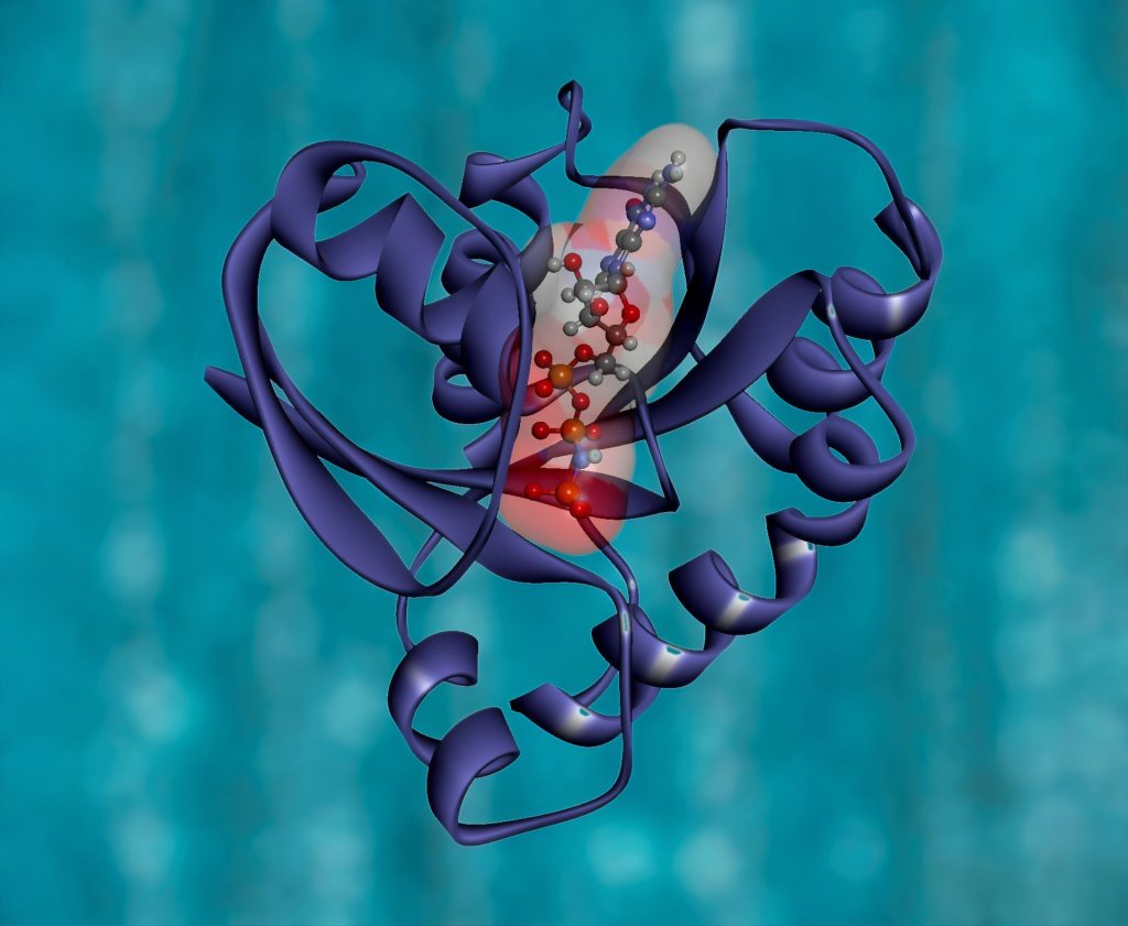
KRAS Protein Structure. RAS is a family of related proteins that is expressed in all animals. KRAS is one of three RAS genes found in humans. RAS genes are mutated in approximately one-third of all human cancers. Photo by National Cancer Institute on Unsplash
Researchers have identified a novel drug that effectively thwarts pancreatic tumours that are addicted to the cancer-causing mutant KRAS gene.
Because early detection of pancreatic cancer is difficult, it has a low survival rate, accounting for just over 3% of all new cancer cases in the US, but leading to nearly 8% of all cancer deaths, according to the National Cancer Institute.
The KRAS gene was recognised more that 25 years ago as the component of Kirsten sarcoma virus responsible for oncogenesis. Since then, mutations of KRAS have been described in a large proportion of solid tumors ranging from more than 90% of pancreatic carcinomas to 20% to 30% of pulmonary adenocarcinomas.
Through a pre-clinical study, Said Sebti, PhD, associate director for basic research at VCU Massey Cancer Center, identified a novel drug that effectively thwarts pancreatic tumors that are addicted to the cancer-causing mutant KRAS gene.
“We discovered a link between hyperactivation of the CDK protein and mutant KRAS addiction, and we exploited this link preclinically to counter mutant KRAS-driven pancreatic cancer, warranting clinical investigation in patients afflicted with this deadly disease,“ said Dr Sebti, who is also the Lacy Family Chair in Cancer Research at Massey and a professor of pharmacology and toxicology at the VCU School of Medicine. “Our findings are highly significant as they revealed a new avenue to combat an aggressive form of pancreatic cancer with very poor prognosis due mainly to its resistance to conventional therapies.”
In 90 percent of pancreatic cancers, KRAS is mutated. Prior studies have shown that some tumours harbouring mutant KRAS are in fact addicted to the mutant gene, meaning they cannot survive or grow without it. Sebti set out to discover if there is a drug that can specifically kill those tumours with a mutant KRAS addiction.
Searching for a suitable drug
Dr Sebti and colleagues used a three-pronged approach to tackle this question.
First of all, they mapped the blueprint of pancreatic cancer cells through global phosphoproteomics, showing them how the addicted and non-addicted tumours differ at the phosphoprotein level. They found two proteins, CDK1 and CDK2, which signalled which cells were addicted to mutant KRAS.
Additionally, they analysed a comprehensive database from the Broad Institute of MIT and Harvard which contains genome-wide CRISPR gRNA screening datasets. They discovered that CDK1 and CDK2 as well as CDK7 and CDK9 proteins were associated with mutant KRAS-addicted tumors.
Finally, they evaluated 294 FDA drugs to selectively kill mutant KRAS-addicted cancer cells over non-KRAS-addicted cancer cells in the lab. They determined the most effective drug in preclinical experiments was AT7519, an inhibitor of CDK1, CDK2, CDK7 and CDK9.
“Using three entirely different approaches, the same conclusion presented itself clearly to us: pancreatic cancer patients whose tumors are addicted to mutant KRAS could benefit greatly from treatment with the CDK inhibitor AT7519,” Dr Sebti said.
To further validate these findings in fresh tumours taken from pancreatic cancer patients the researchers found that AT7519 suppressed the growth of xenograft cells from five mutant KRAS pancreatic cancer patients who relapsed on chemotherapy and/or radiation therapies.
Though AT7519 had previously been tested unsuccessfully in a number of clinical trials, none of the trials were for pancreatic cancer.
“If our findings are correct and translate in humans, then we should be able to see a positive response in pancreatic cancer patients whose tumors are addicted to mutant KRAS,” Dr Sebti said.
As well as pancreatic cancer, the study authors believe these findings may also have clinical implications for colorectal and non-small cell lung cancer patients with prevalent KRAS mutations.
Journal information: Kazi, A., et al. (2021) Global Phosphoproteomics Reveal CDK Suppression as a Vulnerability to KRas Addiction in Pancreatic Cancer. Clinical Cancer Research. doi.org/10.1158/1078-0432.CCR-20-4781.
Chinese researchers have discovered a new regulatory mechanism for the regeneration capacity of skin hair, with important clues for developing treatments for hair loss. Hair loss or alopecia is an extremely common condition, yet there is still no effective therapy for it.
In the skin, activation of hair follicle stem cells (HFSCs) and progenitors by growth factor stimulation is the basis for hair follicle and hair regeneration. Hair regeneration defects can often attribute to blunted responses of HFs to growth stimuli, but it is how the sensitivity of HFSCs or progenitors to growth stimuli is determined is still unclear. Figuring out the answer to this question will provide important clues for the treatment of hair-related diseases such as alopecia.
To this end, Prof Zhang Liang’s group from the Shanghai Institute of Nutrition and Health (SINH) of the Chinese Academy of Sciences, and collaborators uncovered the role of the micro RNA miR-24 and its mechanism in limiting the regenerative ability of hair follicle (HF) progenitors, opening up new therapeutic avenue for hair loss treatment. microRNAs regulate key steps of cell differentiation and development through suppressing gene expression in a sequence-specific manner.
The researchers discovered that that the resting-to-activation transition of HF is associated with significant down-regulation of miR-24 in HF progenitors prior to their activation.
By experimenting with mouse models, they found that miR-24 limits the sensitivity of HF progenitors to growth stimuli. miR-24 over-expression in the skin epithelium significantly delayed HF progenitor activation and hair cycle progression, while its conditional ablation significantly accelerated the hair cycle and increased the HFs’ sensitivity to growth stimuli.
Interestingly, the conditional ablation of miR-24 in skin epithelium significantly improved the effect of Minoxidil lotion on stimulating hair growths without detectable side effects, indicating that miR-24 could be a new potential target for hair regeneration therapies.
Mechanistically, the researchers discovered that Plk3 is a new miR-24 target gene that mediates the function of miR-24 to limit hair growth by regulating CCNE1, a key cell cycle regulator. They also found that miR-24 acts downstream bone morphogenetic protein (BMP), which is a known inhibitory signal for hair growth.
The study revealed that miR-24 is a key factor limiting the regenerative ability of skin HF progenitors. How adult stem cells respond appropriately to environmental stimuli is a question of fundamental importance in stem cell biology.
Journal information: Fengzhen Liu et al, miR-24 controls the regenerative competence of hair follicle progenitors by targeting Plk3, Cell Reports (2021). DOI: 10.1016/j.celrep.2021.109225
Researchers have discovered that a key genetic repair protein also cleans up ‘traps’ left by another protein, its partner in genetic repair.
DNA is constantly getting damaged: the delicate strands that carry life’s genetic code take quite a beating as they jumble about in the course of their work. Errors can accumulate if left untreated, with fatal consequences — such as cancerous tumors — for the cell and the organism.
Two key proteins are involved in preventing the damage from getting out of hand: PARP — or poly ADP ribose polymerase — acts as a marker for a trouble spot, allowing XRCC1 — or X-ray repair cross-complementing protein 1 — to locate the damage and start repairs.
The repair functions of these two proteins have been known for some time. The importance of this has been recognised with the 2015 Nobel prizes for chemistry, as this knowledge allowed the development of anti-cancer drugs, known as PARP inhibitors, that disrupt the growth of certain kinds of tumours.
Although these key proteins had been identified, their precise roles were not well understood. It took a team of scientists at Tokyo Metropolitan University, the University of Sussex, and Kyoto University to revealed how exactly XRCC1 accomplishes its work — and it was a surprising discovery.
“PARP turns out to be something of a villain,” explained Kouji Hirota at Tokyo Metropolitan. “The spots it marks become ‘PARP traps’, which left un-repaired lead to disfunction and cell death.”
It seems that XRCC1 doesn’t just simply repair DNA, it goes about disarming PARP traps. The scientists compared cells without the XRCC1 gene to those without PARP as well as to still others which lacked both proteins. The team found that without XRCC1 on patrol, PARP traps accumulate like landmines.
“PARP exerts toxic effects in the cell and XRCC1 suppresses this toxicity,” Hirota elaborated.
The team aims to further explore these processes, with the goal of aiding development of future cancer treatments.
KyotoU’s Shunichi Takeda said: “These results indicate that XRCC1 is a critical factor in the resolution of PARP traps and may be a determinant of the therapeutic effect of PARP inhibitors used in the treatment of hereditary breast and ovarian cancer syndromes.”
Journal reference: Demin, A. A., et al. (2021) XRCC1 prevents toxic PARP1 trapping during DNA base excision repair. Molecular Cell. doi.org/10.1016/j.molcel.2021.05.009.
Scientists have developed a set of tools that will help create a gene drive to control mosquito-borne diseases such as the West Nile virus, which has received less attention than controlling mosquitoes that transmit malaria.
Since the advent of CRISPR genetic editing revolution, scientists have been working to use the technology to develop gene drives that target pathogen-spreading mosquitoes such as Anopheles and Aedes species, which spread malaria, dengue and other life-threatening diseases.
Much less genetic engineering work has focused on Culex genus mosquitoes, which spread devastating afflictions stemming from West Nile virus, as well as other viruses such as the Japanese encephalitis virus (JEV). Culex mosquitoes are a significant health risk in Africa and Asia, where they transmit the worm causing filariasis, a disease that can lead to a chronic debilitating condition known as elephantiasis.
University of California San Diego scientists have now developed a number of genetic editing tools that will help create a gene drive designed to stop Culex mosquitoes from spreading disease. Gene drives are designed to spread modified genes, in this case those that disable the ability to transmit pathogens, throughout the targeted wild population. The new study is published in the journal Nature Communications,
The researchers developed a Cas9/guide-RNA expression ‘toolkit’ designed for Culex mosquitoes. Since so little genetic engineering work has been done on Culex mosquitoes, the researchers were required to develop their toolkit from scratch, starting with a careful examination of the Culex genome.
“My coauthors and I believe that our work will be impactful for scientists working on the biology of the Culex disease vector since new genetic tools are deeply needed in this field,” said Gantz, an assistant research scientist in the Division of Biological Sciences at UC San Diego. “We also believe the scientific community beyond the gene drive field will welcome these findings since they could be of broad interest.”
The researchers also demonstrated the applicability of their tools in other insects.
“These modified gRNAs can increase gene drive performance in the fruit fly and could potentially offer better alternatives for future gene drive and gene-editing products in other species,” said Gantz.
Gantz and his colleagues have now tested their new tools to ensure proper genetic expression of the CRISPR components and are now on the verge of applying them to a gene drive in Culex mosquitoes. This could be used to stop pathogen transmission by Culex mosquitoes, or alternatively employed to suppress the mosquito population to prevent biting.
Researchers at University of California San Diego School of Medicine have received a grant to conduct a first-in-human Phase 1 clinical trial of a gene therapy for treating Alzheimer’s disease (AD) or Mild Cognitive Impairment (MCI), a condition often preceding dementia.
Gene therapy is an experimental technique that uses genes or gene products for the treatment or prevention of diseases by altering the DNA of living cells. Viral vectors are commonly used to insert the DNA changes into the target cells’ nuclei, but non-viral vectors also exist though they are generally less efficient.
The clinical trial, developed by principal investigator Mark Tuszynski, MD, PhD, professor of neuroscience and director of the Translational Neuroscience Institute at UC San Diego School of Medicine, delivers the brain-derived neurotrophic factor (BDNF) gene into the brains of qualifying trial participants where it is hoped it will stimulate BDNF production in cells.
BDNF belongs to a family of growth factors (proteins) found in the brain and central nervous system that support existing neurons and promote growth and differentiation of new neurons and synapses. BDNF is particularly important in brain regions susceptible to degeneration in AD.
“We found in earlier studies that delivering BDNF to the part of the brain that is affected earliest in Alzheimer’s disease — the entorhinal cortex and hippocampus — was able to reverse the loss of connections and to protect from ongoing cell degeneration,” said Tuszynski. “These benefits were observed in aged rats, aged monkeys and amyloid mice.”
The three-year-long trial seeks to recruit 12 participants with either diagnosed AD or MCI to receive AAV2-BDNF treatment, with another 12 persons serving as a control group over that period.
This will be the first safety and efficacy assessment of AAV2-BDNF in humans. A previous gene therapy trial from 2001 to 2012 using AAV2 and a different protein called nerve growth factor (NGF) found increased growth, axonal sprouting and activation of functional markers in the brains of participants.
“The BDNF gene therapy trial in AD represents an advance over the earlier NGF trial,” said Tuszynski. “BDNF is a more potent growth factor than NGF for neural circuits that degenerate in AD. In addition, new methods for delivering BDNF will more effectively deliver and distribute it into the entorhinal cortex and hippocampus.”
Despite controversial claims, the SARS-CoV-2 virus likely does not integrate its genetic material into the genes of humans, according to a study published in the Journal of Virology.
A prior study reported the virus’s genetic material was found to have integrated into human DNA in cells in petri dishes, though the scientists conducting the newer research now say this was resulted from genetic artefacts in the testing.
Study co-lead author Majid Kazemian, a Purdue University assistant professor of biochemistry and computer science, said that this finding has two important implications.
“Relatively little is known about why some individuals persistently test positive for the virus even long after clearing the infection,” he explained. “This is important because it’s not clear whether such individuals have been re-infected or whether they continue to be infectious to others. So-called ‘human genome invasion’ by SARS-CoV-2 has been suggested as an explanation for this observation, but our data do not support this case.
“If the virus was able to integrate its genetic material into the human genome, that could have meant that any other mRNA could do the same. But because we have shown that this is not supported by current data, this should allay any concerns about the safety of mRNA vaccines,” he concluded.
It is indeed possible for virus genetic material to be incorporated into the DNA of humans and other animals, which are referred to as ‘chimeric events’. These have happened over many millions of years; human DNA contains approximately 100 000 pieces of ‘fossil DNA’ from viruses that have been accumulated throughout our evolution, accounting for nearly 10% of the genetic material in our cells. Some viral fragments even play a role in diseases such as cancer.
Recent scientific journal articles have raised controversy by claiming that the SARS-CoV-2 virus can also cause these chimeric events. Even before this study demonstrated it was not the case, the researchers suspected it was unlikely, said co-lead author Dr Ben Afzali, an Earl Stadtman Investigator of the National Institutes of Health’s National Institute of Diabetes and Digestive and Kidney Diseases.
“While an earlier study suggested that, in cells infected with SARS-CoV-2, genetic material from the virus copied and pasted itself into human DNA, our group thought this seemed unlikely,” Dr Afzali said. “SARS-CoV-2, like HIV, has its genetic material in the form of RNA but, unlike HIV, does not have the machinery to convert the RNA into DNA. SARS-CoV-2 is unlikely to paste itself into the genome and coronaviruses, in general, does not go near human DNA. As our study shows, we find it highly improbable that SARS-CoV-2 could integrate into the human genome.”
Christiane Wobus, associate professor of microbiology and immunology at the University of Michigan Medical School, another co-lead author on the study, said that the collective understanding of RNA viruses is that SARS-CoV-2 integrating into the human genome was extremely unlikely, it was still worth asking the question.
“Unexpected findings in science—when confirmed independently—lead to paradigm shifts and propel fields forward. Therefore, it is good to be open-minded and examine unexpected results carefully, which I believe we did in our study,” she said. “However, we did not find conclusive evidence for SARS-CoV-2 integration, but instead showed that during the RNA sequencing methodology, chimeras are produced at a very low level as an artifact of the laboratory technique.”
To examine any possible integration event, the researchers came up with a novel technique involving extraction the genetic material from infected cells and then boosting the amount of the genetic material 30-fold. With any chimeric events in the host cell DNA, these bits of genetic material from SARS-CoV-2 would have also increased those by 30. The data did not show this.
“We found the frequency of host-virus chimeric events was, in fact, not greater than background noise,” Kazemian stated. “When we enriched the SARS-CoV-2 sequences from the bulk RNA of infected cells, we found that the chimeric events are, in all likelihood, artifacts. Our work does not support the claim that SARS-CoV-2 fuses or integrates into human genomes.”
Journal information: Bingyu Yan et al, Host-virus chimeric events in SARS-CoV2 infected cells are infrequent and artifactual, Journal of Virology (2021). DOI: 10.1128/JVI.00294-21
Researchers are a step closer in the quest to use gene therapy to enable people born deaf to hear, having uncovered a new role for a key protein.
The study, published in Molecular Biology of the Cell, focused on a large gene responsible for an inner-ear protein called otoferlin. Otoferlin mutations are linked to severe congenital hearing loss, a common type of deafness in which patients can hear almost nothing.
“For a long time otoferlin seemed to be a one-trick pony of a protein,” explained Colin Johnson, associate professor of biochemistry and biophysics in the Oregon State UniversityCollege of Science. “A lot of genes will find various things to do, but the otoferlin gene had appeared only to have one purpose and that was to encode sound in the sensory hair cells in the inner ear. Small mutations in otoferlin render people profoundly deaf.”
Because the otoferlin gene is too big as it normally is to package into a delivery vehicle for molecular therapy, Prof Johnson’s team explored the use of a shortened version.
Research led by graduate student Aayushi Manchanda showed the shortened version needed to have part of the gene known as the transmembrane domain, for a surprising reason: without it, the sensory cells matured slowly.
“That was surprising since otoferlin was known to help encode hearing information but had not been thought to be involved in sensory cell development,” Johnson said.
For years, scientists in Prof Johnson’s lab have been working with the otoferlin molecule and in 2017 they identified a shortened form of the gene that can function in the encoding of sound.
To find out if the transmembrane domain of otoferlin needed to be part of the shortened version of the gene, Manchanda shortened the transmembrane domain in zebrafish.
Zebrafish are a small freshwater species that is very popular as a research organism. They grow rapidly, from a cell to a swimming fish in about five days, and share a remarkable similarity to humans at the molecular, genetic and cellular levels due to the conservation of mammalian genes early in their evolution. Embryonic zebrafish are transparent and easily maintained, and are amenable to genetic manipulation.
“The transmembrane domain tethers otoferlin to the cell membrane and intracellular vesicles but it was not clear if this was essential and had to be included in a shortened form of otoferlin,” Manchanda said. “We found that the loss of the transmembrane domain results in the sensory hair cells producing less otoferlin as well as deficits in hair cell activity. The mutation also caused a delay in the maturation of the sensory cells, which was a surprise. Overall the results argue that the transmembrane domain must be included in any gene therapy construct.”
At the molecular level, Manchanda found that a lack of transmembrane domain led to otoferlin not properly linking the neurotransmitter-filled synaptic vesicles to the cell membrane, resulting in less neurotransmitter being released.
“Our study suggests otoferlin’s ability to tether the vesicles to the cell membrane is a key mechanistic step for neurotransmitter release during the encoding of sound,” Manchanda said.
A study published in the journal Cell Reports reveals that a genetic switch that could potentially be targeted to develop new treatments for melanoma by keeping the switch turned off.
Melanoma causes the majority of skin cancer-related deaths, despite only making up roughly 1 percent of skin cancers. The incidence of malignant melanoma is rapidly increasing around the world, and this increase is occurring at a faster rate than that of any other cancer except lung cancer in women. Treatments exist for this serious disease, but the effectiveness of these drugs can vary depending on the individual.
“We’ve been able to correlate the activity of this genetic switch to melanin production and cancer,” said Salk study corresponding author Marc Montminy, a professor in the Clayton Foundation Laboratories for Peptide Biology.
Melanoma develops when melanocytes, the pigment-producing skins in the cell, mutate and begin to multiply out of control. These mutations can cause proteins such as CRTC3 to prompt the cell to produce an abnormal amount of pigment, or to migrate and be more invasive.
While it was known that the CRTC family of proteins (CRTC1, CRTC2, and CRTC3) is involved in pigmentation and melanoma, obtaining precise details about the individual proteins has proven difficult. “This is a really interesting situation where different behaviours of these proteins, or genetic switches, can actually give us specificity when we start thinking about therapies down the road,” said first author Jelena Ostojic, a former Salk staff scientist and now a principal scientist at DermTech.
When the researchers deleted the CRTC3 gene in mice caused a color change in the animal’s coat color, demonstrating that the protein is needed for melanin production. They also found that when melanoma cells lacked the protein, they migrated and invaded less, meaning they were less aggressive, suggesting that inhibiting the protein could help treat the disease.
The team also described the connection between two cellular signalling systems that work on the CRTC3 protein in melanocytes. These two systems tell the cell to either proliferate or make the pigment melanin. Montminy likens this process to a relay race: essentially, a baton (chemical message) is passed from one protein to another until it reaches the CRTC3 switch, either turning it on or off.
“The fact that CRTC3 was an integration site for two signaling pathways—the relay race—was most surprising,” says Montminy, who holds the J.W. Kieckhefer Foundation Chair. “CRTC3 makes a point of contact between them that increases specificity of the signal.”
Next, the team plans to further investigate the mechanism of how CTRC3 impacts the balance of melanocyte differentiation to develop a better understanding of its role in cancer.