Category: Diseases, Syndromes and Conditions

Ancient Y. Pestis DNA Suggests Earlier Start to Black Death

Plague doctor costume
Photo by Peter Kvetny on Unsplash

The origin of the mediaeval Black Death pandemic (AD 1346–1353) has long been studied because of its massive impact on population and society. However, most studies have focused on surviving European records, but they provide little insight into the actual origin of this world-changing pandemic. A new study published in Nature reconstructs the DNA of Yersinia pestis from ancient burial sites, suggesting that 1338 was the date of the first outbreak which would later go on to ravage Eurasia.

Conventional thinking puts the onset of the Black Death at 1346 in the Black Sea region. Recent analysis of historical, genetic and ecological data led to the suggestion that the emergence of Y. pestis branches occurred more than a century before the beginning of the Black Death. According to the proposed model, this initial diversification was linked with territorial expansions of the Mongol Empire across Eurasia during the early thirteenth century. But in this study, the researched present ancient Y. pestis data from central Eurasia supporting a fourteenth-century emergence – putting the emergence a full century later, closer to the conventionally accepted 1346 date.

Until now, the most debated archaeological evidence on the pandemic’s initiation came from cemeteries located near Lake Issyk-Kul in modern-day Kyrgyzstan.

These sites are thought to have housed victims of a fourteenth-century epidemic as tombstone inscriptions directly dated to 1338–1339 state ‘pestilence’ as the cause of death for the buried individuals.

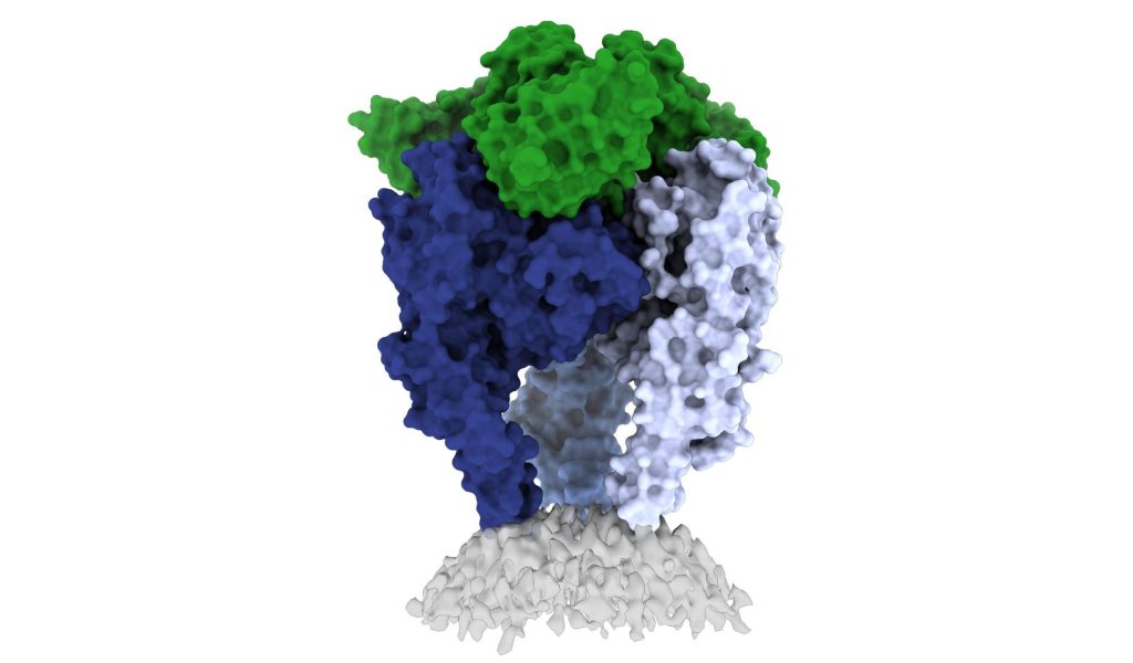
Researchers analysed ancient DNA data from seven individuals exhumed from two of these cemeteries, Kara-Djigach and Burana. The combination of archaeological, historical and ancient genomic data implicates Y. pestis in this epidemic event.

Two reconstructed ancient Y. pestis genomes represent a single strain and are identified as the most recent common ancestor of a major diversification commonly associated with the pandemic’s emergence, here dated to the first half of the fourteenth century. Comparing these ancient genomes present-day diversity from Y. pestis reservoirs in the Tian Shan area where China, Kazakhstan and Kyrgyzstan meet supports a local emergence of the recovered ancient strain.

Exactly how Y. pestis made it to western Eurasia is unknown, but previous research suggested that both warfare and/or trade networks were some of the main contributors in the spread of Y. pestis. However, the lack of any military campaigns in this period and the proximity of trans-Asian networks plus trade items at the site suggest trade playing a role in Y. pestis dissemination.

The authors conclude that “Although the ancient Y. pestis genomes reported in this Article offer biological evidence to settle an old debate, it is the unique historical and archaeological contexts that define our study’s scope and importance. As such, we envision that future synergies will continue to reveal important insights for a detailed reconstruction of the processes that triggered the second plague pandemic.”

Early Intervention in Spinal Muscular Atrophy is Key

Photo by National Cancer Institute on Unsplash

According to the results of a new study published in Developmental Medicine & Child Neurology, early identification and treatment of patients with spinal muscular atrophy (SMA) can greatly reduce the total financial costs associated with the condition. 

A genetic disorder, SMA is characterised by progressive muscle weakness, reduced tone with associated destruction of alpha motor units. There are four main subtypes of spinal muscular atrophy defined by the age of onset and severity with type 0 presenting in utero and causing death within the first months of life and type 4 in adulthood, causing mild weakness and no effect on lifespan. Understanding the underlying pathophysiology, subtypes, and emerging treatments is key to treating patients with spinal muscular atrophy effectively.

Analysing the data of 149 SMA patients, (93 untreated, 42 treated after symptoms arose, and 14 treated after early diagnosis), the total societal cost was lower in untreated patients (due to high drug costs in treated patients), but costs were lower for treated patients who were identified by newborn screening than for treated patients identified due to the development of symptoms. 

“These data are important as they are issued from a real-life prospective collection. They demonstrate clearly that as long as the decision to reimburse treatments for SMA has been made, newborn screening becomes a no-brainer—not only because it gives patients a much better future, but also because it saves a significant amount of money for the taxpayer,” said senior author Laurent Servais, PhD, of the University of Liege, in Belgium and the University of Oxford, in the UK. “Using these data issued from the real world, we are working currently on a model that estimates the lifetime cost of the different strategies.” 

Source: Wiley

A Crystal Clear Look at Rabies Opens up New Vaccines

Scientists from La Jolla Institute for Immunology and the Institut Pasteur have shed light on the structure of the rabies virus glycoprotein, seen here. Credit: Heather Callaway, Ph.D., LJI

In a new study, researchers from La Jolla Institute have unveiled one of the first high-resolution looks at the rabies virus glycoprotein in its vulnerable ‘trimeric’ form. These new images, published in Science Advances, may open up a new vaccine for the deadly virus.

The CDC estimates that 59 000 people die from rabies virus every year, with 40% of those bitten by rabid animals being under 15. Some victims, especially kids, don’t realise they’ve been exposed until it is too late. The intense rabies treatment regimen is not widely available and the average $3800 is out of the reach of less well-off families.

Rabies vaccines, rather than treatments, are much more affordable and easier to administer. But according to Professor Erica Ollmann Saphire, PhD, of the La Jolla Institute, lead researcher of the new study, those vaccines also come with a massive downside.

“Rabies vaccines don’t provide lifelong protection. You have to get your pets boosted every year to three years,” she said. “Right now, rabies vaccines for humans and domestic animals are made from killed virus. But this inactivation process can cause the molecules to become misshapen – so these vaccines aren’t showing the right form to the immune system. If we made a better shaped, better structured vaccine, would immunity last longer?”

“The rabies glycoprotein is the only protein that rabies expresses on its surface, which means it is going to be the major target of neutralising antibodies during an infection,” said LJI Postdoctoral Fellow Heather Callaway, PhD, the study’s first author.

“Rabies is the most lethal virus we know. It is so much a part of our history – we’ve lived with its spectre for hundreds of years,” added Prof Saphire. “Yet scientists have never observed the organisation of its surface molecule. It is important to understand that structure to make more effective vaccines and treatments – and to understand how rabies and other viruses like it enter cells.”

Shapeshifting Rabies virus evades antibodies

Why rabies vaccines don’t provide long-term protection is still unclear, but they do know that its shape-shifting proteins are a problem.

The rabies glycoprotein has sequences that unfold and flip upward when needed, like a Swiss Army knife. The glycoprotein can shift back and forth between pre-fusion (before fusing with a host cell) and post-fusion forms. It can also come apart, changing from a trimer structure (where three copies come together in a bundle) to a monomer (one copy by itself).

This shapeshifting can make rabies invisible to human antibodies, which are built to recognise a single site on a protein. They cannot follow along when a protein transforms to hide or move those sites.

The new study gives scientists a critical picture of the correct glycoprotein form to target for antibody protection.

Capturing the glycoprotein at last

Over the course of three years, Callaway worked to stabilise and freeze the rabies glycoprotein in its pre-fusion form.

Callaway paired the glycoprotein with a human antibody, which helped her pinpoint one site where the viral structure is vulnerable to antibody attacks. The researchers then captured a 3D image of the glycoprotein using cutting-edge cryo-electron microscope equipment at LJI. 

The new 3D structure highlights several key features researchers hadn’t seen before. Importantly, the structure shows the fusion peptides, the way they appear in real life. These two important sequences link the bottom of the glycoprotein to the viral membrane, but project into the target cell during infection. Getting stable image of these sequences is challenging: other rabies researchers have had to cut them off to try to get images of the glycoprotein.

Dr Callaway solved this problem by capturing the rabies glycoprotein in detergent molecules. “That let us see how the fusion sequences are attached before they snap upward during infection,” said Prof Saphire.

Now that scientists have a clear view of this viral structure, they can better design vaccines to create antibodies with a better picture of the targt.

“Instead of being exposed to four-plus different protein shapes, your immune system should really just see one – the right one,” said Dr Callaway. “This could lead to a better vaccine.”

Preventing a family of viruses

More images are needed of rabies virus and its relatives together with neutralising antibodies, and could reveal common antibody targets for lyssaviruses, which can also infect humans and animals. According to Dr Callaway, scientists are working on solving several of these structures, which could reveal antibody targets that lyssaviruses have in common.

“Because we didn’t have these structures of the rabies virus in this conformational state before, it’s been hard to design a broad-spectrum vaccine,” Dr Callaway said.

Source: La Jolla Institute for Immunology

Oil Exploration Software Reveals why Cystic Fibrosis Drugs Fail

Photo by WORKSITE Ltd. on Unsplash

Scientists have harnessed a computational approach usually used in oil exploration to search for cures for rare genetic diseases such cystic fibrosis. By using the method to analyse the spatial relationships between different variants of a protein, instead of the relationships between test wells across an oil field, the researchers can obtain valuable information on how disease affects a protein’s underlying shape and how drugs can restore that shape to normal.

The new method, detailed in the journal Structure, runs with just a few gene sequences collected from people with disease. Then, it determines how the structure of each corresponding variant protein is associated with its function, and how this functional structure can affect pathology and be repaired by therapeutics. To test the techniques, the researchers showed why existing drugs for cystic fibrosis fall short of curing the disease.

“This is an important step forward for treating rare diseases,” said senior author William Balch, PhD, professor of Molecular Medicine at Scripps Research. “The fact that we can get so much information from a few gene sequences is really unprecedented.”

Studies on inherited diseases often rely on the precise three-dimensional shape of a protein affected by disease. But genetic diseases can be caused by thousands of gene variants, some of which destabilise or change the protein shape in ways that make isolating the protein for further investigation much more difficult than usual.

Prof Balch, with Scripps Research senior staff scientist Chao Wang and staff scientist Frédéric Anglés, instead wanted to use natural variation to their advantage. So the group developed a method called variation-capture (VarC) mapping to analyse the natural array of gene sequences which exist in the human population and determine the mechanism by which they each changed a protein’s structure to cause disease.

Among other statistical tools, Prof Balch’s group integrated the methods that oil companies use to draw inferences about the location of an oil reservoir using only a small number of test wells. With only a few gene sequences, this let the researchers determine the most likely structural mechanisms driving function for each variant leading to disease, as well as model how drugs impacted those structural functions.

In the case of cystic fibrosis, disease is caused by genetic variants in the cystic fibrosis transmembrane conductance regulator (CFTR), leading to a buildup of mucus in the lungs. More than 2000 variants of the CFTR gene have been identified, and many of these variants were known to have very different effects on the CFTR protein, but it has been difficult to compare and contrast these variants to guide how patients with different variants should be treated differently in the clinic.

“When you want to treat patients, you really have to appreciate that different therapeutics might target different variants in completely different ways, and that’s why our approach that looks at many different variants all at once is so powerful,” explained Wang. “Our approach not only reveals how these variants contribute to each patient’s biology, but also connects them in a way that each variant can inform how to manage the others.”

The researchers input about 60 genetic variants found in the cystic fibrosis population into their VarC program. The analysis captured how each amino acid residue talks to every other residue to generate function, and revealed that most of the cystic fibrosis patients had the same net effect on the protein: an unstable inner core.

When the program modelled how existing cystic fibrosis drugs impacted the structures, the researchers discovered that, despite the drugs’ effect on CFTR structure, none of them effectively stabilised the protein’s hidden inner core. This was like how the location of an oil reservoir in a complex landscape can be revealed by test wells.

Now that the researchers better understand the structural deficiencies in CFTR in cystic fibrosis patients, they say that the job of developing an effective drug to fix it is much easier. Potential compounds can be modelled in advance of lab experiments for their effect on the inner core of the CFTR protein.

“In most drug discovery, you throw thousands of compounds at a protein and see which ones change it, often without fully understanding the mechanism,” said Prof Balch. “To fix a thing, you must first understand the problem.”

Already, his team is applying the method to other rare genetic diseases, as well as pursuing new drugs to treat cystic fibrosis.

Source: Scripps Research Institute

One in 500 Men Carry an Extra Sex Chromosome, Increasing Disease Risk

Photo by Cottonbro on Pexels

Around one in 500 men could be carrying an extra sex chromosome (X or Y), putting them at increased risk of diseases such as type 2 diabetes, atherosclerosis and thrombosis, according to a study published in Genetics in Medicine.

Researchers from the universities of Cambridge and Exeter analysed genetic data on 200 000 men aged 40 to 70 from UK Biobank. They found 356 men who carried either an extra X chromosome or an extra Y chromosome.

Some men have an extra X or Y chromosome – XXY or XYY, which is usually not obvious without a genetic test. Men with extra X chromosomes, a condition known as Klinefelter syndrome, are sometimes identified during investigations of delayed puberty and infertility; however, most are unaware that they have this condition. Men with an extra Y chromosome tend to be taller as boys and adults, but otherwise they have no distinctive physical features.

In today’s study, the researchers identified 213 men with an extra X chromosome and 143 men with an extra Y chromosome. As the participants in UK Biobank tend to be ‘healthier’ than the general population, this suggests that around one in 500 men may carry an extra X or Y chromosome.

Only a small minority of these men had a diagnosis of sex chromosome abnormality on their medical records or by self-report: fewer than one in four (23%) men with XXY and only one of the 143 XYY men (0.7%) had a known diagnosis.

By linking genetic data to routine health records, the team found that men with XXY have much higher chances of reproductive problems, including a three-fold higher risk of delayed puberty and a four-fold higher risk of being childless. These men also had significantly lower blood concentrations of testosterone. Men with XYY appeared to have a normal reproductive function.

Men with either XXY or XYY had higher risks of several other health conditions: a three-fold higher risk of developing type 2 diabetes, six-fold risk of venous thrombosis, three-fold risk of pulmonary embolism, and four-fold risk of chronic obstructive pulmonary disease (COPD).

It is unclear why an extra chromosome should increase the risk, said the researchers, or why the risks were so similar regardless of which sex chromosome was duplicated.

Yajie Zhao, a PhD student at the Medical Research Council (MRC) Epidemiology Unit at the University of Cambridge, the study’s first author, said: “Even though a significant number of men carry an extra sex chromosome, very few of them are likely to be aware of this. This extra chromosome means that they have substantially higher risks of a number of common metabolic, vascular, and respiratory diseases — diseases that may be preventable.”

Professor Ken Ong, also from the MRC Epidemiology Unit at Cambridge and joint senior author, added: “Genetic testing can detect chromosomal abnormalities fairly easily, so it might be helpful if XXY and XYY were more widely tested for in men who present to their doctor with a relevant health concern.

“We’d need more research to assess whether there is additional value in wider screening for unusual chromosomes in the general population, but this could potentially lead to early interventions to help them avoid the related diseases.”

Professor Anna Murray, at the University of Exeter, said: “Our study is important because it starts from the genetics and tells us about the potential health impacts of having an extra sex chromosome in an older population, without being biased by only testing men with certain features as has often been done in the past.”

Previous studies have found that around one in 1,000 females have an additional X chromosome, which can result in delayed language development and accelerated growth until puberty, as well as lower IQ levels compared to their peers.

Source: University of Cambridge

Amid Measles Spike, WHO Warns of Outbreaks of Vaccine-preventable Diseases

Syringe injection into the upper arm
Image source: NCI on Unsplash

An increase in measles cases in January and February 2022 is a worrying sign of a heightened risk for the spread of vaccine-preventable diseases and could trigger larger outbreaks, particularly of measles affecting millions of children in 2022, warn WHO and UNICEF.

The agencies warn that pandemic-related disruptions, widening vaccine access inequality, and the under-resourcing of routine immunisation are leaving too many children open to measles and other vaccine-preventable diseases.

The risk for large outbreaks has increased as communities relax social distancing practices and other anti-COVID measures. Additionally, the displacement of millions of people due to conflicts and crises including in Ukraine, Ethiopia, Somalia and Afghanistan, is causing disruptions in immunisation services, a lack of clean water and sanitation, and overcrowding, all of which increase the risk of vaccine-preventable disease outbreaks.

Almost 17 338 measles cases were reported worldwide in January and February 2022, compared to 9665 during the first two months of 2021. Measles is highly contagious, so cases tend to show up quickly when vaccinations decline. The agencies are concerned that outbreaks of measles could also forewarn outbreaks of other diseases that do not spread as rapidly.

Apart from its direct, sometimes lethal, effect on the body, the measles virus also weakens the immune system rendering a child more vulnerable for months after to other infectious diseases like pneumonia and diarrhoea.  Most cases occur in settings that have faced social and economic hardships due to COVID, conflict or other crises, and have chronically weak health system infrastructure and insecurity.

“Measles is more than a dangerous and potentially deadly disease. It is also an early indication that there are gaps in our global immunization coverage, gaps vulnerable children cannot afford,” said Catherine Russell, UNICEF Executive Director. “It is encouraging that people in many communities are beginning to feel protected enough from COVID to return to more social activities. But doing so in places where children are not receiving routine vaccination creates the perfect storm for the spread of a disease like measles.”  

In 2020, 23 million children missed out on basic childhood vaccines through routine health services, the highest number since 2009 and 3.7 million more than in 2019.

Top 5 countries with reported measles cases in the last 12 months, until April 2022 1

CountryReported Measles casesRate per million casesFirst dose measles coverage (%), 20192First dose measles coverage (%), 20203
Somalia90685544646
Yemen36291196768
Afghanistan3628916466
Nigeria12 341585454
Ethiopia3039266058

As of April 2022, the agencies report 21 large and disruptive measles outbreaks around the world in the last 12 months. Most of the measles cases were reported in Africa and the East Mediterranean region. The figures are likely higher as the pandemic has disrupted surveillance systems globally, with potential underreporting.

Countries with the largest measles outbreaks since the past year include Somalia, Yemen, Nigeria, Afghanistan and Ethiopia. Insufficient measles vaccine coverage is the major reason for outbreaks, wherever they occur.

“The COVID pandemic has interrupted immunisation services, health systems have been overwhelmed, and we are now seeing a resurgence of deadly diseases including measles. For many other diseases, the impact of these disruptions to immunisation services will be felt for decades to come,” said Dr Tedros Adhanom Ghebreyesus, Director-General of the World Health Organization. “Now is the moment to get essential immunisation back on track and launch catch-up campaigns so that everybody can have access to these life-saving vaccines.”

As of 1 April 2022, 57 vaccine-preventable disease campaigns in 43 countries that were scheduled to take place since the start of the pandemic are still postponed, impacting 203 million people, most of whom are children. Of these, 19 are measles campaigns, which put 73 million children at risk of measles due to missed vaccinations. In Ukraine, the measles catch-up campaign of 2019 was interrupted due to the COVID pandemic and thereafter due to the war. Routine and catch-up campaigns are needed wherever access is possible to help make sure there are not repeated outbreaks as in 2017–2019, when there were over 115 000 cases of measles and 41 deaths in the country – this was the highest incidence in Europe.

Coverage at or above 95% with 2 doses of the safe and effective measles vaccine can protect children against measles. However, COVID pandemic related disruptions have delayed the introduction of the second dose of the measles vaccine in many countries.

Source: World Health Organization

Keratinocytes Play a Role in Stable Vitiligo Disease

Targeting keratinocyte metabolism could be a new method of vitiligo treatment. Photo by Hanen BOUBAHRI on Unsplash

A new study published today in JCI Insight reveals the unique cell-to-cell communication networks that can perpetuate inflammation and prevent repigmentation in patients with stable vitiligo disease, and the particular role that keratinocytes play.

“In this study, we couple advanced imaging with transcriptomics and bioinformatics to discover the cell-to-cell communication networks between keratinocytes, immune cells and melanocytes that drive inflammation and prevent repigmentation caused by vitiligo,” said Anand K. Ganesan, MD, PhD, professor at University of California, Irvine. “This discovery will enable us to determine why white patches continue to persist in stable vitiligo disease, which could lead to new therapeutics to treat this disease.”

Vitiligo is an autoimmune skin disease characterised by the progressive destruction of melanocytes by immune cells called autoreactive CD8+ T cells, resulting in disfiguring patches of white depigmented skin. This disease has shown to cause significant psychological distress among patients. Melanocyte destruction in active vitiligo is mediated by CD8+ T cells, but until now, why the white patches in stable disease persist was poorly understood.

“Until now, the interaction between immune cells, melanocytes, and keratinocytes in situ in human skin has been difficult to study due to the lack of proper tools,” said Jessica Shiu, MD, PhD, assistant professor of dermatology and one of the first authors of the study. “By combining non-invasive multiphoton microscopy (MPM) imaging and single-cell RNA sequencing (scRNA-seq), we identified distinct subpopulations of keratinocytes in lesional skin of stable vitiligo patients along with the changes in cellular compositions in stable vitiligo skin that drive disease persistence. In patients that responded to punch grafting treatment, these changes were reversed, highlighting their role in disease persistence.”

MPM is a unique tool that has broad applications in human skin. MPM is a noninvasive imaging technique capable of providing images with sub-micron resolution and label-free molecular contrast which can be used to characterise keratinocyte metabolism in human skin.

Most studies on vitiligo have focused on active disease, while stable vitiligo remains somewhat of a mystery. Studies are currently investigating when metabolically altered keratinocytes first appear and how they may affect the repigmentation process in patients undergoing treatment.

The study findings suggest the possibility of targeting keratinocyte metabolism in vitiligo treatment. Further studies are needed to improve the understanding of how keratinocyte states affect the tissue microenvironment and contribute to disease pathogenesis.

Source: University of California – Irvine

Mutations that Predispose Patients to Severe Staph Infection

Methicillin-resistant Staphylococcus aureus (MRSA) bacteria. Credit: CDC

A common culprit of skin and respiratory infections, Staphylococcus aureus is highly unpredictable, with the bacteria mostly harmlessly present in the skin of 20–30% of people. However, these bacteria can occasionally cause infections that lead to deadly complications, such as pneumonia, deep skin infections, and sepsis. This was a totally unpredictable outcome – until now.

Now, a new study published in Science identifies a mutated gene common to multiple patients who suffer life-threatening infections and suggests that people living with a genetic condition known as 5p- or Cri-du-chat syndrome may be at similar risk.

“We have characterised severe Staphylococcus aureus infection at the genetic, cellular, immunological, and clinical levels,” said András Spaan, the study’s first author. “By integrating these levels, we have established causality and provided clues for future interventions.”

A first for cell intrinsic immunity

To find out why S. aureus causes disease in some people but not others, scientists examined the protein-coding genomes of more than 100 patients who had suffered from unexplained severe staph infections.

The common genetic thread linking some of these disparate patients were mutations of a gene called OTULIN, which is perched along the short arm of chromosome 5 and codes for an enzyme involved in regulating inflammation. These individuals were not entirely bereft of OTULIN –only one of their two copies of the gene was mutated – but that deficiency appeared to be all it took to render them vulnerable to infections that would scarcely harm other people.

The scientists expected to find that OTULIN deficiency somehow cripples white blood cells or otherwise prevents the immune system from snuffing out S. aureus. But further investigation revealed that these mutations indirectly cause an unrelated protein to aggregate on the surfaces of skin and lung cells, gumming up the tools that those cells use to defend themselves from a toxin produced by S. aureus. This mechanism of defense is known as cell intrinsic immunity.

This finding was particularly surprising because, until then, specific defects in cell intrinsic immunity had only been linked to a predisposition to some viral infections, from COVID to herpes to encephalitis. It had never been shown to play a role in bacterial disease. “This is the first known instance of cell intrinsic immunodeficiency predisposing patients to bacterial infection,” Spaan says.

A larger role for OTULIN

While the individuals whom Spaan and colleagues studied were only missing one copy of OTULIN, people born without either functional copy of this gene face a bevy of early-onset inflammatory diseases, which often prove fatal in the first year of life.

This observation led Spaan to conclude that one functional copy of OTULIN is enough to prevent inflammatory disease, but insufficient to protect against life-threatening staph infections—a genetic mechanism known as haploinsufficiency. “The genetic mechanism was important to pin down,” Spaan says. “People with two functional copies of the gene appear to be healthy, those with no functional copies have autoinflammatory disease, and those with one functional copy are susceptible to severe staph infections.”

Given that general rule, the researchers hypothesized that any population missing only one copy of OTULIN would be similarly predisposed to severe infections. So they then examined a group of volunteers with 5p- syndrome, the most common chromosomal deletion disorder in humans characterized by developmental delays, intellectual disabilities and, in infants, a high-pitched cry. Most 5p- syndrome patients are missing the entire short arm of chromosome 5 and therefore invariably go about their lives with only one functional copy of OTULIN.

Indeed, upon examining six 5p- syndrome patients, the team found that one third were susceptible to lung infections. “We were able to demonstrate that this susceptibility is driven by the fact that they had only one functional copy of OTULIN,” Spaan says. “In many ways, these patients looked genetically similar to the patients we had identified with severe staph infections.”

“Both clinically, and on the cellular level, they could almost be said to have the same disease.”

The findings do not imply that everyone with OTULIN haploinsufficiency or 5p- syndrome will contract severe infections. In fact, the initial results of the study suggested that only 30 percent of individuals with these mutations develop severe disease. Why OTULIN haploinsufficiency appears to cause disease in some patients but not others—a common phenomenon that genetics researchers call “incomplete penetrance”—will be the subject of follow-up studies.

“Many genetic disorders act in this way, but it remains puzzling,” Spaan says. “Why are some people with these mutations perfectly healthy, while others get super ill and may even die?”

One potential answer has already surfaced. Spaan and colleagues found that individuals with OTULIN mutations but no sign of severe disease had high levels of antibodies that neutralise the toxin produced by S. aureus, perhaps due to prior exposure to the common skin bacteria. Individuals with severe disease, on the other hand, had precious few antibodies.

Further investigation into genetic predisposition to diseases, particularly ones as stubborn as staphylococcal infections, may help the development of future treatments. “Studies on these disorders can act as a compass,” Spaan said, “Our research clarifies the interactions between hosts and pathogens, revealing scientific insights into pathogenesis and immunity.”

Source: Rockefeller Institute

80 Cases of Monkeypox Reported in 12 Countries

Close-up of monkeypox lesions on the arm and leg of a female child. Credit: Wikimedia Commons

On Friday, May 20, the World Health Organization has reported that there were 80 cases of monkeypox reported in 12 countries, but has not mentioned which countries those are. However, the National Institute for Communicable Diseases has not reported any cases in South Africa, though there has now been a case reported in Australia.

Update: as of 23 May, the NICD has reported that there are 145 cases in 15 countries, but confirms there are no local cases.

Normally endemic to certain countries where it resides in animal reservoirs, monkeypox is rarely encountered in countries outside those regions. The WHO notes that this is “atypical” for the zoonotic orthopoxvirus, which causes smallpox-like symptoms but with a lower mortality. European public health agencies have so far reported that the UK, Spain, Portugal, Germany, Belgium, France, the Netherlands, Italy and Sweden have seen cases. The first patient in the UK with the virus had returned from a trip to Nigeria, likely catching it there. Cases have been reported in the US and Canada.

The WHO advises that, “As monkeypox spreads through close contact, the response should focus on the people affected and their close contacts. People who closely interact with someone who is infectious are at greater risk for infection: this includes health workers, household members and sexual partners.”

At present, it is unclear why this unusual outbreak is happening now, especially amid the heightened vigilance of the COVID pandemic. One possibility is that some mutation is responsible, though there is little evidence at present to suggest a new variant is responsible.

Another explanation could be that this is simply a matter of the right place and time for the virus. It may also be easier for monkeypox to spread nowadays compared to when there was more widespread use of smallpox vaccine.

Source: BBC News

Methylprednisolone Halves Kidney Failure Risk in IgA Nephropathy

Anatomic model of a kidney
Photo by Robina Weermeijer on Unsplash

A large study has found that treatment with methylprednisolone – a cheap, widely used corticosteroid – halves the risk of losing kidney function and kidney failure in IgA nephropathy. The study, published in the journal JAMA, also found that this can be effectively achieved with fewer side effects if a reduced dose is used.

Researchers say the results of the multi-country study will provide a clear treatment option with definite benefits outweighing well defined and mostly manageable risks. 

IgA nephropathy is a common form of glomerulonephritis caused by the deposition of IgA immunoglobulins in the glomerular basement membrane. Immune-mediated damage to the basement membrane results in haematuria and renal insufficiency progressing to kidney failure in some.

Joint Principal Investigator Professor Vlado Perkovic said that around 10–30% of people with the condition go on to develop kidney failure that requires dialysis or kidney transplantation to prevent death.

“There are few proven treatment options so many treatments including corticosteroids have been used in some patients for decades, despite uncertainty about their effectiveness, as well as the ideal dose. This has led to significant regional variability and clinical uncertainty about this treatment,” he said.

The Therapeutic Evaluation of Steroids in IgA Nephropathy Global (TESTING) study is a double-blinded, randomised, controlled trial that assessed the effects of oral methylprednisolone on major kidney outcomes, kidney failure and safety in patients with IgA nephropathy.

503 patients diagnosed with IgA nephropathy were recruited from centres across Australia, Canada, China (including Hong Kong), India and Malaysia between May 2012 and November 2019. Patients were randomised to receive methylprednisolone or a placebo at:

  1. full dose of 0.6-0.8mg/kg per day of methylprednisolone or placebo for 2 months reducing by 8mg per day each month (262 participants between May 2012 and November 2015), or
  2. reduced dose of 0.4mg/kg per day of methylprednisolone or placebo, also for two months, reducing to 4mg per day each month (241 participants between March 2017 and November 2019), 

for a total treatment period of 6–9 months.

“We found that that treatment with methylprednisolone for six to nine months significantly reduced the risk of losing substantial kidney function, kidney failure requiring dialysis or transplantation, or death from kidney disease compared to placebo,” said Professor Perkovic.

“However, there was an increase in serious adverse events in those who received methylprednisolone, mainly seen in the full dose regimen with fewer in the reduced dose treatment group.”

Joint Principal Investigator Professor Hong Zhang said that with IgA nephropathy being an immune-mediated condition, the benefits seen were likely due to the immune suppressing action of the steroid treatment.

“A well-known side effect of steroid treatment is an increased risk of infections, but we found that this could be mitigated to a degree by using the lower dose and giving the patients antibiotics to prevent infections,” she said. 

“This is the strongest evidence yet for the benefit of any treatment for the prevention of kidney failure in people with IgA nephropathy.

“The results provide a treatment option for clinicians and patients, especially at the lower dose, given the net benefits versus the risk of side effects,” she added.

Associate Professor Muh Geot Wong said that given that the condition develops slowly, and that there was some indication that the effects of treatment appeared to diminish over time, the research team have now extended the study.

“We are now following a significant number of patients from our original study for another five years so we will have a total of around ten years follow up,” he said.

“By then, we hope to have the most comprehensive set of evidence ever collected to help guide the treatment of people with this type of kidney disease.”

Source: EurekAlert!