People with strong legs are less likely to develop heart failure after a heart attack, according to new research. Myocardial infarction is the most common cause of heart failure, with around 6–9% of heart attack patients going on to develop the condition. Previous research has shown that having strong quadriceps is associated with a lower risk of death in patients with coronary artery disease.
Presented at Heart Failure 2023, a scientific congress of the European Society of Cardiology (ESC), this study tested the hypothesis that leg strength is associated with a lower risk of developing heart failure after acute myocardial infarction. The study included 932 patients hospitalised in 2007 to 2020 with acute myocardial infarction who did not have heart failure prior to the admission and did not develop heart failure complications during their hospital stay. The median age was 66 years and 753 participants (81%) were men.
Maximal quadriceps strength was measured as an indicator of leg strength. Patients sat on a chair and contracted the quadriceps muscles as hard as possible for five seconds. A handheld dynamometer attached to the ankle recorded the maximum value in kg. The measurement was performed on each leg and the researchers used the average of both values. Strength was expressed relative to body weight, meaning that quadriceps strength in kg was divided by body weight in kg and multiplied by 100 for a % body weight value. Patients were classified as ‘high’ or ‘low’ strength according to whether their value was above or below the median for their se
The median value for women was 33% body weight and the median value for men was 52% body weight. A total of 451 patients had low quadriceps strength and 481 had high strength. During an average follow-up of 4.5 years, 67 patients (7.2%) developed heart failure. The incidence of heart failure was 10.2 per 1000 person-years in patients with high quadriceps strength and 22.9 per 1000 person-years in those with low strength.
The researchers analysed the association between quadriceps strength (low vs. high) and the risk of developing heart failure. The analysis was adjusted for factors known to be associated with the development of heart failure after myocardial infarction including age, sex, body mass index, prior myocardial infarction or angina pectoris, diabetes, atrial fibrillation, chronic obstructive pulmonary disease, peripheral arterial disease and kidney function. Compared with low quadriceps strength, a high strength level was associated with a 41% lower risk of developing heart failure (hazard ratio [HR]: 0.59; 95% confidence interval [CI] 0.35–1.00; p=0.048).
The investigators also analysed the association between quadriceps strength as a continuous variable and the risk of developing heart failure. Each 5% body weight increment in quadriceps strength was associated with an 11% lower likelihood of heart failure (HR 0.89; 95% CI 0.81–0.98; p=0.014).
Study author Mr. Kensuke Ueno, a physical therapist at the Kitasato University Graduate School of Medical Sciences, Sagamihara, Japan said: “Quadriceps strength is easy and simple to measure accurately in clinical practice. Our study indicates that quadriceps strength could help to identify patients at a higher risk of developing heart failure after myocardial infarction who could then receive more intense surveillance. The findings need to be replicated in other studies, but they do suggest that strength training involving the quadriceps muscles should be recommended for patients who have experienced a heart attack to prevent heart failure.”
New data from the phase III INTERACT3 study demonstrates that a new combination of treatments for stroke due to intracerebral haemorrhage (ICH) significantly improves the chances of favourable outcomes and reduces deaths by one in every 35 patients. The results are important for low- and middle-income countries like South Africa, as the proportion of ICH is much higher in such countries.
The INTERACT3 study is the first-ever randomised controlled trial to show a clearly positive outcome for the treatment of ICH. Timely administration of the new Care Bundle centred on rapid hypertension control, led to improved recovery, lower rates of death, and better overall quality of life in ICH patients. The findings were presented at the European Stroke Organisation Conference in Munich, Germany, and simultaneously published in The Lancet.
Professor Craig Anderson, Director of Global Brain Health at The George Institute and a senior author of the research said, “Despite the high rates of ICH and its severity, there are few proven options for treating it, but early control of high blood pressure is the most promising. Time is critical when treating this type of stroke, so we tested a combination of interventions to rapidly stabilise the condition of these patients to improve their outcomes. We estimate that if this protocol was universally adopted, it could save tens of thousands of lives each year around the world.”
In the INTERACT3 study, over 7000 patients were enrolled across 144 hospitals in 10 countries – nine middle-income countries and one high-income country. The research team evaluated the effectiveness of the new Care Bundle, which included early intensive lowering of systolic blood pressure, strict glucose control, fever treatment, and rapid reversal of abnormal anticoagulation.
They found that using this new treatment protocol compared to usual care reduced the likelihood of a poor functional outcome, including death, after six months. This was estimated to prevent one additional death for every 35 patients treated.
Central to this was a rapid reduction in systolic blood pressure, where target levels were achieved, on average, in 2.3 hours [range 0.8 to 8.0hrs], compared to 4.0 hours [range 1.9 to 16.0hrs] in the control group. The interventional protocol resulted in a statistically significant reduction in mortality, number of serious adverse events, and time spent in hospital, as well as demonstrating an improvement in health-related quality of life.
Patients in the care bundle group had fewer serious adverse events than those in the usual care group (16.0% vs 20.1%).
The burden of ICH is greatest in low- and middle-income countries. In 2019, 30% of all stroke cases in LMICs were ICH, almost double the proportion seen in high-income countries (16%). This is in part due to high rates of hypertension and limited resources for primary prevention strategies, including identification and management of stroke risk factors by healthcare services.
Dr Lili Song, joint lead author and Head of the Stroke Program at The George Institute China, said, “A lack of proven treatments for ICH has led to a pessimistic view that not much can be done for these patients. However, with INTERACT3, we demonstrate on a large scale how readily available treatments can be used to improve outcomes in resource-limited settings. We hope this evidence will inform clinical practice guidelines across the globe and help save many lives.”
June Bellamy’s 83-year-old mother got COVID in March 2022 and was in intensive care for two weeks. She was diagnosed with heart disease. When she was discharged, she was put on oxygen and prescribed medication, including blood-thinning tablets containing rivaroxaban.
During a visit to the doctor earlier this month, Bellamy and her mother were shown a list of different medications containing rivaroxaban. But when they tried to obtain the generic version at the dispensary, which is about 40% cheaper, the supervisor said that because of an ongoing court case they were not allowed to supply it anymore.
“We’ve had to buy the expensive one,” says Johannesburg resident Bellamy, who has been unemployed since 2017. It costs her R1100 a month and she also has to buy other medications. She says her mother is on a basic medical aid plan and the medication is not covered.
“While I’m financially decimated, I’m trying to do what I can,” said Bellamy.
One year and counting
There are a number of court cases dealing with the issue. One of these is between Bayer and Clicks and was heard in the court of the commissioner of patents in Gauteng. The details are complicated and we explain them below. But what is clear is that in April 2022 Judge Colleen Collis reserved judgment in this urgent matter about the sale of blood-thinning tablets. More than a year later, she has failed to hand down her ruling.
In urgent matters judgment is expected almost immediately. It is astonishing for a judge to take a year over any judgment, let alone an urgent one. The judicial norms and standards state that judgments, in non-urgent matters, should be handed down within three months of being reserved.
GroundUp previously reported that the last available list of late judgments on the judiciary’s website is 31 December 2021. The judiciary has stonewalled our requests for an updated list.
We asked for comment from Chief Justice Raymond Zondo and Judge Collis but received no response.
What the case is about
The case deals with the extension of Bayer’s patent on rivaroxaban from December 2020 to January 2026.
In 2000, Bayer obtained a patent on rivaroxaban. Patents are granted for 20 years and so the patent was to expire in December 2020.
The patent-holder of a medicine, in this case Bayer, has exclusive control over it. No other pharmaceutical company may sell the medicine in South Africa during the patent period, at least not without Bayer’s permission. Effectively a patent holder has a monopoly. The point of patents is to create an incentive for pharmaceutical companies to develop new medicines.
In 2007 Bayer obtained a patent for rivaroxaban to be dosed once daily (the original patent was silent on dosing). This extended the patent to 19 January 2026. This kind of patent extension is widely criticised by health activists and is called evergreening.
After the initial patent expired in December 2020, two pharmaceutical companies, Austell and Dr Reddy’s, launched generic versions of rivaroxaban. To cut a long story short, there followed a series of court actions which resulted in Austell and Dr Reddy’s being interdicted from selling their versions of rivaroxaban in South Africa. But the interdicts did not yet stop the big three pharmacy groups, Dis-Chem, Alpha Pharm and Clicks, selling the stock they had of both generic products.
Dis-Chem and Alpha Pharm reached a settlement with Bayer in respect of Dr Reddy’s product, Rivaxored, but Clicks did not. Bayer applied to the court of the commissioner of patents to interdict Clicks from selling rivaroxaban and obtained an urgent interim interdict in March 2022. Shortly after that, in April 2022, the main (and still urgent) hearing for this application took place and Judge Collis reserved judgment. That is where matters stand, over a year later.
As of 13 May, OpenUp’s medicine price website gives the price of a pack of 42 Xarelto (Bayer’s rivaroxaban product) 15mg tablets as R1532. Austell’s equivalent product, Rezalto, is R931.26. Dr Reddy’s product, Rivaxored, is a little higher priced than Rezalto (at 15mg) but considerably lower than Bayer. There’s also iXarola, Bayer’s “authorised generic”, which they brought to market just before the expiry of the 2000 patent. It’s priced at R1285. (These prices exclude the dispensing fee.)
The timeline below contains more detail.
Timeline
2000: Bayer gets patent for rivaroxaban (expires December 2020).
2007: Bayer gets patent for dosing rivaroxaban once daily instead of twice daily (effectively means that the patent expires on 19 January 2026).
2020, December: Initial patent expires.
2021, January: Austell launches its generic version of rivaroxaban, called Rezalto.
2021, April 1: Dr Reddy’s launches its generic version of rivaroxaban, called Rivaxored.
2021, May: Bayer obtains urgent interdicts that stop Austell from selling Rezalto.
2021, December: Bayer obtains interim interdict against Dr Reddy’s, but the interdict does not extend to stopping pharmacies from selling the stock they already had of Dr Reddy’s generic pills.
2022, January: Bayer then launches another urgent application to interdict three pharmacy groups from selling Dr Reddy’s generic pills still in stock. Dis-Chem and Alpha Pharm reach a settlement with Bayer. But Clicks refuses to settle and opposes Bayer’s application.
2022, March 15: Bayer obtains an urgent interim interdict against Clicks at the court of the commissioner of patents in Gauteng, pending a main hearing which takes place at the same court in April.
2022, April: The urgent interdict application between Bayer and Clicks is heard by Judge Colleen Collis in the court of the commissioner of patents in Gauteng. Collis reserves judgment.
2023, May: Judgment has still not been handed down by Judge Collis.
The risk of dying after a heart attack is more than twice as high for women than it is for men, according to research presented at Heart Failure 2023, held by the European Society of Cardiology (ESC).
“Women of all ages who experience a myocardial infarction are at particularly high risk of a poor prognosis,” said study author Dr Mariana Martinho of Hospital Garcia de Orta. “These women need regular monitoring after their heart event, with strict control of blood pressure, cholesterol levels and diabetes, and referral to cardiac rehabilitation. Smoking levels are rising in young women and this should be tackled, along with promoting physical activity and healthy living.”
Previous studies have found that women with ST-elevation myocardial infarction (STEMI) have a worse prognosis during their hospital stay compared to men, and that this may be due to their older age, increased numbers of other conditions, and less use of stents (percutaneous coronary intervention; PCI) to open blocked arteries. This study compared short- and long-term outcomes after STEMI in women and men, and examined whether any sex differences were apparent in both premenopausal (55 years and under) and postmenopausal (over 55) women.
This was a retrospective observational study which enrolled consecutive patients admitted with STEMI and treated with PCI within 48 hours of symptom onset between 2010 and 2015. Adverse outcomes were defined as 30-day all-cause mortality, five-year all-cause mortality and five-year major adverse cardiovascular events (MACE; a composite of all-cause death, reinfarction, hospitalisation for heart failure and ischaemic stroke).
The study included 884 patients. The average age was 62 years and 27% were women. Women were older than men (average age 67 vs 60 years) and had higher rates of high blood pressure, diabetes and prior stroke. Men were more likely to be smokers and have coronary artery disease. The interval between symptoms and treatment with PCI did not differ between women and men overall, but women aged 55 and below had a significantly longer treatment delay after arriving at the hospital than their male peers (95 vs 80 minutes).
The researchers compared the risk of adverse outcomes between women and men after adjusting for factors that could influence the relationship including diabetes, high cholesterol, hypertension, coronary artery disease, heart failure, chronic kidney disease, peripheral artery disease, stroke and family history of coronary artery disease. At 30 days, 11.8% of women had died compared to 4.6% of men, for a hazard ratio (HR) of 2.76. At five years, nearly one-third of women (32.1%) had died versus 16.9% of men (HR 2.33). More than one-third of women (34.2%) experienced MACE within five years compared with 19.8% of men (HR 2.10).
Dr Martinho said: “Women had a two to three times higher likelihood of adverse outcomes than men in the short- and long-term even after adjusting for other conditions and despite receiving PCI within the same timeframe as men.”
The researchers conducted a further analysis in which they matched men and women according to risk factors for cardiovascular disease including hypertension, diabetes, high cholesterol and smoking. Adverse outcomes were then compared between matched men and women aged 55 years and under, and between matched men and women over 55 years old.
There were 435 patients in the matched analysis. In matched patients over 55 years of age, all adverse outcomes measured were more common in women than men. Some 11.3% of women died within 30 days compared with 3.0% of men, for an HR of 3.85. At five years, one-third of women (32.9%) had died compared with 15.8% of men (HR 2.35) and more than one-third of women (34.1%) had experienced MACE compared with 17.6% of men (HR 2.15). In matched patients aged 55 years and below, one in five women (20.0%) experienced MACE within five years compared to 5.8% of men (HR 3.91), while there were no differences between women and men in all-cause mortality at 30 days or five years.
Dr Martinho said: “Postmenopausal women had worse short- and long-term outcomes after myocardial infarction than men of similar age. Premenopausal women had similar short-term mortality but a poorer prognosis in the long-term compared with their male counterparts. While our study did not examine the reasons for these differences, atypical symptoms of myocardial infarction in women and genetic predisposition may play a role. We did not find any differences in the use of medications to lower blood pressure or lipid levels between women and men.”
She concluded: “The findings are another reminder of the need for greater awareness of the risks of heart disease in women. More research is required to understand why there is gender disparity in prognosis after myocardial infarction so that steps can be taken to close the gap in outcomes.”
While HIV and tuberculosis (TB) rates in South Africa are slowly declining, indications are that rates of non-communicable diseases (NCDs) like hypertension and diabetes are on the rise. One response to this shift is to bring some of the strategies used in combatting HIV to NCDs.
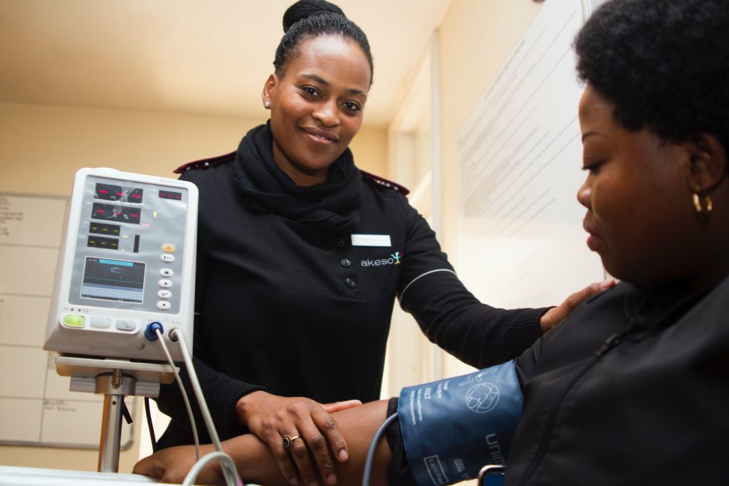
Hypertension, more commonly known as high blood pressure, has been described as a “silent killer” because there are often no symptoms associated with having it. Hypertension is when someone’s blood pressure is consistently higher than normal, which can lead to a host of complications, including stroke, heart attack, and kidney disease. Someone’s risk of developing hypertension is influenced by a number of things, including lifestyle, genetics, age, and family history as well as conditions like diabetes. (Spotlight previously reported on the state of hypertension in South Africa.)
90-60-50
For much of the last decade, UNAIDS’s 90-90-90 targets have been central to how governments have kept track of their HIV responses. The first 90 measured the success of testing programmes, the second 90 measured the success of efforts to get people on to treatment, and the third 90 provided information on how well people are doing once on treatment.
South Africa’s National Strategic Plan (NSP) for the prevention and control of NCDs (2022-2027) sets out similar targets for hypertension and diabetes. As with HIV, the three hypertension indicators will paint a picture of how South Africa is doing on testing, getting people onto treatment, and finally how well people are doing once on treatment.
The hypertension targets are as follows:
90% of people over 18 will know whether they have raised blood pressure.
60% of people with raised blood pressure will receive interventions.
50% of people receiving interventions for hypertension will have controlled blood pressure levels.
Implementation will be key
Local experts interviewed by Spotlight agree that the NSP is a step in the right direction but are clear that much more will be needed.
Professor Brian Rayner, Emeritus Professor in the Division of Nephrology and Hypertension at the University of Cape Town, says he finds the NSP lacking in practical details of how the targets will be achieved. “I’d love for the government to have the plan for how they can achieve this and not another document actually… they need to actually say how are we going do this,” he says.
Professor Angela Woodiwiss of the School of Physiology at the University of the Witwatersrand, and member of the board of the Southern African Hypertension Society has similar concerns. She says the objectives and deliverables in the NSP are sound, but it is short on details when it comes to implementation.
Ways to address this, according to Woodiwiss, is to include “examples of cost-effective practical approaches such as the establishment of cardiovascular screening centres at all district clinics where measurements of blood pressure are done; monthly screening drives at community centres over weekends to increase accessibility to those that work during the week; [and] awareness campaigns at shopping centres”. Another suggestion is for awareness and education campaigns on hypertension to be conducted on media platforms like TV and radio.
“In order to reduce the burden of disease, this target needs to be raised. I would therefore suggest 90-80-70 as the proportions,” she adds.
Professor Andre Kengne, the director of NCD research at the South African Medical Research Council, who was also part of the planning committee for this version of the NSP, says the plan is only a starting point. “The plan says that these [NSP targets] are the entry point, so it’s going to be a catalyst,” he says. “That’s why we just need to start somewhere and then improve on that and again, I think that’s exactly the approach that the plan is taking, This is let’s start small but with the aim of actually progressing.”
Screening: 90% of people over 18 will know whether they have raised blood pressure
A major challenge with NCDs such as hypertension and diabetes is that we don’t have very good epidemiological data in South Africa. Experts referred Spotlight to data from two sources.
Kengne says that based on data collected by the NCD Risk Factor Collaboration, a global network of health scientists that provide data on NCDs, which he is part of, about 40% of adult men and about 42% of adult women in South Africa had hypertension in 2019. Only about 38.5% of men with hypertension were diagnosed at the time and 61.5% of women.
Woodiwiss cites data collected through ‘May Measure Month’ (MMM) South Africa, of where she is a principal investigator. MMM is a global campaign run by the International Society of Hypertension to raise awareness. She cites data collected from screenings conducted from 2017 to 2022.
“The proportion of hypertensive adults aware that they have hypertension ranged from 42.5 to 56.7%,” she says.
When looking at the South African population as a whole, Woodiwiss calculates that this means that only around 13.6 to 19.6% of all people over the age of 18 are aware of whether they have hypertension or not. “We, therefore, have a long way to go in order to achieve the target of 90% of all adults being aware of whether they have raised blood pressure or not,” she adds.
Whichever of the two data sources you look at, South Africa seems to fall well short of the 90% target.
To improve the country’s performance on this measure, experts interviewed by Spotlight agree that there needs to be greater awareness of hypertension (including the importance of checking your blood pressure regularly) and better opportunities for screening.
“There will be no other way of actually improving the numbers without screening people,” Kengne says.
“The current screening approach is essentially hospital-based, and it’s not even yet comprehensive. Meaning only those in contact with the health system are likely, for a proportion, to get their blood pressure measured and then eventually diagnosed with hypertension,” he explains. “The first focus is really to optimise that hospital-based screening, to make sure that everything is in place to measure the blood pressure of whoever gets in contact with the health system.”
Ultimately, Kengne suggests what is needed is to implement community-based approaches to blood pressure screening. One way to do this would be to couple HIV community screening efforts with hypertension screening. As well as to empower community healthcare workers to check blood pressure when doing household visits and then refer people with elevated blood pressure to clinics if needed.
“There need to be national awareness campaigns on TV and radio. These campaigns can be used to encourage individuals to have their blood pressure measured at free screening sites such as community centres, shopping malls, and university campuses as is done as part of the May Measure Month campaign,” Woodiwiss suggests. She adds that a celebrity ambassador would be a great asset for such campaigns.
Treatment: 60% of people with raised blood pressure will receive interventions
“About 85% of those [men] who are diagnosed [with hypertension] are on treatment. And in women it’s about 86%,” Kengne says.
He adds that this is where the NSP targets are maybe not as ambitious as they could be because when you look at the data in the context of everyone who has hypertension (not just those with diagnosed hypertension), only 33% of men and 53% of women are on treatment.
In Woodiwiss’s data, the proportion of hypertensive adults who were receiving medication for hypertension ranged from 36.1 to 49.2%.
Either way, both data sources suggest that one of the biggest challenges to getting people onto treatment is actually diagnosing them in the first place. There is a question, however, whether the health system will be able to cope with the increased treatment load should diagnosis improve.
Kengne suggests that facilities, specifically public health sector facilities, may not be able to cope with the increased demand. “We’re going to need to prepare the health system to cope with the high demand for hypertension care subsequent to increased screening,” he says.
He thinks task-shifting may be part of the solution. Task-shifting was critical to the scaling up of South Africa’s HIV treatment programme, for example, by allowing qualifying nurses to prescribe antiretroviral treatment. Similarly, more healthcare workers, including community healthcare workers, nurses, and field workers can be trained to screen for and treat hypertension.
Woodiwiss stresses the importance of education and awareness when it comes to treatment.
“To facilitate the participation of individuals in the management of their blood pressure, education, and awareness are paramount… An important aspect is to empower individuals to be part of the management of their blood pressure; to re-enforce that hypertension is a chronic problem that requires daily management; and to dispel any notions of stigmatisation due to having high blood pressure,” she says.
Another important practical step would be to reduce the pill burden on hypertension patients in the public sector, according to Rayner. While medication is relatively cheap in this sector, there has not been a move towards combining multiple blood pressure drugs into a single pill, which would make patient adherence easier.
He adds that the process of prescribing blood pressure medication in the private sector needs to be simplified. In line with the idea of task-shifting, Rayner suggests allowing nurses to prescribe medication for straightforward hypertension cases in the public sector as a cost-effective way of treating hypertension.
Control: 50% of people receiving interventions are controlled
About 43% of men in South Africa with hypertension and who are on treatment have controlled blood pressure compared to 54.6% of women, according to Kengne. “Now taken as a proportion of all those with hypertension, I mean our target of 50% controlled will narrow down to about 27% of all people with hypertension [being controlled],” he says. “Using that as the estimate among men currently only 14% of all those with hypertension are controlled and among women, 29% are controlled.”
Data from Woodiwiss suggested that “the proportion of treated individuals with controlled blood pressure ranges from 49.6 to 57.5%.”
For this target then, the country isn’t too far off the 50% target.
But Kengne stresses that blood pressure control is not straightforward. “Diagnosing, it’s not that difficult. Starting treatment it’s not difficult, but actually treating to target it’s a challenge and a number of factors can come into play. Some factors [are] linked to people with hypertension [and] some linked to healthcare providers and the health system,” he says.
For patients, issues like adherence to treatment can be difficult. He suggests using mobile technology, like text messages, to remind patients to take their medication. As well as reducing the pill burden by investing in combination medications.
From the healthcare provider side, Kengne says there needs to be monitoring of patients so that changes to the treatment plan can be made if needed so that the patient can achieve blood pressure control.
“Improving the proportion of treated individuals who have controlled blood pressure requires ongoing monitoring and regular blood pressure checks. As the vast majority of South Africans cannot afford home blood pressure monitors, easy access to blood pressure checks at community clinics, pharmacies, etc. should be provided country-wide,” Woodiwiss says. “It would be ideal if companies could all have corporate wellness days for employees.”
Among a variety of diets a low-calorie ‘green’ Mediterranean diet caused the biggest reduction in aortic stiffness among overweight or dyslipidaemic individuals in a post hoc analysis of a randomised trial. The findings were discussed in Journal of the American College of Cardiology.
As its name suggests, the green Mediterranean diet is rich in plant polyphenols and lower in red or processed meat and simple carbohydrates than a typical low-calorie Mediterranean diet.
Controlling for other variables, the green Mediterranean diet reduced proximal aortic stenosis (PAS) by 15%, better than a typical hypocaloric Mediterranean diet (7.3% reduction) or following standard healthy diet guidelines (4.8% reduction).
The study used the unique environment of a remote Israeli nuclear research facility, where the makeup of the staff meals could be closely controlled and monitored. This also created the limitation of having a predominantly male population for the study group.
More than simple weight loss, the green Mediterranean diet may have greater influence on PAS, which as a measure of “the aortic stiffness from the ascending to the proximal descending thoracic aorta, is a distinct marker of vascular aging and a sensitive early predictor of cardiovascular morbidity and mortality risk,” the researchers noted. “Beyond aging, and similarly to atherosclerosis, PAS is sensitive to obesity-related metabolic conditions, specifically metabolic syndrome.”
“This study provides some of the strongest and most rigorous data thus far to support the connection that better diets maylead to higher fitness,” said study author Dr Michael Mi of Beth Israel Deaconess Medical Center. “The improvement in fitness we observed in participants with better diets was similar to the effect of taking 4000 more steps each day.”
Cardiorespiratory fitness reflects the body’s ability to provide and use oxygen for exercise, and it integrates the health of multiple organ systems, such as the heart, lungs, blood vessels and muscles. It is one of the most powerful predictors of longevity and health. While exercise increases cardiorespiratory fitness, it is also the case that among people who exercise the same amount, there are differences in fitness, suggesting that additional factors contribute. A nutritious diet is associated with numerous health benefits, but it has been unclear whether it is also related to fitness.
This study examined whether a healthy diet is associated with physical fitness in community-dwelling adults. The study included 2380 individuals in the Framingham Heart Study. The average age was 54 years and 54% were women. Participants underwent a maximum effort cardiopulmonary exercise test on a cycle ergometer to measure peak VO2. This is the gold standard assessment of fitness and indicates the amount of oxygen used during the highest possible intensity exercise.
Participants also completed the Harvard semi-quantitative food frequency questionnaire to assess intake of 126 dietary items during the last year ranging from never or less than once per month to six or more servings per day. The information was used to rate diet quality using the Alternative Healthy Eating Index (AHEI; 0 to 110) and Mediterranean-style Diet Score (MDS; 0 to 25), which are both associated with heart health. Higher scores indicated a better quality diet emphasising vegetables, fruits, whole grains, nuts, legumes, fish and healthy fats and limiting red meat and alcohol.
The researchers evaluated the association between diet quality and fitness after controlling for other factors that could influence the relationship, including age, sex, total daily energy intake, body mass index, smoking status, cholesterol levels, blood pressure, diabetes and routine physical activity level. The average AHEI and MDS were 66.7 and 12.4, respectively. Compared with the average score, an increase of 13 points on the AHEI and 4.7 on the MDS was associated with a 5.2% and 4.5% greater peak VO2, respectively.
Dr Mi said: “In middle-aged adults, healthy dietary patterns were strongly and favourably associated with fitness even after taking habitual activity levels into account. The relationship was similar in women and men, and more pronounced in those under 54 years of age compared to older adults.”
To discover the potential mechanism linking diet and fitness, the researchers performed further analyses. They examined the relationship between diet quality, fitness and metabolites, which are substances produced during digestion and released into the blood during exercise. Researchers measured 201 metabolites (eg amino acids) in blood samples collected in a subset of 1154 study participants. Some 24 metabolites were associated with either poor diet and fitness, or with favourable diet and fitness, after adjusting for the same factors considered in the previous analyses. Dr Mi said: “Our metabolite data suggest that eating healthily is associated with better metabolic health, which could be one possible way that it leads to improved fitness and ability to exercise.”
“This was an observational study and we cannot conclude that eating well causes better fitness, or exclude the possibility of a reverse relationship, i.e. that fit individuals choose to eat healthily.”
Dr Mi concluded: “There are already many compelling health reasons to consume a high-quality diet, and we provide yet another one with its association with fitness. A Mediterranean-style diet with fresh, whole foods and minimal processed foods, red meat and alcohol is a great place to start.”
With the administration of the first COVID vaccines two years ago, public health officials found an increase in cases of myocarditis, particularly among young males who had been vaccinated with mRNA vaccines. The underlying cause of these reactions remained a mystery.
Now Yale scientists have identified the immune signature of these heart inflammation cases. Published in the journal Science Immunology, their findings eliminate some of the theorised causes of the heart inflammation and point to the consequences of a slightly over-stimulated immune system.
Myocarditis is a generally mild inflammation of heart tissue which can cause scarring but is usually resolved within days. The increased incidence of myocarditis during vaccination was seen primarily in males in their teens or early 20s, who had been vaccinated with mRNA vaccines, which are designed to elicit immune responses specifically to the SARS-CoV-2 virus.
According to the Centers for Disease Control and Prevention (CDC), among males aged 12 to 17, about 22 to 36 per 100 000 experienced myocarditis within 21 days after receiving a second vaccine dose. The incidence of myocarditis was 50.1 to 64.9 cases per 100 000 after infection with the COVID virus among males in this age group.
For the new study, the Yale research team conducted a detailed analysis of immune system responses in those rare cases of myocarditis among vaccinated individuals. They found that the heart inflammation was not caused by antibodies created by the vaccine, but rather by a more generalised response involving immune cells and inflammation.
“The immune systems of these individuals get a little too revved up and over-produce cytokine and cellular responses,” said team leader Carrie Lucas, associate professor of immunobiology.
Earlier research had suggested that increasing the time between vaccination shots from four to eight weeks may reduce risk of developing myocarditis.
Lucas noted that, according to CDC findings, the risk of myocarditis is significantly greater in unvaccinated individuals who contract COVID than in the vaccinated. She emphasised that vaccination offers the best protection from COVID-related disease.
“I hope this new knowledge will enable further optimising mRNA vaccines, which, in addition to offering clear health benefits during the pandemic, have a tremendous potential to save lives across numerous future applications,” said Anis Barmada, an MD/PhD student at Yale School of Medicine, who is a co-first author of the paper with Jon Klein, also a Yale MD/PhD student.
Postmenopausal women with atherosclerosis are at higher risk of heart attacks than men of similar age, according to research presented at EACVI 2023, a scientific congress of the European Society of Cardiology (ESC), and published in European Heart Journal – Cardiovascular Imaging. Researchers used imaging techniques to examine the arteries of nearly 25 000 patients and followed them for heart attacks and death.
“The study suggests that a given burden of atherosclerosis is riskier in postmenopausal women than it is in men of that age,” said study author Dr Sophie van Rosendael of Leiden University Medical Centre. “Since atherosclerotic plaque burden is emerging as a target to decide the intensity of therapy to prevent heart attacks, the findings may impact treatment. Our results indicate that after menopause, women may need a higher dose of statins or the addition of another lipid-lowering drug. More studies are needed to confirm these findings.”
While young women do have heart attacks, in general, women develop atherosclerosis (narrowing of arteries due to plaque buildup) later in life than men and have heart attacks at an older age than men, in part because of the protective effect of oestrogen. This study examined whether the prognostic importance of atherosclerotic plaques are the same for women and men at different ages as this could be important for selecting treatments to prevent heart attacks.
The study included 24 950 patients referred for coronary computed tomography angiography (CCTA) and enrolled in the CONFIRM registry, which was conducted in six countries in North America, Europe, and Asia. CCTA is used to obtain 3D images of the arteries in the heart.
Total atherosclerotic burden was rated using the Leiden CCTA score, which incorporates the following items for each coronary segment: plaque presence (yes/no), composition (calcified, noncalcified or mixed), location, and severity of narrowing, for a final value of 0 to 42. Patients were divided into three categories previously found to predict the risk myocardial infarction: low atherosclerotic burden (0 to 5), medium (6 to 20) and high (over 20). In addition, obstructive coronary artery disease was defined as 50% narrowing or more.
The primary outcome was the difference in Leiden CCTA score between women and men of similar age. The investigators also analysed sex differences in the rates of major adverse cardiovascular events (MACE), which included all-cause death and myocardial infarction, after adjusting for age and cardiovascular risk factors (hypertension, high cholesterol, diabetes, current smoking and family history of coronary artery disease).
A total of 11 678 women (average age 58.5 years) and 13 272 men (average age 55.6 years) were followed for 3.7 years. Regarding the primary outcome, the study showed an approximately 12 year delay in the onset of coronary atherosclerosis in women: the median Leiden CCTA risk score was above zero at age 64 to 68 years in women versus 52 to 56 years in men (p<0.001). In addition, the overall plaque burden as quantified by the Leiden CCTA score was significantly lower in women, who had more non-obstructive disease.
Dr. van Rosendael said: “The results confirm the previously reported delay in the start of atherosclerosis in women. We also found that women are more likely to have non-obstructive disease. It was formerly thought that only obstructive atherosclerosis caused myocardial infarction but we now know that non-obstructive disease is also risky.”
The burden of atherosclerosis was equally predictive of MACE in premenopausal women (aged under 55 years) and men of the same age group. However, in postmenopausal women (age 55 years and older), the risk of MACE was higher than men for a given score. In postmenopausal women, compared to those with a low burden, those with a medium and high burden had 2.21-fold and 6.11-fold higher risks of MACE. While in men aged 55 years and older, compared to those with a low burden, those with a medium and high burden had 1.57-fold and 2.25-fold greater risks of MACE.
Dr van Rosendael said: “In this study, the elevated risk for women versus men was especially observed in postmenopausal women with the highest Leiden CCTA score. This could be partly because the inner diameter of coronary arteries is smaller in women, meaning that the same amount of plaque could have a larger impact on blood flow. Our findings link the known acceleration of atherosclerosis development after menopause with a significant increase in relative risk for women compared to men, despite a similar burden of atherosclerotic disease. This may have implications for the intensity of medical treatment.”
Using new genetic tools to study statins in human cells and mice, researchers have uncovered how these drugs protect the cells that line blood vessels. Published in Nature Cardiovascular Research, the findings provide new insight into statins’ curiously wide-ranging benefits, for conditions ranging from arteriosclerosis to diabetes, that have long been observed in the clinic.
“The study gives us an understanding, at a very deep mechanistic level, of why statins have such a positive effect outside of reducing LDL [low-density lipoprotein],” said professor of medicine Joseph Wu, MD, PhD, the study’s senior author. “Given how many people take statins, I think the implications are pretty profound.”
Developed in the 1980s from compounds found in mould and fungi, statins target an enzyme that regulates cholesterol production in the liver. But clinical trials have shown that they also seem to safeguard against cardiovascular disease beyond their ability to lower cholesterol.
Heart failure patients who take statins, for example, are less likely to suffer a second heart attack. They have also been shown to prevent the clogging of arteries, reduce inflammation and even lower cancer risk. Yet these underlying mechanisms are poorly understood.
“Statins were invented to lower cholesterol by targeting the liver. But we didn’t know the targets or the pathways in the cardiovascular system,” said Chun Liu, PhD, an instructor at the Stanford Cardiovascular Institute and co-lead author.
Mesenchymal cells are poor substitutes
To take a closer look at statins’ effect on blood vessels, Liu and colleagues tested a common statin, simvastatin, on lab-grown human endothelial cells derived from induced pluripotent stem cells. Endothelial cells make up the lining of blood vessels, but in many diseases they transform into a different cell type, known as mesenchymal cells, which are poor substitutes.
“Mesenchymal cells are less functional and make tissues stiffer so they cannot relax or contract correctly,” Liu said.
The researchers suspected that statins could reduce this harmful transition. Indeed, endothelial cells treated with simvastatin in a dish formed more capillary-like tubes, a sign of their enhanced ability to grow into new blood vessels.
RNA sequencing of the treated cells offered few clues. The researchers saw some changes in gene expression, but they “didn’t find anything interesting,” Liu said.
It was not until they employed a newer technique called ATAC-seq that the role of statins became apparent. ATAC-seq reveals what happens at the epigenetic level, meaning the changes to gene expression that do not involve changes to the genetic sequence.
They found that the changes in gene expression stemmed from the way strings of DNA are packaged inside the cell nucleus. DNA exists in our cells not as loose strands but as a series of tight spools around proteins, together known as chromatin. Whether particular DNA sequences are exposed or hidden in these spools determines how much they are expressed.
“When we adopted the ATAC-seq technology, we were quite surprised to find a really robust epigenetic change of the chromatin,” Liu said.
ATAC-seq revealed that simvastatin-treated cells had closed chromatin structures that reduced the expression of genes that cause the endothelial-to-mesenchymal transition. Working backward, the researchers found that simvastatin prevents a protein known as YAP from entering the nucleus and opening chromatin.
The YAP protein is known to play important roles in development, such as regulating the size of our organs, but also has been implicated in the abnormal cell growth seen in cancer.
A look at diabetes
To see the drug in context, the researchers tested simvastatin on diabetic mice. Diabetes causes subtle changes to blood vessels that mimic the damage commonly seen in people who are prescribed statins — older patients who do not have a cardiovascular condition, Liu said.
They found that after eight weeks on simvastatin, the diabetic mice had significantly improved vascular function, with arteries that more easily relaxed and contracted.
“If we can understand the mechanism, we can fine-tune this drug to be more specific to rescuing vascular function,” Liu said.
The findings also provide a more detailed picture of the vascular disease process, which could help doctors identify and treat early signs of vascular damage.