Category: Cancer

Excellent Outcomes for Shrinking Liver Cancer Tumours Before Transplant

Photo by Piron Guillaume on Unsplash

Shrinking liver cancer tumours in order to allow the patient to qualify for a liver transplant leads to excellent 10-year post-transplant outcomes, according to the findings of a new study published in JAMA Surgery. The results validate current US policies around transplant eligibility.

Selection of patients with hepatocellular carcinoma (HCC), the most common form of liver cancer, for transplant has been guided for more than two decades by standards known as the Milan criteria. The Milan criteria state that transplantation should be performed in those with a single tumour of 5cm or less in diameter or three tumours that are each 3cm or less in diameter, have no macrovascular invasion, and no metastasis. Over time, the rising incidence of HCC and mortality rates in the United States have led to refinements to the selection policy, shifting the focus to guidelines that also incorporate tumour biology, response to bridging therapies, and waiting times for patients within and beyond the Milan criteria.

One aspect of the current criteria is known as downstaging: the process of applying liver directed therapy to tumours too big for the Milan criteria with the hope of reducing them to the suggested size. Downstaging is now an option in selecting suitable liver transplant candidates with initial tumors that exceed the criteria. However, liver cancer can recur after transplantation, either within or outside the liver. The treatment options of patients who have recurrence post transplantation is limited and prognosis is poor.

In this cohort study, a retrospective multicentre analysis of prospectively collected data was conducted for 2645 adults who had undergone liver transplant for HCC at five centres between January 2001 and December 2015. The outcomes of 341 patients whose disease was downstaged to fit within the Milan criteria were compared with those in 2122 patients whose disease always fit within the criteria and 182 patients whose disease was not downstaged.

The 10-year post-transplant survival and recurrence rates were, respectively, 52.1% and 20.6% among those whose disease was downstaged; 61.5% and 13.3% in those always within the criteria; and 43.3% and 41.1% in those whose disease was not downstaged.

“Our study validates national policy on downstaging prior to transplantation and shows the clear utility benefit for transplantation prioritisation decision-making,” said Parissa Tabrizian, MD, co-lead author on the study. “These results can increase the level of recommendations for the downstaging policy on a global basis. It also demonstrates that surgical management of HCC recurrence after transplantation is associated with improved survival in well-selected patients and should be pursued. The study also supports expanding the policy of downstaging applied to guidelines in Europe and Asia.”

“Our study represents a solid confirmation that HCC patients effectively downstaged to Milan criteria have an outstanding median survival of 10 years, thus providing the rationale to adopt this policy on a global basis,” said Josep Llovet, MD, PhD, co-lead author on the study. “With this study clinical practice guidelines of management of HCC can recommend our approach with an acceptable level of evidence.”

Source: The Mount Sinai Hospital

Hormone Therapy does not Increase Breast Cancer Recurrence Risk in Survivors

Research published in the Journal of the National Cancer Institute found that menopausal hormone therapy for breast cancer survivors is not associated with breast cancer reoccurrence, despite worries among some researchers and physicians.

Hot flashes and night sweats, as well as vaginal dryness and urinary tract infections, are common in breast cancer survivors, worsening quality of life and can lead patients to discontinue therapy. These symptoms may be alleviated by vaginal oestrogen therapy or menopausal hormone therapy (MHT). However, the safety of systemic and vaginal oestrogen use among breast cancer survivors, particularly those with oestrogen receptor-positive disease, has been unclear.

Many doctors caution breast cancer survivors against using MHT following the demonstration of an increased risk of breast cancer recurrence in two trials in the 1990s. Though later studies have not shown increased recurrence, they were seriously limited, with small sample sizes and short follow-up periods.

This study compared hormonal treatment with the risk of breast cancer recurrence and mortality in a large cohort of Danish postmenopausal women treated for early-stage oestrogen receptor-positive breast cancer.

Participants were diagnosed between 1997 and 2004 with early-stage breast cancer who received no treatment or five years of hormone therapy.

Among 8461 women, 1957 and 133 used vaginal oestrogen therapy or MHT, respectively, after diagnosis. No increase was seen in the risk of recurrence or mortality for those who received either vaginal oestrogen therapy or MHT.

“This large cohort study helps to inform the nuanced discussions between clinicians and breast cancer survivors about the safety of vaginal oestrogen therapy,” said Elizabeth Cathcart-Rake, writing in an accompanying editorial. “These results suggest that breast cancer survivors on tamoxifen with severe genitourinary symptoms can take vaginal estrogen therapy without experiencing an increase in their risk for breast cancer recurrence. However, caution is still advised when considering vaginal oestrogen for breast cancer survivors on aromatase inhibitors, or when considering menopausal hormonal therapy.”

Source: EurekAlert!

Cancer Cells Seek their ‘Goldilocks’ Zone of Tissue Softness

Source: National Cancer Institute on Unsplash

Research into the mechanisms of cell migration and the impact of tissue rigidity on cell positioning and steering has found that cancer cell migrate towards and opens new possibilities for stopping and directing it.

An international team of scientist have uncovered for the first time how tissue stiffness determines cell positioning and regulates all types of cell migration ranging from the neuronal growth cone turning to dissemination of malignant cancer cells in brain tumors and breast cancer.

Each cell in the body has a specific task and carefully determined position within a tissue. Cell positioning is regulated by many factors, including tissue rigidity. Cells are capable of probing and sensing their environment, and different cell types have different preferences for optimal conditions. A little bit like Goldilocks in the story trying out the different beds of the bear family and finding one bed to be too soft, the other one two hard and one to be just right. While this has been well-known for a long time, it has remained a mystery to researchers how cells are able to steer themselves to the optimal environment.

“The prevailing view among scientists was that all cell types prefer high-rigidity environments and migrate towards increasing stiffness. This process has been coined the term ‘durotaxis‘ – migration towards hard from Greek and Latin,” said Aleksi Isomursu, a doctoral researcher.

“I was visiting the University of Minnesota for a research project and noticed that brain cancer cells grown on engineered substrates with alternating stiffness show the opposite behaviour they turned towards soft,” Isomursu continues.

This observation launched an interdisciplinary research project involving cancer cell biology, computational modelling and engineering and involving researchers from three continents. As the outcome, the researchers uncovered the basic mechanism all cell types use to steer themselves towards their optimal environment.

These results will be important for future research in stopping and directing cancer cell migration.

“I experimented with different types of drugs and identified ones that could make brain cancer cells stop moving or change direction,” explains Postdoctoral Researcher Mathilde Mathieu.

Identification of the mechanism of cell steering provides explanations for many thus far mysterious steps in cancer dissemination, for example how cancer cells migrate out from the stiff core of a breast tumour.

“These findings have been gaining a lot of interest in researchers and we have even played around with the idea of launching a new term – ‘mollitaxis‘, migration towards soft,” says the Principal Investigator of the laboratory at the University of Turku, Professor Johanna Ivaska.

Source: University of Turku

In Liver Cancer, Protective p53 Protein Turns Traitor

Genetics
Image source: Pixabay

p53 is one of the most important proteins in cancer biology. Often referred to as a “guardian of the genome”, p53 activates in response to cellular stressors such as DNA damage. Its activation induces processes such as cell death that prevent cancer development. Because of this, p53 mutations are extremely common in cancers, including hepatocellular carcinoma.

However, a study published in Cancer Research, revealed that the constant activation of p53 in liver cells of patients suffering from chronic liver disease (CLD) could actually promote the development of liver cancer.

CLD can be brought on by different factors including viruses, alcohol use, and fat accumulation, all of which can induce p53 activation. Previous studies have shown that p53 is in a constant state of activation in the liver cells of CLD patients. Yet, it is not clear what role this plays in CLD pathophysiology.

“Clinical data clearly show that p53 is activated in the hepatocytes of individuals with CLD,” says Yuki Makino, lead author of the study at Osaka University. “Because p53 is such a vital part of how the human body prevents tumor formation, its role in CLD became even more intriguing.”

To create a mouse model with p53 accumulation in hepatocytes, researchers deleted Mdm2, the protein responsible for regulating p53 expression by targeting it for degradation. These mice developed liver inflammation with higher amounts of hepatocyte apoptosis and senescence-associated secretory phenotype (SASP), a phenomenon where cells produce signals within the microenvironment that can cause nearby cells to become cancerous. In fact, mice with p53 accumulation did have increased liver tumour development.

“We also observed an expanded population of hepatic progenitor cells (HPCs), which have stem cell-like characteristics,” explained senior author Tetsuo Takehara. “When the HPCs were isolated, grown in culture, and then injected under the skin of lab mice, these animals developed tumors. This suggested that HPCs played a key part in the liver tumor formation seen in the animals with p53 accumulation.”

Interestingly, acceleration of liver tumour development and the other observed phenotypes did not occur when p53 was deleted in addition to Mdm2 in the hepatocytes. These results demonstrated the significance of constant p53 activity in the tumorigenesis.

“We then compared samples from 182 CLD patients with 23 healthy liver samples,” said Dr Makino. “The CLD liver biopsy samples showed activated p53 was positively correlated with apoptosis levels, SASP, HPC-associated gene expression, and later cancer development.”

The authors concluded that constitutively activated p53 in hepatocytes of CLD patients can create a microenvironment that is supportive of tumour formation from HPCs. Their work proposes a novel and paradoxical mechanism of liver tumorigenesis because p53 is one of the most well-known tumor suppressor genes. These data could highlight p53 as a potential cancer-prevention treatment target for CLD patients.

Source: Osaka University

Topping up Mitochondrial Content to Fight Kidney Cancer

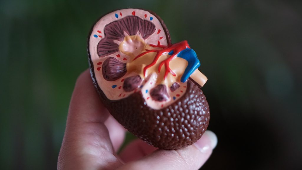
Anatomic model of a kidney
Photo by Robina Weermeijer on Unsplash

Researchers at Karolinska Institutet in Sweden have linked resistance to treatment for VHL syndrome-induced kidney cancer to low mitochondrial content in the cell. When the researchers increased the mitochondrial content with an inhibitor, the cancer cells responded to the treatment. Their findings, which are published in Nature Metabolism, may lead to more targeted cancer drugs.

Mitochondria are the most oxygen-demanding component of the cell, but it was not known how mitochondria adapt in a low-oxygen environment and how they are linked to cancer therapy resistance.

“We’ve shown for the first time how the formation of new mitochondria is regulated in cells that lack oxygen and how this process is altered in cancer cells with VHL mutations,” explained Associate Professor Susanne Schlisio, group leader at the Karolinska Institutet.

A gene called von Hippel-Lindau (VHL) prevents healthy cells from turning cancerous. The 2019 Nobel Prize in Physiology or Medicine was awarded to the discovery that VHL was part of the cell’s oxygen detection system. Normally, VHL breaks down another protein called HIF – but when VHL is mutated, HIF accumulates and causes a disease called VHL syndrome in which the cells react as if they were lacking oxygen. This syndrome greatly increases the risk of tumours, both benign and malignant. VHL syndrome-induced kidney cancer has a poor prognosis, with a five-year survival rate of just 12%.

Researchers analysed the protein content of cancer cells from patients with different variants of VHL syndrome, to see how they differed from another group of individuals with a special VHL mutation called Chuvash, a mutation involved in hypoxia-sensing disorders without any tumour development. Those with the Chuvash VHL-mutation had normal mitochondria in their cells, while those with VHL syndrome mutation had few.

To increase the amount of mitochondrial content in VHL related kidney cancer cells, the researchers treated these tumours with an inhibitor of a mitochondrial protease called “LONP1.” This resulted in the cells becoming susceptible to the cancer drug sorafenib, which they had previously resisted. In mouse studies, this combination treatment led to reduced tumour growth.

The study’s first author Shuijie Li, postdoctoral researcher in the Schlisio’s group, suggested that the findings could be applied to more than just VHF syndromic kidney cancers.

“We hope that this new knowledge will pave the way for more specific LONP1 protease inhibitors to treat VHL-related clear cell kidney cancer,” Dr Li said. “Our finding can be linked to all VHL syndromic cancers, such as the neuroendocrine tumours pheochromocytoma and paraganglioma, and not just kidney cancer.”

Source: Karolinska Institutet

PET/CT Scans Fail to Beat MRI for Prostate Cancer Detection

Credit: Darryl Leja / National-Human-Genome Research Institute / National Institutes of Health

Researchers found that MRI scans, the current gold standard, can still detect prostate cancer more accurately than the newer, prostate-specific -PSMA PET/CT scanning technique.

The findings were presented at the European Association of Urology’s annual congress (EAU22).

Prostate-specific membrane antigen (PSMA) PET/CT scans, approved by the US FDA in 2020, use a radioactive dye to highlight areas of PSMA, which is overexpressed on the surface of prostate cancer cells. Presently, these scans are used to manage prostate cancer, as they can accurately measure the progression or recurrence of the disease. The researchers set out to find if they could be used to diagnose prostate cancer as well.

The PEDAL trial recruited 240 patients at risk of prostate cancer, with each patient given both an MRI scan and a PSMA PET/CT scan. If imaging suggested the presence of prostate cancer, a biopsy was performed by the patient’s urologist.

The MRI scans picked up abnormalities in 141 patients, while the PSMA PET/CT scans picked up abnormalities in 198 patients. A total of 181 patients (75%) underwent a prostate biopsy, and subsequently 82 of those patients were found to have clinically significant prostate cancer.

The MRI scans were significantly more accurate at detecting any grade of prostate cancer than the PSMA PET scans.

The research team was led by Associate Professor Lih-Ming Wong, who explained: “Our analysis found that MRI scans were better than PSMA-PET for detecting any grade of prostate cancer. When we looked only at clinically significant prostate cancers, there was no difference in accuracy.  As this study is one of the first to explore using PSMA-PET to diagnose cancer within the prostate, we are still learning and adjusting how to improve using PSMA-PET in this setting.

Although detection thresholds will be  fine-tuned as diagnostic use develops,  Prof Wong believes the trial has important lessons for clinicians. 

He said: “This study confirms that the existing ‘gold standard’ of pre-biopsy detection – the MRI – is indeed a high benchmark. Even with fine-tuning, we suspect PSMA PET/CT won’t replace the MRI as the main method of prostate cancer detection. But it will likely have application in the future as an adjunct to the MRI, or for people for whom an MRI is unsuitable, or as a single combined “diagnostic and staging” scan for appropriately selected patients.”

Source: EurekAlert!

‘Goldilocks’ Window for Immunotherapy Without Side Effects

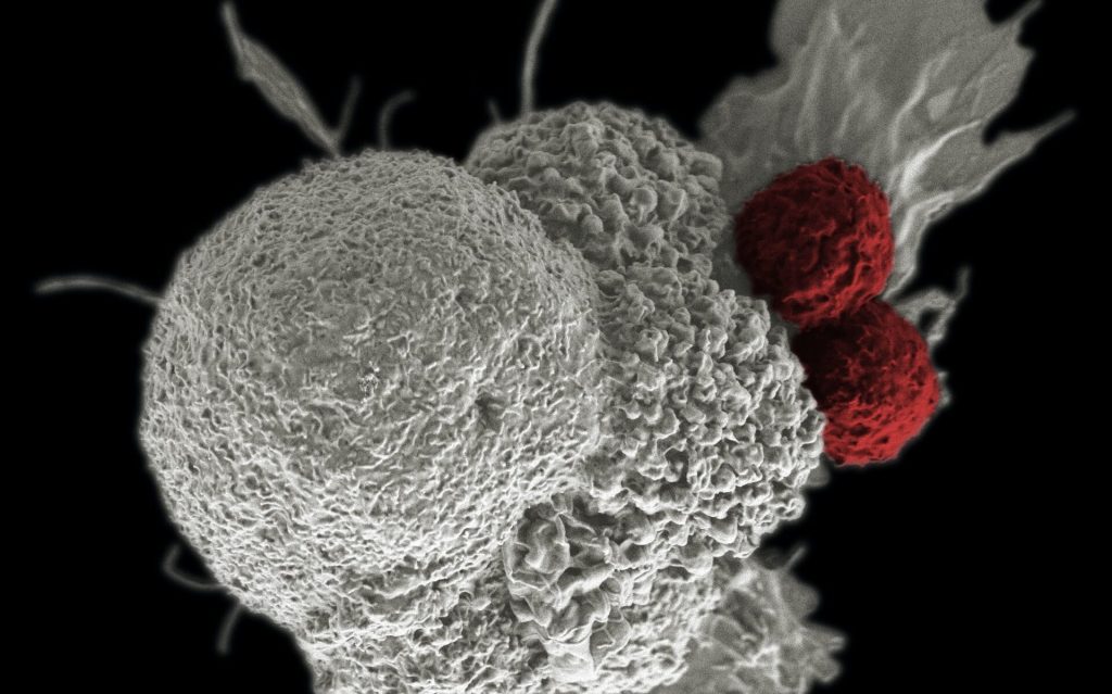
Shown here is a pseudo-colored scanning electron micrograph of an oral squamous cancer cell (white) being attacked by two cytotoxic T cells (red), part of a natural immune response. Photo by National Cancer Institute on Unsplash

Researchers have developed a way to potentially reduce the toxic side-effects of CAR T cell immunotherapy, in findings that could overcome the pioneering treatment’s biggest limitation.

Their new study, reported in eLife, has come up with a way to identify a ‘goldilocks’ window that fine-tunes the cells used in the immunotherapy so that their activity is strong enough to eliminate the cancer but not so strong that they generate toxic side-effects.

Therapy provokes ‘perfect storm’

CAR T cell therapies involve collecting T cells from a cancer patient and supercharging the cells by individually re-engineering them in the laboratory. These enhanced cells are then put back into patients.

CAR T cell immunotherapy can be up to 90% effective in certain blood cancers, even curing some patients. But the treatment has harmful side-effects, with about 50% of patients experiencing dangerous complications.

The T cells are engineered to produce proteins on their surface called chimeric antigen receptors (CARs), which enable T cells to recognise and bind to specific proteins on the surface of cancer cells more efficiently.

Associate Professor Matthew Call said this synthetic sensor is what gives T cells the enhanced ability to attack and eliminate threats, like cancer cells.

“While putting these supercharged T cells into a patient with a high tumour burden can swiftly eradicate cancer cells, it also creates the perfect storm for an ongoing toxic response that can be harmful,” Associate Prof Call said.

There is currently no way of reliably predicting how strong CAR T cell therapy will be for a patient.

While previous studies have attempted to fine-tune T cells by targeting the end sections of the sensor, which either bind to the cancer cell or instruct the T cells to kill, the new research is the first to look at completely redesigning the middle part.

Researchers leveraged the computational expertise of the Weizmann Institute of Science to stitch together pieces of natural immune sensors with custom-designed synthetic elements, to generate new circuits that could be used to tune and assess variations of potency.

“Focusing on the connector fragment in the middle allows us to generate different versions of CARs that we know are stronger or weaker, enabling us to customise them to a patient’s potency requirements,” Associate Prof Call said.

“Being able to predictably tune this T cell activity significantly broadens our research, contrary to previous studies, because we are targeting something that exists in every immunotherapy scenario.

“For the first time, we can establish rules that will be applicable to any cancer where CAR T cell immunotherapy is being used.”

Enhanced treatment

Associate Prof Call said the ability to fine-tune T cells would dramatically reduce the number of patients experiencing severe side-effects from the treatment, which can include fever, high blood pressure and respiratory distress.

“CAR T cell therapy has proven effective in eradicating very advanced leukaemias and lymphomas, while also keeping the cancer at bay for many years – even after a patient has stopped taking cancer medication,” Associate Professor Call said.

“The therapy has incredible potential for cancer patients, but is currently used as a last resort due to these potentially severe side-effects.

“Our tools could lead to a fundamental rethink of the way CAR T cell therapy is offered by reducing a patient’s exposure risk to harmful side-effects. This would allow patients with a broad range of cancers to be given CAR T cell therapy far earlier in the treatment process.”

There are currently over 600 clinical trials of CAR T cell immunotherapy, with the treatment already being used for several blood cancers.

Researchers hope their new tool could be used to triage immunotherapy patients according to the potencies required in the early treatment phases, bringing the field closer to hitting that ‘goldilocks’ treatment window for many different cancers.

The next research phase will focus on progressing these findings into a clinical setting to see CAR T cell therapy used as a safer, first-line treatment.

Source: Walter and Eliza Hall Institute of Medical Research

Erectile Dysfunction Drugs Repurposed for Cancer Treatment

Killer T cells about to destroy cancer cell
Killer T cells about to destroy cancer cell (centre). Credit: NIH

Researchers report repurposing an unusual class of drugs to combat oesophageal cancer – PDE5 inhibitors, which are mainly used as erectile dysfunction treatments.

Tumours are surrounded by a microenvironment made up of blood vessels, immune cells, enzymes and a variety of other cells the tumour needs to survive, including cells called fibroblasts that are essential in building connective tissue.

Research suggests that this microenvironment is key to a cancer’s development, and now researchers have found that an erectile dysfunction drug that targets the tumour microenvironment that could improve treatment for certain cancers.

Finding a new target

In some cancers, the tumour microenvironment allows tumours to resist treatment, preventing chemotherapy from having a beneficial effect.

This is the case in oesophageal cancers, which, though rare, currently has poor survival outcomes.

To try to overcome this resistance, a team of researchers led by Professor Tim Underwood at the University of Southampton, wanted to identify the cells in the tumour microenvironment that protect the tumour from treatment so they could target them.

“Where targeting cancer cells with one specific treatment can be difficult because they differ between patients, targeting the microenvironment cells may be more likely to have traction because they are similar across patients,” said Prof Underwood.

“Rather than going after the cancer cells, actually, if we take away their ‘soil’ and go after the environment they live in, we might have more success.

“The plants might be different, but if you poison the soil, they’ll all die.”

New uses for old drugs

By examining cells from oesophageal cancers called adenocarcinomas, the team found that levels of an enzyme called PDE5 are higher in these cancer cells than in health oesophageal tissues.

Specifically, the high levels of PDE5 were found in cells called cancer associated fibroblasts (CAFs), which are important for tumour growth. They also found that the more PDE5 a tumour contained, the worse the prognosis was, suggesting that PDE5 would be an effective target for treatment.

Luckily, PDE5 inhibitors already exist, commonly used to treat erectile dysfunction.

The researchers discovered that in addition to its usual function of relaxing muscles to allow increased blood flow, PDE5 inhibitors were able to suppress CAF activity, and make them behave like normal fibroblasts again.

Improving treatment safely

Once Prof Underwood’s team had found that PDE5 inhibitors worked, a collaborating team took samples of tumour cells from 15 tissue biopsies from 8 patients and used them to create artificial lab-grown tumours. With these tumours, the researchers could test a combination of PDE5 inhibitions and standard chemotherapy in the lab.

Twelve of these samples were taken from people whose tumours had shown a poor response to chemotherapy in the clinic. Of these, 9 were made sensitive to chemotherapy following the addition of the PDE5 inhibitor targeting CAFs.

They also tested the treatment on mice implanted with chemotherapy resistant oesophageal tumours and found that there were no adverse side effects to the treatment, and that chemotherapy combined with PDE5 inhibitors shrunk the tumours more than chemotherapy alone.

Repurposing existing drugs like PDE5 inhibitors takes advantage of well-established safety profiles.

However giving PDE5 inhibitors to people with oesophageal cancer would be extremely unlikely to cause erections without the appropriate stimulation.

“The chemotherapy resistant properties of oesophageal tumours mean that many patients undergo intensive chemotherapy that won’t work for them,” said Prof Underwood.

“Finding a drug, which is already safely prescribed to people every day, could be a great step forward in tackling this hard-to-treat disease.”

Source: Cancer Research UK

Study Reveals That Breast Cancer Spreads at Night

Sleeping woman
Photo by Cottonbro on Pexels

Researchers previously assumed that metastasising tumours release cells continuously. However, a new study has reached a surprising conclusion: circulating cancer cells that later form metastases mainly arise during the sleep phase of the affected individuals. This may have implications for oncologists, as timing of samples may affect their results. The study findings have just been published in Nature.

Circadian rhythm-regulated hormones control metastasis

“When the affected person is asleep, the tumour awakens,” said study leader Professor Nicola Aceto at ETH Zurich. During their study, which included 30 female cancer patients and mouse models, the researchers found that the tumour generates more circulating cells when the organism is asleep. Cells that leave the tumour at night also divide more quickly and therefore have a higher potential to form metastases, compared to circulating cells that leave the tumour during the day.

“Our research shows that the escape of circulating cancer cells from the original tumour is controlled by hormones such as melatonin, which determine our rhythms of day and night,” said Zoi Diamantopoulou, the study’s lead author and a postdoctoral researcher at ETH Zurich.

An accidental discovery led to the study

In addition, the study indicates that the time in which tumour or blood samples are taken for diagnosis may influence the findings of oncologists. It was an accidental finding along these lines that first put the researchers on the right track, “Some of my colleagues work early in the morning or late in the evening; sometimes they’ll also analyse blood at unusual hours,” Prof Aceto said with a smile. The scientists were surprised to find that samples taken at different times of the day had very different levels of circulating cancer cells.

Another clue was the surprisingly high number of cancer cells found per unit of blood in mice compared to humans. The reason was that as nocturnal animals, mice sleep during the day, which is when scientists collect most of their samples.

“In our view, these findings may indicate the need for healthcare professionals to systematically record the time at which they perform biopsies,” Prof Aceto said. “It may help to make the data truly comparable.”

The researchers’ next step will be to figure out how these findings can be incorporated into existing cancer treatments to optimise therapies. As part of further studies with patients, Prof Aceto wants to investigate whether different types of cancer behave similarly to breast cancer and whether existing therapies can be made more successful if patients are treated at different times.

Source: ETH Zurich

Parabens in Hair Products May Increase Breast Cancer Risk for Black Women

Parabens, chemicals which are found in widely used hair and personal care products, cause harmful effects in breast cancer cells from Black women, according to a new study presented at ENDO 2022, the Endocrine Society’s annual meeting.

In the US, the lifetime risk of breast cancer is one in eight, and Black women are at a higher risk of getting breast cancer under the age of 40 than any other racial or ethnic group in that country. Breast cancer rates among Black South African women are also on the increase, but the cause remains unexplained and research in this area has been lacking.

“One reason for the higher risk of breast cancer may be exposure to harmful chemicals called endocrine-disrupting chemicals in hair and personal care products,” said lead researcher Lindsey S. Treviño, PhD. “These chemicals mimic the effects of hormones on the body.”

Parabens are endocrine-disrupting chemicals that are commonly used as preservatives in hair and other personal care products. Parabens cause breast cancer cells to grow, invade, spread, and express genes linked to cancer and to hormone action. Research showed that fewer paraben-free options are marketed to Black women.

“Black women are more likely to buy and use hair products with these types of chemicals, but we do not have a lot of data about how parabens may increase breast cancer risk in Black women,” Dr Treviño said. “This is because Black women have not been picked to take part in most research studies looking at this link. Also, studies to test this link have only used breast cancer cell lines from White women.”

The new study tested the effects of parabens on breast cancer cells from Black women. Parabens were found to increase the growth of a Black breast cancer cell line but not in the White breast cancer cell line. Parabens increased expression of genes linked to hormone action in breast cancer cell lines from both Black and White women. Parabens also promoted the spread of breast cancer cells, with a bigger effect seen in the Black breast cancer cell line.

“These results provide new data that parabens also cause harmful effects in breast cancer cells from Black women,” Dr Treviño said. 

The study is a part of a community-led project called the Bench to Community Initiative (BCI), which brings together scientists and community members (including breast cancer survivors) to create ways to reduce exposures to harmful chemicals in hair and personal care products in Black women with breast cancer. 

“While this project focuses on Black women, the knowledge we gain about the link between exposure to harmful chemicals in personal care products and breast cancer risk can be used to help all women at high risk of getting breast cancer,” Dr Treviño said.

Source: Endocrine Society