Category: Cancer

Reaffirming Commitment to the Fight Against Cancer This World Cancer Day

Photo by Rfstudio on Pexels

4 February 2025 – As the world unites to mark World Cancer Day, L’Oréal South Africa reaffirms its unwavering commitment to supporting individuals affected by cancer through groundbreaking initiatives in research, patient care, and workplace inclusion. Over the past three years, efforts have been strengthened through a series of global and local programmes designed to ease the journey of cancer patients and survivors while advancing scientific innovation in supportive care.

The L’Oréal Dermatological Beauty team leads this commitment with two major initiatives. Save Your Skin focuses on skin cancer prevention and early detection, promoting safe sun habits and strong photoprotection. Fight with Care addresses the dermatological side effects experienced by over 80% of cancer patients undergoing treatment, advancing scientific research in supportive care, training healthcare professionals, and providing direct support through educational resources and partnerships with 50 NGOs worldwide, including the Union for International Cancer Control (UICC) and the Multinational Association of Supportive Care in Cancer (MASCC).

Recognising the profound impact of cancer on employees and their families, the Share & Care programme, now in its tenth year, has been expanded to provide financial assistance to employees diagnosed with cancer, psychological and emotional support services, and practical aid to ease their journey through treatment and workplace reintegration. This programme aligns with our leadership in the Working with Cancer charter, advocating for workplace inclusion, care, and long-term support.

Believing in the transformative power of beauty, L’Oréal has introduced dedicated wellness therapy programmes in hospitals and specialised care centres. These include skincare treatments to alleviate the side effects of cancer therapies, beauty services such as make-up sessions to restore confidence and enhance well-being, and therapeutic massages and wellness sessions to improve overall quality of life.

In South Africa, L’Oréal has partnered with the Look Good, Feel Better campaign for over 20 years. This initiative has been a beacon of hope for women undergoing cancer treatment, providing beauty workshops designed to help them regain confidence and a sense of normality during their journey. By offering expert guidance on skincare, make-up, and emotional well-being, the programme empowers participants to face their treatment with renewed strength.

As part of ongoing efforts to support underserved communities, L’Oréal is introducing the Healing Pouch initiative, which donates full-size oncology skincare essentials—including cleansers, emollients, sun care, and wound creams, to hospitals, NGOs, and patient associations. These products, specifically formulated for sensitive and oncology-affected skin, provide essential relief while reinforcing the belief that touch, and tenderness are powerful agents of healing.

“As we mark World Cancer Day, we stand in solidarity with cancer patients, survivors, healthcare professionals, and our own employees,” said Thandi Kunene, Corporate Affairs Lead at L’Oréal South Africa. “Through continued investment in research, support programmes, and longstanding partnerships such as Look Good, Feel Better, we remain steadfast in our mission to make a meaningful difference in the lives of those affected by cancer.”

Through these global and local initiatives, L’Oréal remains committed to advancing cancer awareness, fostering inclusion, and delivering tangible support, because care is at the heart of everything we do.
New Combination Immunotherapy for Melanoma and Breast Cancer

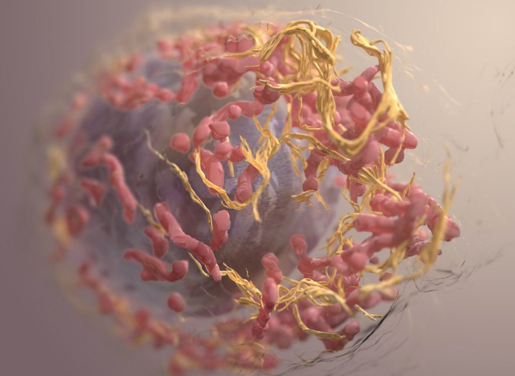
3D structure of a melanoma cell derived by ion abrasion scanning electron microscopy. Credit: Sriram Subramaniam/ National Cancer Institute

A research team at the Medical University of Vienna led by Maria Sibilia has investigated a new combination therapy against cancer. This therapy employs systemic administration of the tissue hormone interferon-I combined with local application of Imiquimod. Promising results were seen in topically accessible tumours like melanoma and breast cancer models: the therapy led to the death of tumour cells at the treated sites and simultaneously activated the adaptive immune system to fight even distant metastases. The findings, published in Nature Cancer, could improve the treatment of superficial tumours such as melanoma and breast cancer.

In recent years, immunotherapies have had significant success in the treatment and cure of a wide range of cancers. However, for some patients, these agents are still not sufficiently effective. As part of a preclinical study, Maria Sibilia, Head of the Center for Cancer Research at the Medical University of Vienna, therefore investigated the effects of a combination immunotherapy consisting of systemic administration of the tissue hormone interferon (IFN)-I and local imiquimod therapy. Imiquimod is an active substance that activates the innate receptors TLR7/8 and used to treat basal cell carcinomas. The researchers employed various preclinical mouse tumour models of melanoma and breast cancer. What both tumours have in common is that they are accessible to local therapy and often form distant metastases.

Effective for local tumours and distant metastases

Immunotherapies use the body’s own immune system to fight cancer cells. Plasmacytoid dendritic cells (pDCs), which are activated by Imiquimod via TLR7/8, play an important role in this process. The study showed that oral imiquimod stimulates pDCs to produce the tissue hormone IFN-I. This sensitised other dendritic cells and macrophages in the tumour environment to topical imiquimod therapy, which inhibited the formation of new blood vessels via the cytokine IL12 leading to the death of tumour cells.
The combination immunotherapy not only had an effect on the treated tumours, but also on distant metastases. It reduced the formation of new metastases thus preventing tumour relapses and increasing the sensitivity of melanomas to checkpoint inhibitors.

“These findings illustrate that the combination of systemic treatment with imiquimod or IFN-I and topical therapy with imiquimod has the potential to expand treatment options for patients and improve therapy outcomes in locally accessible tumors such as melanoma or breast cancer,” emphasizes Maria Sibilia.
“Topical treatment of the primary tumor with imiquimod is essential for this combination therapy with systemic IFN-I to be effective at the treated site and also to clear distant metastases,” adds Philipp Novoszel, MedUni Vienna, one of the first authors of the study.

The results suggest that this therapeutic strategy has the potential to improve treatment outcomes in superficial and thus locally accessible tumors such as melanoma and breast cancer – on the one hand through therapy-associated cancer cell death at the locally treated tumors, but also through the induction of a T cell-induced anti-tumor immune response at distant metastases, which is further enhanced by checkpoint inhibitors.

“Our aim is to continue developing immunotherapeutic strategies in order to improve the long-term prospects for patients who are not yet responding well to these agents,” says Maria Sibilia, who is also Deputy Head of the Comprehensive Cancer Center of MedUni Vienna and University Hospital Vienna.
“As systemic interferon is a well-known cancer therapy and dendritic cells are activated in a similar way to our preclinical models, we believe that the new combination therapy can show an effect in patients,” adds Martina Sanlorenzo, dermato-oncologist at MedUni Vienna and co-first author of the study.

Publication: Nature Cancer
Systemic IFN-I combined with topical TLR7/8 agonists promotes distant tumor-suppression by c-Jun-dependent IL-12 expression in dendritic cells
Sanlorenzo M, Novoszel P, Vujic I, Gastaldi T, Hammer M, Fari O, De Sa Fernandes C, Landau AD, Göcen-Oguz BV, Holcmann M, Monshi B, Rappersberger K, Agnes Csiszar A, Sibilia M
DOI: 10.1038/s43018-024-00889-9; https://www.nature.com/articles/s43018-024-00889-9

Source: Medical University of Vienna

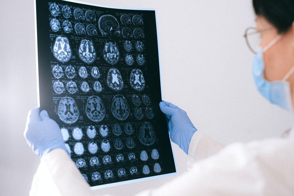
Routine Brain MRI Screening Urged in Asymptomatic Late Stage Breast Cancer

Photo by Anna Shvets on Pexels

A new study led by researchers at Moffitt Cancer Center shows that asymptomatic brain metastasis is more common in stage 4 breast cancer patients than previously believed. The study, published in Neuro-Oncology, suggests that doctors may need to rethink current screening guidelines for detecting brain metastasis in patients without symptoms.

Researchers examined 101 asymptomatic patients diagnosed with stage 4 breast cancer, including triple-negative, HER2-positive and hormone receptor-positive/HER2-negative breast cancer. These patients underwent MRI scans to check for brain metastasis, with a follow-up MRI six months later if the initial scan showed no signs of cancer spread.

Of the patients who completed the initial MRI, 14% had brain metastasis. The rates by subtype were:

  • 18% in triple-negative breast cancer
  • 15% in HER2-positive breast cancer
  • 10% in hormone receptor-positive/HER2-negative breast cancer

After the second MRI, the number of patients with brain metastasis grew to about 25% in each subtype. Following diagnosis, patients went on to receive early treatment for their brain metastases, including changes in systemic therapy and local therapies.

“Our study suggests that asymptomatic brain metastasis is quite common in stage 4 breast cancer,” said Kamran Ahmed, MD, associate member and section chief for Breast Radiation Oncology at Moffitt and principal investigator of the study. “Although larger studies are needed to confirm our findings, given the improvements in systemic and local therapies for breast cancer brain metastasis, the time may be appropriate to reconsider current guidelines that recommend against routine MRI surveillance in late stage breast cancer.”

Source: H. Lee Moffitt Cancer Center & Research Institute

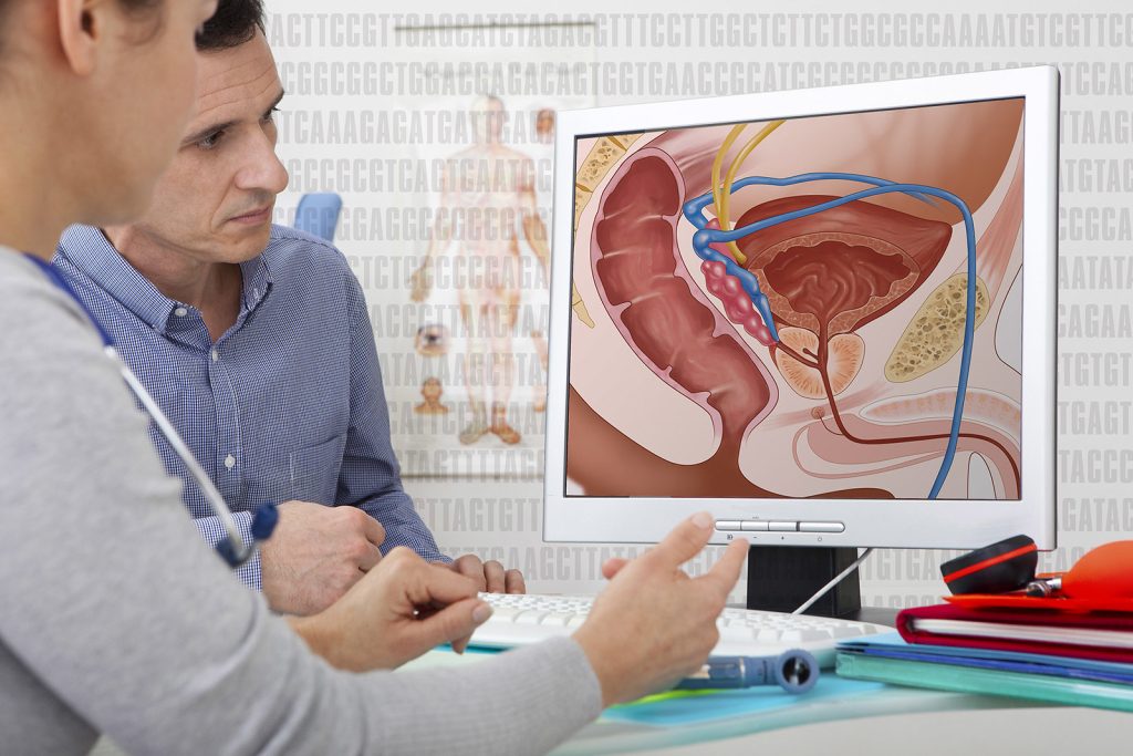
Urine-based Test Detects Aggressive Prostate Cancer

Credit: Darryl Leja National Human Genome Research Institute National Institutes Of Health

Traditional approaches to prostate cancer screening involve blood tests, MRI, and biopsies. Besides being uncomfortable, some of these procedures result in overdiagnosis of low-grade cancers.

In a new study, researchers at the University of Michigan Health Rogel Cancer Center have clinically validated a previously developed urine test, which can potentially bypass these invasive procedures among men who are unlikely to benefit.

Prostate cancers are categorised based on their Gleason Grade or Grade Group. Those with Gleason 3+4=7, or Grade Group 2, or higher are more likely to grow and cause harm in comparison with Gleason 6 or Grade Group 1 prostate cancers, which are considered non-aggressive.

The urine test, called MyProstateScore 2.0, or MPS2, looks at 18 different genes linked to high-grade prostate cancer. 

The researchers had previously demonstrated that the test was effective in identifying GG2 or higher cancers, helping patients avoid unnecessary biopsies. But in that study, urine samples were obtained after a digital rectal examination.

“The process requires the prostate to be compressed, causing the release of cellular debris into a urine sample that the patient provides after the rectal exam,” said Ganesh S. Palapattu, MD, a professor of urology.

Its primary benefit is that the test can accurately predict your probability of developing aggressive prostate cancer, putting both the patient and physician at ease.”

-Ganesh Palapattu, MD

Such an examination may not be practical for many and is associated with some discomfort.

Developing a potential at-home test

In the study, the team modified the urine collection approach so that the MPS2 test could detect markers for prostate cancer, without requiring a prior rectal exam.

Using urine samples from a cohort of 266 men who did not undergo a rectal exam, they found that the test could detect 94% of GG2 or higher cancers and was more sensitive than blood tests.

Further, the team used mathematical models to demonstrate that the use of MPS2 would have avoided up to 53% of unnecessary biopsies.

“These results show that MPS2 has promise as an at-home test,” Palapattu said. 

“Its primary benefit is that the test can accurately predict your probability of developing aggressive prostate cancer, putting both the patient and physician at ease.”

MPS2 can also help patients save on healthcare costs since it is significantly cheaper than an MRI.

The team is interested in repeating the study and corroborating their results with a larger, diverse population of men.

They’re also hoping to study the test’s performance in men as a surveillance screen for low-risk prostate cancer.

“MPS2 could potentially improve the health of our patients by avoiding overdiagnosis and overtreatment and allowing us to focus on those who are most likely to have aggressive cancers,” Palapattu said.

Source: Michigan Medicine – University of Michigan

New Viral Treatment for Basal Cell Carcinoma Shines in Trials

Dermoscopy of an approximately 16mm nodular basal cell carcinoma showing telangiectatic vessels. Source: Wikimedia CC4.0

Basal cell carcinomas, the most common form of skin cancer, occur in chronically sun-exposed areas such as the face.  Locally advanced tumours in particular can be difficult to treat surgically. A research team from MedUni Vienna and University Hospital Vienna has now investigated the effectiveness of a new type of therapy and achieved promising results: The active substance TVEC led to a reduction in the size of the basal cell carcinoma in all study participants, which not only improved surgical removal, but also led to a complete regression of the tumour in some of the patients. The study was published in the top journal Nature Cancer.

In the study Talimogene Laherparepvec (TVEC) was used, which has so far only been approved for the treatment of superficial melanoma metastases. TVEC is a genetically modified herpes simplex virus that specifically destroys tumour cells and simultaneously activates the immune system. The aim of the study was to reduce the size of the tumour before a planned operation so that patients would not suffer any functional or cosmetic restrictions after the procedure. The study included 18 patients who would have required a flap or skin graft due to the size and localisation of their basal cell carcinoma. They each received six intralesional injections of TVEC over a period of 13 weeks before the tumour was surgically removed.

“This enabled the tumour to be reduced in size in half of the patients to such an extent that surgery with direct wound closure was possible. In a third of the cases, the subsequent histological examination even showed no more living tumour cells. All treated tumours at least became smaller, and no tumour grew further under the therapy. The treatment was well tolerated by the patients,” says the principal investigator Christoph Höller, Head of the Skin Tumour Centre at the Department of Dermatology, summarising the results. “The new treatment option for basal cell carcinoma can not only simplify surgery, but also help to avoid disfiguring operations and functional limitations,” adds first author Julia Ressler, also from the Department of Dermatology. In addition to the clinical studies, the researchers from the Department of Dermatology, in cooperation with the St. Anna Children’s Hospital, carried out comprehensive analyses that show that the immune defence in the tumour tissue is strengthened in the course of therapy with TVEC.  

These results suggest that TVEC could be a promising option for the neoadjuvant, i.e. pre-surgical, treatment of basal cell carcinoma, particularly in patients for whom major surgery should be avoided. Further studies are to follow in order to confirm the benefits of this new option in a larger patient population.

Source: Medical University of Vienna

Radiology’s Role in Managing Pain in Cancer Patients

SCP – Dr Winter performing a CT-guided interventional procedure

World Cancer Day, observed every 4 February, aims to raise awareness about cancer, encourage prevention and look at ways of improving a cancer patient’s quality of life. Interventional radiology plays a significant role in pain management for cancer patients.

Traditionally, radiology was used for diagnosing the cause of the pain but interventional radiology has changed this paradigm. Since American radiologist Charles Dotter, first used a guidewire and catheter to perform an interventional procedure in 1964, radiologists have become actively involved in intervention, including interventions for pain management. Today, many minimally invasive procedures are routinely performed in busy radiology departments.

Dr Arthur Winter, a radiologist at SCP Radiology says, ‘Interventional radiology has developed rapidly. Pain management procedures are becoming a daily part of busy radiology departments and play a crucial role in managing pain for cancer patients.

‘Cancer-related pain can be a significant burden, affecting patients’ quality of life and hindering their ability to carry out daily activities,’ says Dr Winter. ‘Radiology offers various techniques and treatments that help alleviate this pain effectively. These therapeutic interventions in pain management include image-guided interventional radiology procedures and radiation therapy.’

Understanding pain

Pain is a signal from the nervous system to let you know that something is wrong in your body. It is transmitted in a complex interaction between specialised nerves, the spinal cord and the brain. It can take many forms, be localised to one part of the body or appear to be widespread.

The nature of cancer pain

Cancer pain can arise from multiple sources, including the tumour itself, which may invade or compress surrounding tissues, organs or nerves. Metastases, the spread of cancer to other parts of the body, can also cause significant pain. Additionally, pain can result from the treatment of cancer, such as chemotherapy and radiation therapy.

Multidisciplinary approach to pain management

Effective pain management for cancer patients requires a comprehensive, multidisciplinary approach. Oncologists, radiologists, pain specialists and other healthcare providers collaborate to develop individualised care plans. Radiology is essential in both the diagnostic and therapeutic phases of this process, providing crucial insights and treatment options.

These personalised care plans, tailored to each patient’s needs, ensure:

  • Accurate diagnosis and identification of pain source or sources
  • Targeted and effective treatment interventions
  • Ongoing monitoring and adjustment of pain management strategies

Imaging techniques

Diagnostic radiology initially uses various imaging techniques to identify the source and extent of pain in cancer patients. These techniques include: X-rays, CT scans, MRI, PET scans and ultrasound.

By identifying the precise location and cause of pain, radiology can help:

  • Determine the most appropriate interventions, such as surgery, radiation therapy or minimally invasive interventional procedures
  • Monitor the effectiveness of pain management strategies and make necessary adjustments
  • Avoid unnecessary treatments that may not address the underlying cause of pain

Interventional radiology

Interventional radiology uses minimally invasive techniques to diagnose and treat various conditions and, for cancer patients experiencing pain, it offers several effective treatments:

  • Radiofrequency ablation (RFA): This uses heat, generated by radiofrequency energy to destroy cancerous tissues – often to treat painful bone metastases or tumours that are difficult to reach surgically
  • Cryoablation: Involves freezing cancerous tissues to destroy them. It is particularly useful for treating painful bone or soft tissue tumours, providing rapid pain relief
  • Nerve blocks: Involve the injection of anaesthetic agents or steroids near specific nerves to block pain signals. They can provide significant pain relief for patients with nerve-related pain

Palliative radiation therapy

In this instance, radiologists are involved with planning imaging only. The actual radiotherapy is performed by the radiation therapist, who works under the supervision of a radiation oncologist. Palliative radiation therapy is specifically designed to relieve symptoms and improve the quality of life for cancer patients. It focuses on pain control and symptom management rather than curing the disease.

Radiation oncologists deliver targeted doses of radiation to cancerous tissues, this palliative radiation therapy can help:

  • Reduce tumour size, alleviating pressure on surrounding tissues and nerves
  • Control bleeding or ulceration caused by tumours
  • Provide rapid pain relief, often within days to weeks of treatment

Improving quality of life

Dr Winter highlights that chronic pain can significantly diminish quality of life and contribute to depression, particularly in patients with underlying cancer. ‘These patients, in particular, should be considered for interventional procedures. For instance, there are highly effective treatments available to manage pain associated with pancreatic and pelvic cancers’.

‘Specialists, such as oncologists and neurologists, acknowledge the significant role of interventional radiology in pain management and collaborate closely with us to support their patients. As a rapidly advancing branch of radiology, it provides minimally invasive solutions and it is incredibly rewarding to witness patients regain their quality of life through effective symptom relief.’

Does Obesity Affect Children’s Chances of Survival after Cancer Diagnosis?

Photo by Patrick Fore on Unsplash

A recent population-based study indicates that among children with cancer, those with obesity at the time of diagnosis may face an elevated risk of dying. The findings are published by Wiley online in CANCER, a peer-reviewed journal of the American Cancer Society.

The retrospective study was based on information from the Cancer in Young People in Canada (CYP-C) database, including all children with newly diagnosed cancer aged 2 to 18 years across Canada from 2001 to 2020. Obesity was defined as age and sex-adjusted body mass index at or above the 95th percentile.

Among 11 291 children with cancer, 10.5% were obese at the time of diagnosis. Investigators assessed 5-year event-free survival (survival free of cancer relapse), as well as overall survival.

Compared with patients without obesity at the time of initial cancer diagnosis, those with obesity had lower rates of 5-year event-free survival (77.5% versus 79.6%) and overall survival (83.0% versus 85.9%).

After adjusting for factors including age, sex, ethnicity, neighbourhood income quintile, treatment era, and cancer categories, obesity at diagnosis was linked with a 16% increase in the risk of relapse and a 29% increase in the risk of death. The negative impact of obesity on prognosis was especially pronounced in patients with acute lymphoblastic leukaemia and brain tumours.

“Our study highlights the negative impact of obesity among all types of childhood cancers. It provides the rationale to evaluate different strategies to mitigate the adverse risk of obesity on cancer outcomes in future trials,” said co–senior author Thai Hoa Tran, MD, of the Centre Hospitalier Universitaire Sainte-Justine, in Montreal. “It also reinforces the urgent need to reduce the epidemic of childhood obesity as it can result in significant health consequences.”

Source: Wiley

New Blood Test for Pancreatic Cancer Exceeds Gold Standard

Pancreatic cancer. Credit: Scientific Animations CC BY-SA 4.0

A recent double-blinded, peer-reviewed analysis published in Cancer Letters revealed that an experimental test for pancreatic cancer correctly identified 71% of lab samples compared to only 44% correctly identified by the current gold-standard test.

An experimental blood test for pancreatic cancer that was developed by teams led by VAI Professor Brian Haab, PhD, and Randall E. Brand, MD, a physician-scientist and professor of medicine at the University of Pittsburgh, created the test. This evaluation by a commercial laboratory is an important milestone toward making the test available for patients.

Before the new test can be used by doctors to diagnose cancer, it must undergo clinical validation. During this process, a CLIA-accredited diagnostics laboratory adapts the experimental test into a version that reliably works under the strict conditions in a clinical lab. CLIA is a rigorous federal standard that ensures lab quality.

“Validation studies are essential for transforming a test developed in an academic lab into one that is used to diagnose real people,” Haab said. “For a person being evaluated for pancreatic cancer, the stakes are high. Validation studies ensure that new tests work as intended.”

The new test works by detecting two sugars — CA199.STRA and CA19-9 — that are produced by pancreatic cancer cells and escape into the bloodstream. CA19-9 is the current gold-standard biomarker for pancreatic cancer. Haab’s lab identified CA199.STRA as a cancer biomarker and developed the technology to detect it.

The new test also greatly reduced the number of false negatives while maintaining a low false positive rate, according to the recent analysis. Low rates of false positives and false negatives are important because they reflect the test’s ability to correctly identify the presence or absence of cancer.

Clinical validation of the test will be conducted by ReligenDx, a CLIA-accredited diagnostics lab based in Pennsylvania. The process is expected to take two years.  

If successful in clinical validation, Haab envisions the test being used in two main ways: 1. Catching pancreatic cancer more quickly in people at high risk of the disease, which would enable earlier treatment and 2. Monitoring progression and treatment response in people diagnosed with pancreatic cancer.

Source: Van Andel Research Institute

High-fat Diets are ‘Ticking Time Bombs’ for Liver Cancer

These images show slices of mouse liver under the microscope, with tumours outlined in yellow and green indicating the expression of different proteins within cells. The left column is a control group. In the centre column, the protein detected (TBG-Cre) is expressed in all liver cells, so the entire image appears green. In the right column, the protein detected (p21-Cre) is only expressed in senescent liver cells. Because green is only visible within the tumour area, the results show that liver tumours originate from previously senescent liver cells. Photo credit: UC San Diego Health Sciences

A new study on the development of liver cancer reveals a complex interplay between cellular metabolism and DNA damage that drives the progression of fatty liver disease to cancer. The findings, published in Nature, suggest new paths forward for preventing and treating liver cancer and have significant implications on our understanding of cancer’s origin and the effects of diet on our DNA.

The incidence of the most common form of liver cancer, hepatocellular carcinoma (HCC), has grown by 25-30% in the past two decades, with much of the growth attributed to the dramatic rise in fatty liver disease. About 20% of individuals with fatty liver disease have a severe form of the disease, called metabolic dysfunction-associated steatohepatitis (MASH), that greatly increases the risk of HCC. However, how MASH transitions to liver cancer is not well understood.

“Going from fatty liver disease to MASH to liver cancer is a very common scenario, and the consequences can be deadly,” said Michael Karin, PhD, Distinguished Professor in the Department of Pharmacology at UC San Diego School of Medicine. MASH ends up destroying the liver, or leading to often-fatal liver cancer, but little is know of the process at the subcellular level.

The researchers used a combination of mouse models and human tissue specimens and databases to demonstrate that MASH-inducing diets, which are rich in fat and sugar, cause DNA damage in liver cells that causes them to go into senescence, a state in which cells are still alive and metabolically active but can no longer divide. Senescence is a normal response to a variety of cellular stressors. In a perfect world, senescence gives the body time to repair damage or eliminate the damaged cells before they’re allowed to proliferate more widely and become cancerous.

“A poor, fast-food diet can be as dangerous as cigarette smoking in the long run. People need to understand that bad diets do far more than just alter a person’s cosmetic appearance. They can fundamentally change how our cells function, right down to their DNA.”

Michael Karin, PhD

However, as the researchers discovered, this isn’t what happens in liver cells, also known as hepatocytes. In hepatocytes, some damaged cells survive this process.

These cells are, according to Karin, “like ticking time bombs that could start proliferating again at any point and ultimately become cancerous.”

“Comprehensive genomic analyses of tumour DNA indicate that they originate from liver cells damaged by MASH, emphasising a direct link between diet-induced DNA damage and the development of cancer,” added study co-author Ludmil Alexandrov, PhD, associate professor of cellular and molecular medicine and bioengineering at UC San Diego and member of UC San Diego Moores Cancer Center.

The findings suggest that developing new drugs to prevent or reverse DNA damage could be a promising therapeutic approach for preventing liver cancer, particularly in people with MASH.

“There are a few possibilities for how this could be leveraged into a future treatment, but it will take more time and research to explore these ideas,” said Karin. “One hypothesis is that a high-fat diet could lead to an imbalance in the raw materials our cells use to build and repair DNA, and that we could use drugs or nutri-chemicals to correct these imbalances. Another idea is developing new antioxidants, much more efficient and specific than the ones we have now, and using those could help block or reverse the cellular stress that causes DNA damage in the first place.”

In addition to opening these new avenues of treatment for liver cancer, the study also offers new insight into the relationship between aging and cancer.

“We know that aging increases the risk of virtually all cancers and that aging is associated with cellular senescence, but this introduces a paradox since senescence is supposed to guard against cancer,” said Karin. “This study helps reveal the underlying molecular biology that allows cells to re-enter the cell cycle after undergoing senescence, and we believe that similar mechanisms could be acting in a wide range of cancers.”

The findings also help directly quantify the detrimental effects of poor diet on cellular metabolism which, according to Karin, could be used to help guide public health messaging related to fatty liver disease.

“A poor, fast-food diet can be as dangerous as cigarette smoking in the long run,” said Karin. “People need to understand that bad diets do far more than just alter a person’s cosmetic appearance. They can fundamentally change how our cells function, right down to their DNA.”

Source: University of California – San Diego

How Checkpoint Immunotherapy also Increases the Risk of Cardiovascular Disease

Source: Wikimedia CC0

Cancer immunotherapy is known to also make patients more vulnerable to heart attack and stroke. A new study led by researchers at NYU Langone Health points to a possible explanation for this side effect: the treatment interferes with immune regulation in the heart’s largest blood vessels.

This new work focused on immune checkpoint inhibitors, which work by blocking molecules embedded on the surface of cells – immune checkpoints – which normally serve as “brakes” that prevent excess immune activity, or inflammation. Some tumours are known to hijack these checkpoints to weaken the body’s defences, so by blocking the checkpoints, the treatments enable the immune system to kill tumour cells.

Unfortunately, this treatment type may also trigger damaging levels of inflammation in the heart, brain, stomach, and other organs, the researchers say. For example, past studies have shown that about 10% of those with atherosclerosis have a heart attack or stroke following cancer treatment. Until now, the specific mechanisms behind this issue had remained unclear.

To address this question, the research team explored at a cellular level how immune checkpoint inhibitors interact with immune cells within arterial plaques. A genetic analysis by the study authors showed that the same type of immune checkpoints targeted by cancer therapies also appear in arterial immune cells, establishing a link between checkpoint inhibitors and cardiovascular events.

“Our findings provide new insight into how a drug intended to target tumours can also prompt a heightened immune response in arteries and increase risk of heart disease,” said study co-senior author Chiara Giannarelli, MD, PhD. “Cancer patients and their physicians should be aware that they may need to monitor for new heart concerns following cancer treatment,” added Dr Giannarelli.

For the current study, published in Nature Cardiovascular Research, the researchers analysed the genetic activity of thousands of immune cells collected from the plaques of 50 men and women undergoing a surgical procedure to address atherosclerosis.

The investigators also explored how type 2 diabetes, a known risk factor for both cancer and heart disease, may make those with atherosclerosis even more vulnerable to the ill effects of checkpoint inhibitors, adds Dr Giannarelli, also an associate professor in the Department of Pathology at NYU Grossman School of Medicine. As part of the study, the team assessed immune checkpoint activity in arterial tissue collected from eight patients with diabetes and four healthy volunteers. Notably, none had a history of atherosclerosis. The results showed that the diabetes patients had less measurable communication between checkpoints, which in turn can prompt increased inflammation.

Other experiments further revealed that taking immune checkpoint inhibitors might make it harder to combat atherosclerosis. Under normal circumstances, physicians typically prescribe low-fat diets to reduce plaque buildup and inflammation. Indeed, the researchers’ experiments in rodents confirmed that such diets boost communication between immune checkpoints within arteries. However, cancer patients may be at a disadvantage because their therapy, by blocking these same checkpoints, may counteract the anti-inflammatory benefits of fat reduction.

“Our findings highlight how cancer, diabetes, and heart disease do not exist in a vacuum, and that it is critical to consider how targeting one of these conditions can affect the others,” said study co-senior author Kathryn J. Moore, PhD. “Now that experts have a better understanding of the interplay between these diseases, they can begin to explore new strategies to lower the risk of unintended health concerns caused by their treatment,” added Dr Moore. She cautions that the study did not directly assess immune checkpoint behaviour in cancer patients. The team plans to do so in future investigations, she adds.

Source: NYU Langone Health