Author: ModernMedia

Psilocybin Byproduct Delays Aging and Extends Lifespan, New Study Suggests

Photo by Marek Piwnicki

As revenues from the anti-aging market – riddled with hope and thousands of supplements – surged past $500 million last year, Emory University researchers identified a compound that actively delays aging in cells and organisms.

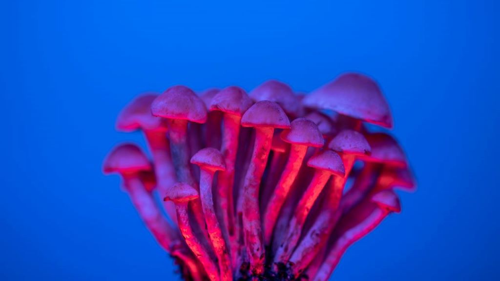
A newly published study in Nature Partner Journals’ Aging demonstrates that psilocin, a byproduct of consuming psilocybin, the active ingredient in psychedelic mushrooms, extended the cellular lifespan of human skin and lung cells by more than 50%.

In parallel, researchers also conducted the first long-term in vivo study evaluating the systemic effects of psilocybin in aged mice of 19 months, or the equivalent of 60–65 human years. Results indicated that the mice that received an initial low dose of psilocybin of 5mg/kg, followed by a monthly high dose of 15mg/kg for 10 months, had a 30% increase in survival compared to controls. These mice also displayed healthier physical features, such as improved fur quality, fewer white hairs and hair regrowth.

While traditionally researched for its mental health benefits, this study suggests that psilocybin impacts multiple hallmarks of aging by reducing oxidative stress, improving DNA repair responses, and preserving telomere length. Telomeres are the structured ends of a chromosome, protecting it from damage that could lead to the formation of age-related diseases, such as cancer, neurodegeneration or cardiovascular disease. These foundational processes influence human aging and the onset of these chronic diseases.

The study concludes that psilocybin may have the potential to revolutionize anti-aging therapies and could be an impactful intervention in an aging population.

“Most cells in the body express serotonin receptors, and this study opens a new frontier for how psilocybin could influence systemic aging processes, particularly when administered later in life,” says Louise Hecker, PhD, senior author on the study, and former associate professor at Emory, where the research was initiated and funded.

While much of what researchers know about psilocybin relates to the brain, few studies have examined its systemic impacts. Many people associate psilocybin with the hallucinogenic impacts, but the majority of the cells in the body express serotonin receptors.

“Our study opens new questions about what long-term treatments can do. Additionally, even when the intervention is initiated late in life in mice, it still leads to improved survival, which is clinically relevant in healthy aging,” adds Hecker, currently an associate professor at Baylor College of Medicine.

Not just a longer life, but a healthier life

“This study provides strong preclinical evidence that psilocybin may contribute to healthier aging – not just a longer lifespan, but a better quality of life in later years,” says Ali John Zarrabi, MD, director of psychedelic research at Emory’s Department of Psychiatry. “As a palliative care physician-scientist, one of my biggest concerns is prolonging life at the cost of dignity and function. But these mice weren’t just surviving longer – they experienced better aging,” adds Zarrabi, co-investigator of the study. 

Zarrabi emphasises the importance of further research in older adults, as well as the well-documented overlap between physical and mental health.

“Emory is actively involved in Phase II and III clinical trials of psilocybin-assisted therapy for depression, and these results suggest we also need to understand psilocybin’s systemic effects in aging populations,” says Zarrabi. “My hope is also that if psilocybin-assisted therapy is approved as an intervention for depression by the FDA in 2027, then having a better quality of life would also translate into a longer, healthier life.”

Source: Emory Health Sciences

Do Claimed Past-life Memories Affect Mental Health?

Photo by jesse orrico on Unsplash

In Brazil, belief in reincarnation is common, with up to a third of people believing it is possible. Some claim to have memories of their past lives. Researchers analysed the profile of adults who claim to have past-life memories, the features of these memories, and their associations with mental health, happiness, and religiosity/spirituality. The results indicated a high prevalence of mental disorder symptoms, with religiosity and spirituality being protective factors.

Most religious and spiritual traditions worldwide share a belief in a transcendental realm and/or the continuity of life after physical death. This belief is common among the adult population of 35 countries across six continents, with percentages ranging from 38% in Sweden to 85% in Indonesia.

A recent survey, involving nationally representative samples in 22 countries across all continents, concluded that there are no countries where most of the population said they did not believe in life after death, with the majority answering “yes” or “unsure” to this question.

While there are studies of claims of past-life memories (PLM) in children, there is still little knowledge about cases in adults and the impact of PLM on claimers. To fill this gap, Sandra Maciel de Carvalho and her team analysed the profile of adults who claim to have PLM, including sociodemographic data, the features of these memories, and their associations with mental health, happiness, and religiosity/spirituality.

With support from the BIAL Foundation, researchers from the Federal University of Juiz de Fora (Brazil) and the University of Virginia (USA) performed an online survey for the first time among a sample of 402 adults living in Brazil who reported having memories of past lives. In this country, 66% of citizens say that there is probably, or definitely, life after death, and 33% believe that people will be reborn in this world.

Most of the sample participants were middle age 41.6 years, female (79%), with higher education (68%), Spiritists (54.5%), and very/moderately spiritual (91%). PLM started spontaneously in 82%, on average, at 19.9 years old. Birthmark/defect (54%), unusual philia (intense and unusual attractions or interests; 30%), and phobia in childhood (71%), and persistent phobia (71%) were commonly associated features.

The results were published in the article “Who Does Report Past-Life Memories? Claimers’ Profile, Religiosity/Spirituality and Impact on Happiness and Mental Health”, published in July in the scientific journal The International Journal for the Psychology of Religion and indicated that the PLM features in sample participants are similar to those usually reported among children, except for the female prevalence.

In this sample, childhood philias and phobias were associated with lower happiness and more symptoms of mental disorders (46%), which may indicate a lasting psychological impact associated with PLM. Post-traumatic stress disorder (36%) was associated with phobias. Religion/spirituality was associated with greater happiness and lower mental disorder outcomes, potentially acting as a protective factor.

According to Sandra Maciel de Carvalho, “this study demonstrated that PLM in adults may be more prevalent than previously thought and may be associated with significant suffering and distress”. PLM may constitute a “relevant issue in mental health, and further studies are needed on its prevalence, impact, and proper clinical management”, the researcher emphasises.

Learn more about the project “89/18 – National survey of “Cases of Reincarnation Type” in Brazil” here.

Source: BIAL Foundation

New CANSA and ALVI ARMANI Partnership Offers Hair Restoration Support for Cancer Survivors

Restorative programme helps post-cancer treatment patients regain hair, confidence, and quality of life after facing cancer 

Photo by Natasha Brazil on Unsplash

The Cancer Association of South Africa (CANSA) has partnered with internationally renowned hair restoration clinic Alvi Armani South Africa, with head offices in Beverly Hills Los Angeles, to launch an initiative offering complimentary consultations and assessments to those recovering or recovered from cancer.

For many, completing cancer treatment is an experience that brings immense relief. However, it doesn’t always mark the end of the emotional journey. While chemotherapy and radiation often save lives, they can leave lasting reminders – and hair loss is among the most visible.

Cindy Pretorius, a cancer survivor who was diagnosed with basal cell carcinoma, an invasive skin cancer knows firsthand how the impact of the disease affects not just self-confidence but self-worth. After the cancer was removed, the surgery left lasting and visible scarring on her hairline. A hairline that was subsequently treated and restored through a minimally invasive hair transplant at Alvi Armani South Africa. “The team at Alvi Armani restored not only my hairline, but also my confidence,” said Pretorius.

Launching in August 2025, the initiative will offer CANSA-affiliated patients in recovery access to complimentary, in-depth, and personalised consultations. This may include scalp density and mapping assessments, as well as checks for lingering treatment effects. Where needed, survivors will receive advice and support with restorative hair treatments or transplants at Alvi Armani South Africa – offering significant financial relief and a renewed sense of hope.

“This isn’t about vanity. It’s about healing the whole person,” notes Dr Kashmal Kalan, Medical Director of Alvi Armani South Africa. “Unfortunately, even when cancer treatments end, the physical and emotional recovery continues. Many individuals in remission are confronted with reminders every time they look in the mirror and see someone who still looks like a patient, often making it difficult to reconnect with the person they were before cancer.”

For those recovering from cancer, the devastation of hair loss can continue to weigh heavily on their mental well-being. Studies show that persistent thinning, patchiness, or recession after treatment can fuel anxiety, depression, and social withdrawal. Even when remission is achieved, hair regrowth can be slow, and this gap between survival and self-image can take a heavy toll.

“Hair plays an important role in how we express identity; by restoring it, we help people feel like themselves again – more confident to re-enter public life, apply for jobs, or socialise without feeling marked by illness,” he explains.

In cases where hair loss is permanent, transplants using Alvi Armani’s minimally invasive Vitruvian or Maximus follicular unit extraction (FUE) technique may also be performed. Recognised as global leaders in hair transplant procedures, Alvi Armani’s network – spanning Beverly Hills, Salt Lake City, Phoenix, San Diego, Buenos Aires, Montevideo, and Johannesburg – all use state of the art protocols, ensuring that South African patients receive the same world-class standard of care they would get at any other Alvi Armani clinic globally.

“People who’ve overcome cancer deserve more than just a life saved. They deserve the chance to live it fully, with confidence and joy. We’re extremely proud to walk this journey with them, and to help them reclaim their full sense of self.”

Alvi Armani are committing extensive financial and medical resources to support the initiative. A patient referral and screening process is in place to ensure clinical suitability, but any CANSA-affiliated person in remission may apply directly and will be guided accordingly.

CANSA and Alvi Armani will also collaborate at national events such as CANSA Relay For Life, and the CANSA High Tea, where participants will receive expert advice on scalp health, treatment options, and realistic expectations around regrowth.

“When you’ve fought so hard to stay alive, the last thing you want is to be reminded daily of what you lost. This partnership is ultimately about giving people that final piece of the puzzle back, so they can look in the mirror and not only see what they’ve overcome, but truly see themselves again,” concludes Dr Kalan.

“At CANSA, we understand that the cancer experience doesn’t end with treatment – healing also means restoring dignity, self-confidence, and quality of life. Our partnership with Alvi Armani South Africa reflects our commitment to holistic survivorship care. By offering complimentary consultations and access to world-class restorative hair solutions, we’re helping survivors reclaim not only their appearance but also their sense of self,” says Makoma Raolane, CANSA’s Sustainability Manager.

Individuals affected by cancer who are interested in the initiative can contact Alvi Armani South Africa directly, referencing their affiliation with CANSA, to schedule a complimentary consultation.

For more information, visit https://cansa.org.za/ and https://alviarmani.co.za/

Eggs are Sunny-side up for Cholesterol Levels

Photo by Annie Spratt on Unsplash

From poached to panfried, when it comes to eggs, it’s all sunny side up, as new research from the University of South Australia confirms that this breakfast favourite won’t crack your cholesterol.

Long blamed for high cholesterol, eggs have been beaten up for their assumed role in cardiovascular disease (CVD). Now, UniSA researchers have shown definitively that it’s not dietary cholesterol in eggs but the saturated fat in our diets that’s the real heart health concern.

In a world-first study, researchers examined the independent effects of dietary cholesterol and saturated fat on LDL cholesterol (the ‘bad’ kind), finding that eating two eggs a day – as part of a high cholesterol but low saturated fat diet – can actually reduce LDL levels and lower the risk of heart disease.

CVD is the leading cause of death worldwide, responsible for nearly 18 million deaths each year. In Australia, one person dies from CVD every 12 minutes, accounting for one in four of deaths nationwide.

Lead researcher, UniSA’s Professor Jon Buckley, says it’s time to rethink the reputation of eggs.

“Eggs have long been unfairly cracked by outdated dietary advice,” Prof Buckley says.

“They’re unique – high in cholesterol, yes, but low in saturated fat. Yet it’s their cholesterol level that has often caused people to question their place in a healthy diet,” Prof Buckley says.

“In this study, we separated the effects of cholesterol and saturated fat, finding that high dietary cholesterol from eggs, when eaten as part of a low saturated fat diet, does not raise bad cholesterol levels.

“Instead, it was the saturated fat that was the real driver of cholesterol elevation.

“You could say we’ve delivered hard-boiled evidence in defence of the humble egg.”

“So, when it comes to a cooked breakfast, it’s not the eggs you need to worry about – it’s the extra serve of bacon or the side of sausage that’s more likely to impact your heart health.”

Source: University of South Australia

Astrocytes Identified as Hidden Culprit Behind PTSD

Excessive astrocytic GABA impairs fear extinction in PTSD, new drug target offers hope for treatment

Figure 1. Astrocyte-Derived GABA and Therapeutic Effects of KDS2010 in PTSD. Brain imaging of PTSD patients revealed unusually high levels of GABA and reduced cerebral blood flow in the prefrontal cortex, showing that changes strongly correlated with symptom severity. In animal models, this excess GABA was traced to reactive astrocytes producing it abnormally due to increased MAOB and reduced levels of the GABA-degrading enzyme ABAT. This disrupted normal brain function and impaired the ability to extinguish fear. Treatment with KDS2010, a selective MAOB inhibitor, successfully lowered astrocytic GABA, restored brain activity, and rescued fear extinction, highlighting its potential as a therapeutic option. Credit: Institute for Basic Science

Why do patients with post traumatic stress disorder (PTSD) often struggle to forget traumatic memories, even long after the danger has passed? This failure to extinguish fear memories has long puzzled scientists and posed a major hurdle for treatment, especially since current medications targeting serotonin receptors offer limited relief for only a subset of patients.

In a new discovery, scientists at the Institute for Basic Science (IBS) and Ewha Womans University have uncovered a new brain mechanism driving PTSD – and a promising drug that may counteract its effects. The research is reported in Signal Transduction and Target Therapy.

Led by Dr C. Justin Lee at the IBS Center for Cognition and Sociality and Professor Lyoo In Kyoon at Ewha Womans University, the team has shown that excessive GABA (gamma-aminobutyric acid) produced by astrocytes, which are star-shaped support cells in the brain, impairs the brain’s ability to extinguish fear memories. This deficit is a core feature of PTSD and helps explain why traumatic memories can persist long after the threat has passed.

Crucially, the researchers found that a brain-permeable drug called KDS2010, which selectively blocks the monoamine oxidase B enzyme responsible for this abnormal GABA production, can reverse PTSD-like symptoms in mice. The drug has already passed Phase 1 safety trials in humans, making it a strong candidate for future PTSD treatments.

PTSD remains difficult to treat, with current medications targeting serotonin pathways providing limited relief for many patients. The new study focused on the medial prefrontal cortex (mPFC), a region of the brain critical for regulating fear, and found that PTSD patients had unusually high levels of GABA and reduced cerebral blood flow in this area. These findings emerged from brain imaging studies of more than 380 participants. Importantly, GABA levels decreased in patients who showed clinical improvement, pointing to the chemical’s central role in recovery.

To uncover the origin of this excess GABA, the researchers examined postmortem human brain tissue and used PTSD-like mouse models. They discovered that astrocytes, not neurons, were producing abnormal amounts of GABA via the enzyme monoamine oxidase B (MAOB). This astrocyte-derived GABA impaired neural activity, blocking the brain’s ability to forget traumatic memories.

When the researchers administered KDS2010, a highly selective, reversible MAOB inhibitor developed at IBS, the mice showed normalized brain activity and were able to extinguish fear responses. The drug reduced GABA levels, restored blood flow in the mPFC, and re-enabled memory extinction mechanisms. The study thus confirms astrocytic MAOB as a central driver of PTSD symptoms, and MAOB inhibition as a viable therapeutic path.

A major challenge of the study was linking clinical findings in humans with cellular mechanisms in the lab. The researchers addressed this by applying a “reverse translational” strategy: they began with clinical brain scans and moved backward to identify the cellular source of dysfunction, then confirmed the mechanism and tested drug effects in animal models. This approach led to a new understanding of how glial cells – long thought to be passive – actively shape psychiatric symptoms.

“This study is the first to identify astrocyte-derived GABA as a key pathological driver of fear extinction deficit in PTSD,” said Dr Won Woojin, a postdoctoral researcher and co-first author of the study. “Our findings not only uncover a novel astrocyte-based mechanism underlying PTSD, but also provide preclinical evidence for a new therapeutic approach using an MAOB inhibitor.”

Director C. Justin LEE, who led the study, emphasized that “This work represents a successful example of reverse translational research, where clinical findings in human guided the discovery of underlying mechanisms in animal models. By identifying astrocytic GABA as a pathological driver in PTSD and targeting it via MAOB inhibition, the study opens a completely new therapeutic paradigm not only for PTSD but also for other neuropsychiatric disorders such as panic disorder, depression, and schizophrenia.”

The researchers plan to further investigate astrocyte-targeted therapies for various neuropsychiatric disorders. With KDS2010 currently undergoing Phase 2 clinical trials, this discovery may soon lead to new options for patients whose symptoms have not responded to conventional treatments.

Source: Institute for Basic Science

No Benefits Seen from Conservative Oxygen in the ICU

Photo by engin akyurt on Unsplash

A UK trial involving 16 500 mechanically ventilated intensive care unit (ICU) patients found no 90-day survival benefit for conservative supplemental oxygen over usual oxygen therapy. Nevertheless, the study, published in JAMA, did demonstrate the accuracy and cost-effectiveness of conducting a large trial with a simple intervention.

Oxygen is one of the most commonly administered treatments to patients in ICUs, but liberal oxygen therapy to avoid the risks of hypoxaemia may lead to harm, so finding the right level could optimise outcomes. Trials to date have shown mixed results.

For COVID patients admitted to the ICU with severe hypoxaemia, survival without life support was extended with conservative oxygen therapy. In a paediatric ICU study, conservative oxygen therapy resulted in a reduction in a composite of organ support at 30 days or death. A meta-analysis of 13 trials showed no differences between liberal and conservative oxygen therapy.

Even with just a small difference in survival benefit, with tens of millions of patients mechanically ventilated in the ICU would still mean significant numbers of lives saved. Other tests of new drugs and procedures in the ICU are hampered by high cost, as Seitz et al. noted in an accompanying editorial, so this sort of trial comparing two approaches to a common therapy is much more affordable.

The UK Intensive Care Unit Randomised Trial Comparing Two Approaches to Oxygen Therapy (UK-ROX) trial was initiated to determine if there was a difference between conservative and usual oxygen therapy.

The trial randomised 16 500 patients across 97 ICUs in the UK to either conservative oxygen therapy or usual oxygen therapy, in adults receiving mechanical ventilation and supplemental oxygen in the ICU. The primary outcome was mortality at 90 days. Conservative oxygen therapy targeted a peripheral oxygen saturation (Spo2) of 90% (range, 88%-92%), while usual oxygen therapy was at the discretion of the treating clinician.

Patients were early in mechanical ventilation (median, 5 hours), were severely ill (median predicted mortality risk, 35%), had a range of critical illnesses (eg, > 5000 patients with sepsis and > 1500 patients with hypoxic-ischaemic encephalopathy) and with significant hypoxaemia (eg, > 11 000 patients with a Pao2:Fio2 ratio, consistent with acute respiratory distress syndrome). Obtaining informed consent from the patients was, of course, largely not feasible, so this requirement was waived for the study.

Exposure to supplemental oxygen was 29% lower for those in the conservative oxygen therapy group compared with the usual oxygen therapy group. Of the patients randomised to conservative oxygen therapy, 35.4% died by 90 days compared with 34.9% of patients receiving usual oxygen therapy.

No differences were seen for secondary outcomes, including ICU stay, days free of life support and mortality at various time points. No interactions for confirmed or suspected COVID, ethnicity or other illnesses were observed.

Post hoc analysis showed weak evidence of increased harm from conservative oxygen therapy among the first 10 patients in each site but no difference for the random enhanced data collection sample compared with standard data collection.

Seitz et al. pointed out that the high level of adherence to the conservative target resulted in a mean oxygen saturation of 93.3%, versus 95.1% for usual care. The differences in oxygen saturation (1.9%) and Fio2 (0.04) between the trial groups in UK-ROX were about half the magnitude of some prior trials, due to not aiming for widely separated targets, and usual care varies considerably depending on location and clinical considerations.

Therefore, the researchers concluded that the findings do not support an approach of reducing oxygen exposure by targeting an Spo2 of 90% in mechanically ventilated adults receiving oxygen in the ICU. They suggest that future research may involve using AI to determine specific situations where conservative or liberal oxygen therapy may have beneficial outcomes.

References:

Martin DS, Gould DW, Shahid T, et al. Conservative Oxygen Therapy in Mechanically Ventilated Critically Ill Adult Patients: The UK-ROX Randomized Clinical Trial. JAMA. 2025;334(5):398–408. doi:10.1001/jama.2025.9663

Seitz KP, Casey JD, Semler MW. Patient, Treatment, Outcome—Large Simple Trials of Common Therapies. JAMA. 2025;334(5):395–397. doi:10.1001/jama.2025.9657

Oral Cancer Risk is Greatly Elevated in Cannabis Use Disorder

Photo by Elsa Olofsson on Unsplash

A recent study by researchers at the University of California San Diego School of Medicine has found that individuals with cannabis use disorder (CUD) are more than three times more likely to develop oral cancer within five years compared to those without CUD. The study, published in Preventive Medicine Reports, highlights the potential long-term health risks associated with problematic cannabis use.

In 2022, 17.7 million people in the US reported daily or near-daily cannabis use. Though CUD requires a formal diagnosis and not all cannabis users develop the disorder, recent research suggests that as many as 3 in 10 cannabis users will develop CUD.

“Cannabis smoke contains many of the same carcinogenic compounds found in tobacco smoke.”

As cannabis becomes more widely available and socially accepted, it is essential to understand its potential health risks. While many consider cannabis to be safer than other drugs, such as tobacco and alcohol, there are still many unknowns about the health impacts of cannabis, particularly how the drug influences cancer risk. The new study sought to determine the relationship between CUD and oral cancer, for which tobacco smoking is known to be a significant risk factor.

“Cannabis smoke contains many of the same carcinogenic compounds found in tobacco smoke, which have known damaging effects on the epithelial tissue that lines the mouth,” said Raphael Cuomo, PhD, associate professor in the Department of Anesthesiology at UC San Diego School of Medicine and member of UC San Diego Moores Cancer Center. “These findings add to a growing body of evidence suggesting that chronic or problematic cannabis use may contribute to cancer risk in tissues exposed to combustion products.”

By analysing the electronic health records from over 45 000 patients, of whom 949 developed CUD, Cuomo found:

  • After adjusting for age, sex, body mass index and smoking status, people had a 325% higher likelihood of contracting oral cancer within five years compared to those without CUD.
  • Tobacco smokers with CUD were 624% more likely to contract oral cancer within five years compared to tobacco smokers without CUD.

Because the association between CUD and oral cancer remained even after controlling for smoking status, and because CUD was associated with greater oral cancer risk even when the analysis was restricted to smokers, the researchers hypothesise that there may be other factors underlying this risk in addition to smoke inhalation. For example, THC, the active compound in cannabis, is known to have immune-suppressing effects, which may contribute to increased cancer risk.

While more research is needed to fully explain the association between cannabis and oral cancer, the study’s results have immediate implications for cancer screening practices and public health messaging. In particular, the findings emphasise the need for further research on the long-term effects of cannabis use and the importance of integrating oral health awareness into substance use disorder treatment and counselling.

Source: University of California – San Diego

Flawed Advice on Drug Safety is Pushing Women to Stop Breastfeeding – New Study

The evidence supporting the health benefits of breastfeeding is overwhelming, yet many women taking medicines are advised to stop, often unnecessarily.

Photo by Lucy Wolski on Unsplash

The evidence supporting the health benefits of breastfeeding is overwhelming, yet many women taking medicines are being advised to stop, often unnecessarily, according to a new study from the University of Bath.

The research, led by scientists in the Department of Life Sciences at Bath, reveals that concerns about drug safety during breastfeeding are a significant factor in the decision to stop, yet many medications prompting women to stop breastfeeding have either been found to cause no harm in limited studies, or the potential risks to the infant are considered minimal and outweighed by the benefits to the mother.

The new study, published today in the International Breastfeeding Journal, found that up to 18% of women who need a medication stopped breastfeeding due to that medication, rising to up to 58% among those with chronic illnesses.

These findings were supported by a Public Health England survey of 500 UK mothers, where 71% of respondents agreed with the statement, “It (breastfeeding) could prevent me from taking medication.”

The Bath study reviewed research from seven countries across Europe and the USA spanning the past two decades. It was inspired by the researcher’s personal experience as a breastfeeding mother and pharmacist, which led her to undertake a PhD at the University of Bath.

“I suspected the advice I was getting wasn’t right,” said Rachel Pilgrim. “I felt quite nervous challenging the doctor – even though, as a pharmacist, it’s usually part of my job to do that, it felt very different as a patient. But the experience made me think – what it is like for women who don’t feel able to speak up?”

Breastfeeding is widely recommended by health authorities the world over. In the UK, exclusive breastfeeding is advised for the first six months of life, with continued breastfeeding alongside solid foods until at least one year.

Yet, despite widespread promotion of breastfeeding, the UK continues to report some of the lowest breastfeeding rates in Europe. While nearly half of mothers (49%) are breastfeeding at 6-8 weeks after giving birth, only 1% breastfeed exclusively to six months, according to a 2010 UK survey.

International surveys have found that exclusive breastfeeding at six months is reported in 27.2% of infants in the USA, 38% in Canada, and 37.5% in Australia.

The evidence gap

The maternal use of medication during breastfeeding does require caution, as some drugs can pass into breastmilk and affect the baby. However, for many medications, the amount transferred is minimal and unlikely to cause harm.

One problem lies in the lack of clinical trial data, as breastfeeding women are typically excluded from pharmaceutical trials due to the ethical implications of exposing a baby to an untested drug. Information on the effects of a medicine during breastfeeding builds slowly once a new drug starts to be used.

However, key medicines information resources for clinicians, such as the British National Formulary (BNF), often advise against drug use during breastfeeding, not because the medicine is definitely unsafe, but because its effects are not known with certainty. This conservative stance can lead to overly cautious or even incorrect advice. The study found that even for common medications such as antidepressants and painkillers – where safety data is well-established – women are stopping breastfeeding.

“The BNF is the first place most professionals look, but it doesn’t always reflect the full picture,” said Ms Pilgrim.

“As a result, the advice given to women is often incorrect. And even when the safety data is more limited, women need to be given accurate, balanced information that considers both the risks of the medication and the benefits of continued breastfeeding.”

Real-world impact

An evaluation of the Drugs in Breastmilk Service, run by the UK-based Breastfeeding Network, has found that 21% of mothers who contacted the service had been advised to stop breastfeeding by a healthcare professional. In 98% of these cases, this advice was found to be incorrect, and the women could have safely continued breastfeeding.

Alarmingly, fewer than 6% of these women were referred to the service by a healthcare provider – most found it through social media or peer support groups.

This disconnect between available evidence and clinical practice is contributing to unnecessary breastfeeding cessation, says Ms Pilgrim.

Both Ms Pilgrim and Dr Matthew Jones – who co-authored and supervised the study – are sympathetic towards clinicians who err on the side of caution when prescribing to breastfeeding mothers.

Dr Jones, an academic and pharmacist in the Department of Life Sciences at Bath, said: “Double checking the ‘conservative’ advice found in the BNF is highly time consuming.

“In my previous job as an NHS medicines information specialist, I might spend two hours verifying the safety of a drug using more specialist resources. Most people don’t have that kind of time.”

Study co-supervisor Dr Sarah Chapman from King’s College London, added: “We know breastfeeding women frequently choose not to take safe, beneficial medications because of misinformation and worries about effects on their baby; this study shows women also stop breastfeeding unnecessarily.”

Plans to improve safety awareness

Ms Pilgrim will build on her study through a three-phase PhD research project due to start in September and funded by Pharmacy Research UK.

As part of this work, she will develop a resource to make it easier for people to access specialist advice on breastfeeding. This will either be aimed at healthcare professionals or at women themselves.

Her goal is to improve communication between healthcare professionals and women, raise awareness that most drugs are safe to take when breastfeeding and ultimately support more women to continue feeding their babies while taking essential medications.

Source: University of Bath

How the Brain Controls Its Blood Volume

Inhibitory neurons and astrocytes hold the key to sharper functional brain imaging –

Image of an astrocyte, a subtype of glial cells. Glial cells are the most common cell in the brain. Credit: Pasca Lab, Stanford University NIH support from: NINDS, NIMH, NIGMS, NCATS

Researchers at the Center for Neuroscience Imaging Research within the Institute for Basic Science (IBS) have uncovered a two-step mechanism by which the brain regulates blood volume – a breakthrough with significant implications for how scientists understand and interpret functional magnetic resonance imaging (fMRI).

At the core of this study are somatostatin-expressing interneurons (SST neurons), a specialised type of inhibitory neuron. Among all neurons in the brain, approximately 15% are inhibitory neurons, but the role of these neurons in regulating cerebral blood volume has not been clearly elucidated in previous studies, with most of earlier research mainly focusing on the excitatory neurons. The new study by the IBS team sheds light on how inhibitory neurons interact with astrocytes, a type of support cell, to precisely control blood vessel dilation in the brain.

To unravel this complex process, the research team developed mouse models (e.g., SST-ChR2, SST-hM4Di) that allowed selective activation or inhibition of SST neurons. The researchers also used advanced neuroscience techniques in these mice. Specifically, they applied optogenetics and chemogenetics to precisely control the activity of these neurons. To observe the cellular and vascular responses, the team employed calcium imaging to visualise astrocyte activity, electrophysiology to measure neural signals, and intrinsic optical imaging (OIS) alongside ultra-high-field functional MRI (fMRI) to track blood volume dynamics with high spatial and temporal resolution.

Through these experiments, the team uncovered a two-step mechanism of vasodilation. In the early phase, SST neurons, when activated, released nitric oxide (NO) – a powerful vasodilator that triggered rapid and widespread expansion of nearby blood vessels. This was followed by a late phase, in which astrocytes became activated and induced slower, more localised vasodilation, especially during periods of prolonged sensory stimulation.

Figure 1. Somatostatin neurons regulate astrocytes surrounding cerebral blood vessels, enabling more precise delivery of blood volume to specific brain regions, thereby enhancing the spatial accuracy of neurovascular responses. Credit: Institute for Basic Science

Notably, when SST neurons were silenced, the layer-specific precision of blood volume signals observed in fMRI was lost – a finding that links cellular activity directly to spatial specificity in imaging data.

“This two-step mechanism involving inhibitory neurons and astrocytes helps explain why high-resolution fMRI can distinguish activity between different layers of the brain cortex,” the researchers explained. “Our study fills a major gap in understanding how neural signals are translated into the blood volume changes we measure in brain imaging.”

The team also overcame several technical challenges – including distinguishing overlapping signals and timing differences – by integrating multiple imaging techniques and running extensive repeat experiments to validate the causal link between SST neuron activity and vascular responses.

Their findings not only advance basic knowledge of neurovascular coupling, but also open new avenues for investigating neurological and psychiatric conditions in which SST neuron function is impaired, such as Alzheimer’s disease, depression, and autism. The group plans to study disease-model mice to identify how disruptions in this neuron–astrocyte–vascular pathway affect blood volume, brain function, and fMRI signals.

Ultimately, this research lays the groundwork for improving the accuracy of brain imaging interpretation and may lead to better diagnostic and therapeutic strategies for brain disorders.

Source: Institute for Basic Science

Study Reveals Trained Immunity May Cause Lung Damage

Discovery could help explain why some people are more vulnerable to lung damage during severe inflammation

Photo by Anna Shvets

Trained immunity – a process being explored in vaccine and therapy development to boost immune defences – appears to be counterproductive in certain contexts, researchers at McGill University and the Research Institute of the McGill University Health Centre (The Institute) have found.

Trained immunity is when the body’s first line of defence remembers past threats and becomes more reactive, responding more strongly to future infections even if they are different, by changing immune cells’ behaviour.

In an earlier study, the researchers had determined beta-glucan, a molecule found in the cell walls of fungi like yeast and mushrooms, can reduce lung damage during influenza infection. That study had focused on beta-glucan’s impact on neutrophils.

However, in a new study, published in the journal eLifethe team found exposure to beta-glucan can reprogram alveolar macrophages in a way that worsens lung damage during severe inflammation caused by viral or bacterial products. These cells help keep the lungs clean by clearing out dust, debris and pathogens.

“To date, most trained immunity research has focused on circulating immune cells that arise from the bone marrow,” said lead author Renaud Prével, a postdoctoral fellow at the Meakins-Christie Laboratories at The Institute. “We wanted to explore whether beta-glucan could induce trained immunity in alveolar macrophages, and whether that might be helpful or harmful.”

The researchers exposed mice to beta-glucan, which is known to trigger trained immunity and is found in some health supplements. A week later, the mice were exposed to signals that mimic severe viral or bacterial infections with sepsis-like phenotype. Using high-resolution microCT scans and fluid analysis, they found that mice given beta-glucan developed significantly more severe lung damage compared to the untreated control group.

To confirm the immune cells were causing the damage, researchers removed them from the mice, and the inflammation went away. When they put trained alveolar macrophages into other mice, the inflammation came back. The cells showed signs of immune training, but surprisingly, this didn’t happen through the usual immune pathways. It needed signals from infections and help from other immune cells.

“Our study shows that immune memory in the lungs is more dynamic than previously thought,” said senior author Maziar Divangahi, Professor of Medicine at McGill and Associate Director of the Meakins-Christie Laboratories. “This could help explain why some individuals develop more severe lung inflammation, especially in settings like sepsis.”

Source: McGill University