Current clinical guidelines stress that lower LDL cholesterol levels significantly reduce the risk of major cardiac events. Essential strategies for treatment include heart-healthy lifestyle changes and pharmacological interventions using statins, ezetimibe, and PCSK9 inhibitors. Early intervention is vital, as the cumulative exposure to high cholesterol over time – often termed “LDL years” – determines the onset of vascular disease. But a major question has remained as to whether more aggressive lip-lowering targets is worth the potential side effects such as kidney damage.
Now, a new clinical trial published in NEJM provides evidence that an intensive target of less than 55mg/dL is superior for preventing secondary complications. In the Ez-PAVE trial, researchers in South Korea investigated whether this more intensive provided better protection than the conventional goal of less than 70mg/dL. The study found that patients in the intensive group experienced a significant reduction in cardiovascular events over a three-year period. The researchers conclude that their findings support stricter lipid-lowering guidelines, which can safely and effectively improve long-term patient outcomes.
In the analysis of data from the Health and Retirement Study, a nationally representative sample of older US adults, optimism was assessed using the validated Life Orientation Test-Revised in 9071 cognitively healthy individuals within 2 years of obtaining each person’s first measure of cognitive function. Dementia was assessed during up to 14 years of follow-up.
A 1-standard deviation increase in optimism was associated with a 15% lower risk of developing dementia, after adjusting for age, sex, race/ethnicity, education, depression, and major health conditions.
“Identifying optimism as a protective psychosocial factor highlights the potential value of optimism in supporting healthy aging,” the authors wrote.
Strong evidence ties early language difficulties to later adjustment challenges. Can environmental factors make these problems worse? In a new study, FAU researchers, in collaboration with Aarhus University in Denmark, find that unsupervised or “solo” screen time worsens the behavioural and emotional challenges confronting young children with limited language skills. A total of 546 4- to 5-year-old children from 24 childcare centres in Denmark were followed for six months. Investigators assessed their language abilities, behavioural adjustment and the amount of time the children spent watching screens alone.
The study findings, published in the journal Research on Child and Adolescent Psychopathology, found that solitary screen time acts as an amplifier, exacerbating conduct problems in children with poor communication skills and low productive vocabulary. The results highlight the critical role of the home learning environment in early childhood development. For children who struggle with language skills, time spent alone with a screen is time not spent mitigating risks through healthy social engagement with parents or friends.
Early problems with language can have a lasting negative impact on social and emotional development. Building on this foundation, a new groundbreaking study from Florida Atlantic University and Aarhus University in Denmark tests the hypothesis that unsupervised, solitary screen time during early childhood increases the likelihood that language difficulties will lead to socioemotional difficulties.
The study, published in Research on Child and Adolescent Psychopathology , found that pathways from poor communication skills and low productive vocabulary to later adjustment problems were particularly strong among preschool- and kindergarten-aged children who averaged at least 10 to 30 minutes of solitary screen time per day across the course of a week.
Study participants were 546 4- and 5-year-olds (264 girls, 282 boys) attending 24 population-based childcare centers across 13 municipalities in Denmark. Teachers completed assessments twice of child adjustment difficulties, such as conduct and emotional problems, over the course of about six months during a single school year. At the outset, teachers administered standardized tests of child language abilities, including communication skills and productive vocabulary. Parents reported on solitary screen time, which was defined as the average number of hours per week that children spent alone viewing handheld devices or television, excluding screen time supervised by or consumed with an adult.
Consistent with several previous studies, there were longitudinal associations from oral language problems to later adjustment difficulties. Across the six-month period, poor communication skills and high levels of solitary screen time separately predicted escalating emotional difficulties.
Unique to this study was the finding that solo screen time magnified problems arising from language difficulties. Associations from low productive vocabulary and poor communication skills to increases in conduct problems were strongest among children whose parents reported that their children were well above average in solitary screen time exposure.
“Unsupervised screen time forecloses opportunities for social engagement that might mitigate the behavioral risks that follow from language problems,” said Brett Laursen, Ph.D., senior author and a professor of psychology in FAU’s Charles E. Schmidt College of Science.
Laursen uses an economics model to explain the results. Economists define opportunity costs as losses attached to a choice. If an adult stays up late with a book, the opportunity cost of reading is a good night’s sleep.
“The opportunity costs of solitary screen time can be particularly steep for vulnerable youth. Children have a finite number of free time hours in a day,” said Laursen. “Every hour a child spends alone with a device is an hour they aren’t engaged in social interactions that boost language skills. It is an hour not spent practicing the social and emotional skills required to build friendships. Screens don’t demand compromise, sharing or dialogue – the exact skills that children with communication difficulties need to practice.”
Young children learn language from in-person interactions – very little is acquired from video screens. Further, electronic media cannot replace the rich social experiences children gain from play and engagement with peers.
“Young children with limited language skills are already at risk for social and emotional challenges,” said Molly Selover, lead author and an FAU doctoral student in psychology. “There is little reason to expect that screens help children overcome the adaptive challenges posed by oral language problems and many reasons to suspect that they make matters worse.”
Excessive screen use by young children is widespread: the World Health Organization recommends no more than one hour per day for children ages 2 to 5, yet a global review found that two-thirds of households exceed this limit. In the United States, about half of young children spend more than two hours a day on screens during the week, with even higher use on weekends. Of course, both content and supervision matter.
For children ages 2 to 5, the American Psychological Association encourages parents to limit screen time to no more than one hour per day and to co-view and interact with their children during this time rather than using the screen as a babysitter. They also note that the quality of the content on screens is extremely important, perhaps more important than the total amount of time spent viewing.
The authors say that high caliber content has documented benefits for children, especially as children get older. Unfortunately, when left to their own devices, many young children prefer fast-paced, brief and highly stimulating content, some of which may be age-inappropriate.
“Electronic media is as an integral component of the home learning environment; many children spend more time with tablets and phones than with toys, books and friends,” said Selover. “Like other home environment risks, solitary screen time poses a unique peril to young children with heightened vulnerabilities. Adults tend to think of screens as pleasant distractions and may use them as convenient babysitters. But for preschool children with language vulnerabilities, unsupervised screen time is not benign – it can be an active barrier to well-being.”
The authors acknowledge that their findings may not be popular. Screens are a ubiquitous part of everyday life. Nevertheless, they encourage parents to carefully scrutinize how young children engage screens.
“The findings matter because they show that an all-too-common environmental risk – elevated solitary screen time – can worsen behavioural and conduct challenges for children who face an already difficult developmental path,” Selover said.
Targeting immune cells with cold plasma to speed healing and enhance surgical outcomes
A handheld cold atmospheric plasma device. Frontiers in Dermatology, 2022. https://doi.org/10.3389/fonc.2022.918484
Cold plasma devices are increasingly used across surgical procedures, including skin rejuvenation, scar remodeling, liposuction and diabetic wounds. A recent study from Thomas Jefferson University found that using an FDA-approved cold plasma device can enhance tissue healing after surgery by activating a wound-healing response.
“Anecdotally, after receiving cold plasma treatment for dermatology procedures, patients have reported firmer and ‘younger’ feeling skin in the treatment area,” according to senior author Theresa Freeman, PhD. While several published reports support the idea that cold plasma could activate healing in cells, there was little evidence in living organisms. This motivated Dr Freeman and her team to figure out what was happening when injured muscle tissue was treated with a cold plasma device.
“We found that cold plasma produces bursts of ‘reactive species,’ which are molecules that can directly communicate with the immune cells and trigger them to start the healing process,” says Carly Smith, a recently graduated doctoral student in Dr Freeman’s lab and first author on this study.
Researchers treated rat surgical wounds with cold plasma, and within six hours, neutrophils increased in number and began repairing the wound. Cold plasma seemingly uses the natural wound-healing response to its advantage.
To understand how this spike in neutrophils could affect healing, the researchers compared cold plasma-treated to untreated rat muscle tissue at different time points. Repairing injured muscle tissue involves replacing it with new muscle or fat. Dr Freeman notes, “After six hours, plasma-treated tissue increased the expression of pathways and genes related to repairing and restoring muscle tissue. Fourteen days after treatment, plasma reduced the accumulation of fat in the healing muscle tissue. This could explain why patients said their skin feels firmer after cold plasma treatment.”
In addition to promoting healing, cold plasma can kill bacteria. In future studies, Dr Freeman hopes to combine cold plasma with standard-of-care antibiotics used in surgery to boost the healing process and prevent infections. “If we can show this combined treatment is effective, it can be used by clinicians to improve surgical outcomes,” says Dr Freeman.
Some healthcare workers in the public sector are allowed to moonlight in the private sector to earn extra money, subject to certain conditions. Photo by CDC on Unsplash
By Joan van Dyk
The Department of Health allows some public sector doctors and nurses to moonlight in the private sector, but the relevant policy and its implementation caused much controversy over the years. Set against the wider management dysfunction in several provincial health departments, the issue is now coming to a head.
Professional nurse Nomsa Dlamini* has been picking up extra shifts in Gauteng’s private health sector for years, without the required approval from her public sector managers.
The health department has no record of this work, a breach of the rules meant to regulate “moonlighting” among state employees.
She says the benefits of keeping her extra shifts off-book far outweigh the risks of getting caught. If that ever happens, she’s happy to face the consequences, such as disciplinary action. For her, that’s still preferable compared to the cost of following the rules.
Over the course of her 20-year career, Dlamini says she has watched retaliation against her complying colleagues, often in the form of a punishing shift schedule that makes rest unlikely and private sector shifts impossible.
Losing the extra income would be the worst-case scenario, she says.
Dlamini is not the only one bending the rules to avoid backlash.
Moonlighting often not declared
A survey of 1 397 health workers in Gauteng and Mpumalanga found that among public sector employees who were moonlighting, just 20% of professional nurses said they had permission, compared with 85% of doctors and 13% of rehabilitation therapists. The results were published in the South African Medical Journal in 2025.
The fear that managers would refuse permission, or that the act of asking would be met with hostility were high on nurses’ list of reasons for side-stepping the system.
The policy that allows moonlighting – usually called Remunerative Work Outside of the Public Service (RWOPS) – started in the 1990s as a retention strategy with few official rules. The government has gradually layered oversight roles and overtime limits into the system to stem abuse, with mixed success.
The latest policy guideline includes compulsory quarterly reporting to the Department of Public Service and Administration and tighter consequence management. Circulars and job adverts suggest the government is in the process of further beefing up its moonlighting monitoring systems but for now there is little detail about their plans on the public record.
A broader overhaul of South Africa’s health system staffing strategy is on its way too. A ministerial advisory committee (MAC), set up by Health Minister Dr Aaron Motsoaledi in April 2025, hosted an indaba in November 2025 and has sent out questionnaires to gauge health workers’ expectations and concerns about issues including moonlighting, overtime, and community service.
But for some nurses, the details of how their work is regulated has become less important than the everyday task of making a living. Dlamini says she and her colleagues understand why the government needs to make these rules, but they feel the health system no longer has the legitimacy to enforce them. They suggest that years of corruption has gutted the system by draining resources, stripping services, and eroding trust.
Over at Tembisa Hospital, for instance, the Special Investigating Unit (SIU) found that medical supply spending dropped by nearly three-quarters in the year after massive graft was uncovered there. This suggests that money was being spent on ghost stock and overpriced consumables, not the supplies nurses need to do their work. Health workers and patients often flagged medicine shortages at the hospital and were reportedly still borrowing food and drugs from other facilities late in 2025.
Dlamini herself says she has had to push her aching body through understaffed shifts with stretched resources for years, and now she’s being asked to help restore what others have taken.
Worst of all, she says, is an ethics course the higher ups want staff to complete. The request feels alien and disconnected from the realities of a department that has allowed syndicate-linked health workers to siphon millions away from patients. A professional nurse at Tembisa allegedly pocketed nearly R28 million by approving appointments and managing the illicit flow of one of the three syndicates described by the SIU. According to the SIU, a nurse assistant made at least R7.3 million, the equivalent of well over two decades of legitimate salary.
So until Dlamini hears that her pay will be withheld if she doesn’t do the ethics course, she simply refuses. “It’s a slap in the face,” she says.
Standoffs and moonlight mistakes
In 2023, City Press reported that more than 8 700 Gauteng health employees meant to file disclosures had failed to report their financial interests. Nearly two-thirds of the province’s health staff were facing suspension.
The health department’s risk office sent an email saying the rule breakers should “make themselves available at the MEC’s boardroom … to explain themselves”. City Press reported that at least one hospital told its staff not to go.
Whether it is such standoffs between governmental leadership and public servants or the state’s inability to effectively regulate moonlighting, it is patients who ultimately pay the price.
Sometimes, patients aren’t being monitored because their nurse is selling cosmetics for a multi-level marketing scheme in the tea room, Dlamini says. Or a nurse has called in sick when they’re really working in the private sector while still being paid by the government.
There’s also a gruelling cycle that begins after a nurse spends their day at a private facility and then reports for night duty at a public hospital. At some point in the night, they might disappear to get some sleep, leaving an even smaller team to make sure dozens of patients are clean, comfortable and medicated by morning.
Jacky James and Isaac Rabotapi, both Gauteng shop stewards for the Democratic Nursing Organisation of South Africa (Denosa) say they know of many night shift tragedies. The pair regularly represent nurses during disciplinary hearings.
In one instance, they say a six-month-old baby needed a drip. The ward was short staffed and the nurses in attendance were exhausted. Nobody was monitoring the infant once the drip was in. By the time somebody checked up several hours later, the infusion had leaked into the surrounding tissue, causing irreversible damage. Surgeons had to amputate the infant’s entire hand.
The two shop stewards say this is one of many instances they believe are linked to exhaustion and compromised judgement of nurses who work non-stop.
In one nationally representative study from 2015 just over half of surveyed nurses said that they are too tired to work while they’re on duty. This study found no statistically significant link between moonlighting and medico-legal claims but South Africa’s action plan for health sector staffing acknowledges that burnout and clinical mistakes probably contribute to the health department’s sky high malpractice bill.
In a submission to Motsoaledi’s advisory committee, the South African Medical Association (SAMA) describes a health system trapped in a destructive loop in which low base salaries and chronic understaffing feed off each other. Clinicians rely on excessive overtime and side jobs as a financial lifeline. While this keeps services running 24/7, they say extreme burnout and fatigue triggers medical errors and drives overextended staff to quit. When people leave, SAMA says, the staffing gap widens, forcing those who remain to work even more hours. This restarts a cycle that ultimately relies on overworking clinicians to prevent the system from collapsing, SAMA maintains.
The high cost of low salaries
Dlamini, James and Rabotapi are all professional nurses. Among them, they have about 85 years of experience in South Africa’s public hospitals.
“I love my job,” Dlamini says. “For me, it’s about the patients. But the workplace has become unbearable.”
It is worth pointing out here that, even while much of what we describe in this article is negative about the state of nursing in South Africa, we have in the course of our reporting over the years come across scores of nurses who are deeply committed to serving their patients. We have profiled some of these nurses – see here, here, here, and here.
James and Rabotapi say they also used to love nursing, but they both switched to union work in an effort to help patients by improving the system in which they’re treated.
Rabotapi’s view of the system is even worse now that he’s on the road for Denosa because he can see the full extent of poor nursing care. “The lack of empathy is shocking. I’ve seen nurses addressing their patients by conditions instead of their names. That’s a violation of their right to privacy and confidentiality.”
Harsh treatment seems to have become a rite of passage, passed on from older nurses to young recruits, says James. This is especially visible in maternity wards where nurses can be judgemental or cruel towards young mothers, she says.
Obstetric violence, which includes verbal or physical abuse, humiliation or forced medical procedures is widespread. A 2025 report estimates that 1.79 million people who gave birth in KwaZulu-Natal and Gauteng experienced some form of obstetric violence in the past decade.
In February, a coalition of local human rights organisations including Embrace and the Centre for Applied Legal Studies sent Motsoaledi a memorandum demanding change.
By August, they want legal recognition of this abuse and for respectful maternity care to be added to district performance dashboards. They also demand an explicit ban on hiring freezes in sexual and reproductive health services to ensure good staff levels and an adequately funded budget to upgrade dilapidated infrastructure.
“We wouldn’t have any of these problems if nurses were paid well,” Dlamini says.
It’s a sentiment that was repeated by everyone Spotlight interviewed, and in line with the findings of multiple studies conducted over the past decade.
A 2023 study published in BMJ Open found low baseline government pay, the desire for financial freedom, and the need to pay off debts were the biggest drivers of moonlighting among doctors, rehabilitation therapists and professional nurses.
Today, nurses are caught in a financial squeeze. According to our analysis of DSPA data, below-inflation wage increases cumulatively wiped out about 8 percentage points of public sector nurses’ buying power between 2021 and 2023. After three years of losses, their pay has started to recover thanks to lower inflation and wage increases but ultimately, they’re still worse off than they were before the COVID-19 pandemic.
Dlamini says many nurses also earn too much to qualify for government housing subsidies or NSFAS funding for their children’s education, yet they don’t earn enough to afford a bond or expensive university fees on their own.
Professional nurses typically progress through three tiers of seniority as they gain experience. They also get annual salary increases based on performance. The upper limit for the most experienced professional nurse (who isn’t a manager) is about R50 000 per month before tax, according to the DPSA’s latest salary data. This amount includes benefits such as pensions so take-home pay is lower.
Civil servants’ contributions to the state’s medical aid, the Government Employees Medical Scheme (GEMS), are outpacing their earnings. In two years, monthly contributions have jumped 23% in total, and members say they’re paying more for less.
There are reasons for hope. For the first time in two years, Treasury is adjusting tax rules so that inflation doesn’t eat into raises, helping people keep more of their take-home pay.
It’s hard to get a representative picture of what nurses are paid in the private sector. Leading public health researcher Laetitia Rispel, who chaired the process that led to government’s 2030 staffing strategy, explained that private sector partners are not obliged to share this information. They wouldn’t disclose what they paid nurses during the drafting of the staffing plan and withheld this information as confidential during the Competition Commission’s Health Market Inquiry (HMI).
According to the government’s staffing plan, reimbursement data shows that junior nurses tend to have higher salaries in the private sector, while private sector senior nurses may earn less than their counterparts in the public sector.
The coming retirement wave
A retirement crisis now looms over South Africa’s nursing profession, which remains the heart of the public healthcare system.
The latest data from the South African Nursing Council shows nearly half (48%) of the country’s nurses and midwives are aged 50 or older, with about a fifth already in the 60-69 year age bracket.
This exodus will be a massive loss of the nursing expertise and institutional knowledge essential for high-quality care. Their retirement could also exacerbate the existing nurse shortages, which already force nurses to the brink and often, out of public service.
This is more pronounced in rural areas, where exhausted nurses have described stress-related headaches, sleep disturbances and chest pains to researchers. One nurse at a psychiatric hospital in Limpopo told researchers she was responsible for 40 patients on a single night shift. Another collapsed in the ward while she was pregnant. “It’s a prison sentence,” a third nurse told the researchers.
The researchers at the University of Venda argued that low wages could explain why some nurses steal and resell hospital supplies, and why they don’t consider it outright theft.
South Africa is also battling a critical shortage of nurse educators, an unintended consequence of the Occupational Specific Dispensation, which favoured clinical practice over teaching, and thereby created a pay gap that pushed faculty to transition into better paid clinical roles within government hospitals.
The health department’s staffing strategy until 2030 admits that South Africa needs to view nursing as an investment rather than an expense. It describes the many benefits of investing in nursing care which include economic growth and improved health services.
The document, drawn up in 2020, included measurable goals to address workforce issues by 2025, including a plan to meet a shortage of nurse educators and to train and employ up to 34 000 professional nurses and midwives.
The government hasn’t yet tracked progress against these targets, says spokesperson Foster Mohale, but a review by the Department of Planning, Monitoring and Evaluation is in the pipeline to guide the strategy’s remaining period.
In the meantime, the government is building a Human Resources for Health information system and registry and rolling out systems to track workforce indicators, he says. Coordination structures are also being strengthened, and occupational health and safety committees are coming to facilities around the country.
Money isn’t everything
In her 2024 presentation to a panel of experts tasked with getting buy-in from the broader health sector, called the Health Workforce Consultative Advisory Forum, Rispel warned that the 2030 human resource strategy could not be rolled out with an austerity mindset.
Research published in the journal PLOS One in 2025 backs this up. It suggests that professional nurses would give up moonlighting in exchange for a minimum 20% pay increase. That’s much lower than doctors (46%) and rehabilitation specialists (43%).
Modelling suggests however that if the government banned moonlighting, the state would need to bump salaries up by 50% to counteract an exodus among all three cadres.
The study found that a well-resourced environment is worth more than money to many nurses. Nurses would trade a large portion of their pay checks if it means finally having the resources to provide quality care.
Bitter laughter
Dlamini says she became a nurse to continue her mother’s legacy. “I saw how passionate she was. People would come up to her in the streets and say ‘sister, do you remember me, you helped me give birth’, she was so loved.”
She knows that she’s operating in the shadows of the system her mother served and recognises the danger of her own exhaustion. “We really should all be declaring,” she says.
But the feeling fades when she thinks of all the nurses who remain jobless on the one hand, and those who joined syndicates on the other.
It hurts to think about those moonlighting to pay for their children’s education or basic needs while others have opted to “order their skinny jeans through Tembisa hospital”, she says referring to rigged tender contracts that the hospital is mired in.
The two shop stewards laughed when Spotlight relayed Dlamini’s disgust with the hypocrisy of the system. That particularly South African, absurd kind of laughter that sits on the edge of anger and resignation.
“She’s right,” says Rabotapi. “How many more nurses could we have hired with that money?”
*Dlamini is not her real name. Spotlight has agreed to withhold her real name since we believe there is a risk she will be persecuted for speaking to the media.
EthiQal cordially invites you to an ethics webinar on Thursday, 16th April.
During this webinar, the HPCSA Booklet 7: Guidelines for the Withholding and Withdrawing of Treatment and HPCSA Booklet 17: Ethical Guidelines on Palliative Care will be explored from a South African legal and ethical practice perspective. The webinar will offer insights into the complexities of withholding, treating, withdrawing and palliating patients while focusing on offering ethical and compassionate care and support to both patients and their loved ones.
The audience will have an opportunity to listen and engage with clinical, legal and medical malpractice insurance subject matter experts. During the webinar, a range of learning opportunities will be offered, including short lectures, interactive case studies with a series of multiple choice questions, panel discussions, and audience Q&A.
Date: Thursday 16th April 2026
Time: 18h00 – 19h45
2 CPD Ethics Points
Speakers
Dr Shetil Nana
Dr Shetil Nana is a Paediatrician and Paediatric Critical Care Consultant with experience across the South African state and private healthcare sectors. Currently based at Mowbray Maternity Hospital, she works as a sessional consultant in the neonatal intensive care unit – where her clinical expertise is matched by a deep commitment to patient-centred advocacy.
Dr Hlombe Makuluma
Hlombe is a general medical practitioner with a Masters in Medical Law and Ethics. He is currently a PhD Candidate in Medical Law with the University of Pretoria.
Assoc Prof Zainab Mohamed
Associate Professor Zainab Mohamed is a Clinical and Radiation Oncologist in the Department of Radiation Oncology, Groote Schuur Hospital, and the University of Cape Town. She is the head of the clinical unit, runs the Lymphoma, Kaposi’s sarcoma and Thyroid cancer clinics and provides radiotherapy services to Clinical Haematology.
Dr Luyanda Mtukushe
Luyanda is a practicing advocate and a member of the Johannesburg Society of Advocates. He has 10 years’ experience as an Advocate and his areas of practice being medical related litigation, and general commercial litigation.
Amy Wolfe
Amy is a healthcare continuing professional development (CPD) professional with more than 15 years’ experience of building, delivering and evaluation programmes for South African healthcare professionals.
This webinar will focus on specialist practices. Administrative staff working in these practices are welcome to join the discussion.
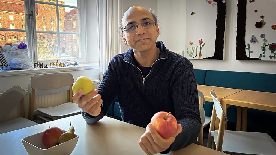
Our dopamine learning system helps us make choices, some as simple as whether to eat a green or red apple, says Arvind Kumar (pictured). He co-authored a new study showing what happens when this system breaks down, and how misalignment contributes to two symptoms of schizophrenia. (Photo: David Callahan)
Scientists have long known that dopamine helps the brain learn from rewards, but a new computational model shows how for people with schizophrenia this learning system can break down and simultaneously produce two very different symptoms – delusions and a loss of motivation.
Publishing in the Journal of Neuroscience, researchers at Stockholm’s KTH Royal Institute of Technology and University of Tokyo, Japan found that problems with motivation and the formation of delusional beliefs may both be linked to a single underlying problem: when an overactivated cortex disrupts the brain’s ability to link actions and consequences.
Arvind Kumar, associate professor in computational neuroscience at KTH, says the study offers a computational neuroscience model that attempts to unify several known roles of the brain’s dopamine system: learning from rewards, controlling motivation and building an internal picture of what’s going on.
Why a unified explanation?
A unified explanation would make it easier to study how these symptoms develop together and may guide future research into treatments, the lead author Kenji Morita says. “If the suggested root cause is validated, then mechanistically grounded therapies could be developed.”
The model in the study shows what happens when this internal cause-and-effect tracking system breaks down. The model suggests that two simultaneous learning processes in the cortico-basal ganglia-midbrain circuits need to align for a person to realize what is rewarding and why.
Deep within the brain, the striatum is a control center that enables the brain to learn which reward is which and selectively increase motivation for right one, such as food when hungry or water when thirsty.
The other dopamine alignment takes place in the cortex. This is the part that enables the brain to essentially follow what is happening. It enables the brain to assign credit correctly: for example, a smell of baked bread predicts food, or the sound of liquid predicts drink.
The researchers found that both reduced motivation and delusion‑like beliefs could arise when an overstimulated cortex disrupts alignment between these processes.
“It causes the brain’s learning system to mix up association between motivation and reward,” Kumar says, “leading to both low motivation and delusion‑like ideas, such as assigning the wrong reasons for things happening.”
“The brain needs to align motivation, reward identity and their causes together to make a suitable choice,” he says.
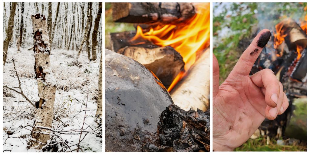
Experiments confirm anti-biotic properties of birch bark extract
The bark of birch trees has been used to produce tar for more than 150 000 years. The centre photo shows birch bark tar condensed onto a rock that borders a hearth. When scraped off the rocks, the viscous tar can be used as both an adhesive and antibiotic.
Neanderthals probably used birch tar for multiple functions, including treating their wounds, according to a study published March 18, 2026 in the open-access journal PLOS One by a team of researchers led by Tjaark Siemssen of the University of Cologne, Germany, and the University of Oxford, UK.
Birch tar is commonly found at Neanderthal archaeological sites, and in some cases this tar is known to have been used as an adhesive to assemble tools. Recently, some researchers have raised the question of whether Neanderthals had multiple uses for this substance. For instance, Indigenous communities in northern Europe and Canada use birch tar to treat wounds, and there is growing evidence that Neanderthals also employed a variety of medical practices.
To investigate the medicinal potential of birch tar, Siemssen and colleagues extracted tar from modern birch tree bark, specifically targeting species known from Neanderthal sites. They used multiple extraction methods, including distillation of tar in a clay pit and condensation of tar against a stone surface, both of which would have been methods available to Neanderthals. When exposed to different strains of bacteria, all of the tar samples were found to be effective at hindering the growth of Staphylococcus bacteria known to cause wound infections.
These experiments not only support the efficacy of Indigenous medicinal practices, but also reinforce the possibility that Neanderthals used birch tar to treat wounds. The authors note that there are other potential uses of birch tar, such as insect repellent, as well as other plants to which Neanderthals had access. Further exploration of the multiple potential uses of these natural ingredients will enable a more thorough understanding of Neanderthal culture.
The authors add: “We found that the birch tar produced by Neanderthals and early humans had antibacterial properties. This has important implications for how Neanderthals may have mitigated disease burden during the last Ice Ages, and adds to a growing set of evidence on healthcare in these early human communities.”
“By bringing together research on indigenous pharmacology and experimental archaeology, we begin to understand the medicinal practices of our distant human ancestors and their closest cousins. Additionally, this study of ‘palaeopharmacology’ can contribute to the rediscovery of antibiotic remedies whilst we face an ever more pressing antimicrobial resistance crisis.”
“The messiness of birch tar production deserves a special mention. Every step of the production is a sensory experience in itself, and getting the tar off our hands after spending hours at the fire has been a challenge every time.”
Image caption: The bark of birch trees has been used to produce tar for more than 150,000 years. The centre photo shows birch bark tar condensed onto a rock that borders a hearth. When scraped off the rocks, the viscous tar can be used as both an adhesive and antibiotic.
Citation: Siemssen T, Oludare A, Schemmel M, Puschmann J, Bierenstiel M (2026) Antibacterial properties of experimentally produced birch tar and its medicinal affordances in the Pleistocene. PLoS One 21(3): e0343618. https://doi.org/10.1371/journal.pone.0343618
Author countries: U.K., Canada, Belgium, Germany
Funding: Tjaark Siemssen is funded by the Boise Trust of the University of Oxford and the German Academic Scholarship Foundation (Studienstiftung). Aderonke Oludare was funded by Canadian Institutes of Health Research (CIHR) Project Grant 420598 awarded to Matthias Bierenstiel. The funders had no role in study design, data collection and analysis, decision to publish, or preparation of the manuscript.
Competing interests: The authors have declared that no competing interests exist.
A new study has found that metformin may mimic one of exercise’s core biological effects in men with prostate cancer, raising levels of a molecule tied to energy balance and weight control even when patients are inactive. The findings suggest that metformin could help counter the metabolic strain of hormone therapy, when fatigue and other side effects often limit physical activity.
Led by physician-scientists at Sylvester Comprehensive Cancer Center, part of the University of Miami Miller School of Medicine, the study appears in the journal EMBO Molecular Medicine.
Exercise is one of the most reliable ways to support health during cancer treatment. It helps regulate weight, blood sugar, and cardiovascular health – factors that shape how patients feel during therapy and how well they recover afterward.
For many people with cancer, however, regular exercise isn’t always feasible. Fatigue, hormone therapy, pain, or advanced disease can limit physical activity precisely when metabolic health becomes most important.
That reality has led researchers to ask a practical question: If exercise confers its benefits through specific biological signals, could some of those signals be activated in other ways?
According to the research, the answer may be yes. Sylvester investigators report that metformin raises levels of a naturally occurring molecule involved in how the body manages energy and weight in prostate cancer patients.
The finding does not suggest that a pill can replace physical activity. Instead, it offers insight into the internal pathways that underlie exercise’s metabolic benefits – and how those pathways might still be engaged when movement is limited.
“This study reflects what’s possible when laboratory science, metabolic biology, and clinical investigation are intentionally brought together for transdisciplinary studies,” said Sylvester researcher and first author, Marijo Bilusic, MD, PhD, genitourinary medical oncologist and professor of medicine and medical oncology at the Miller School. “The result isn’t a new cancer biomarker, but a clearer understanding of how a widely used drug may support metabolic health during prostate cancer treatment – an outcome that matters to patients and clinicians alike.”
The molecule at the heart of the study
At the center of the collaborative, team-science study is a molecule called N-lactoyl-phenylalanine (Lac-Phe). While its name is technical, its role is relatively simple.
Lac‑Phe is produced when the body is under metabolic demand. It forms when lactate, which accumulates during exertion, combines with phenylalanine, a basic building block of protein. Scientists first took notice of Lac‑Phe because its levels spike after intense exercise, coinciding with shifts in energy use and appetite regulation.
In preclinical and early human studies, higher Lac‑Phe levels have been associated with reduced appetite and improved weight control – two effects commonly linked to regular physical activity.
Lac-Phe does not rise only with exercise. Scientists observed elevated Lac-Phe levels in people taking metformin, even in the absence of physical activity. That overlap raised an important question for cancer care: Could a pathway typically associated with exercise be activated pharmacologically in patients whose treatments limit movement?
Why prostate cancer patients are a focus
To explore that question, the Sylvester team focused on prostate cancer, where hormone-based therapies are known to disrupt metabolism, contributing to weight gain, insulin resistance, and cardiovascular risk.
Notably, Lac-Phe levels in patients treated with metformin approximated those previously reported after strenuous exercise. This occurred even though patients were not exercising at the time of blood collection, and the effect persisted after hormone therapy began.
“From a clinical standpoint, seeing a metabolic signal that mirrors what we associate with intense exercise was striking,” said Bilusic. “For patients whose treatments or symptoms limit physical activity, that kind of effect could be especially meaningful.”
Higher Lac-Phe levels were not associated with anti-tumour response to metformin. The metabolite did not correlate with changes in prostate-specific antigen (PSA), a standard marker used to monitor prostate cancer.
What Lac-Phe might mean for patients
That distinction is central to the study’s interpretation. While more expanded studies are needed to determine the utility of Lac-Phe as a marker of anticancer efficacy, it appears to reflect how the body manages energy, weight and metabolic strain during treatment. These results were confirmed to ensure the findings were not limited to one clinical setting. In fact, increases were also observed in patients receiving other metabolic therapies, suggesting Lac-Phe may reflect a broader metabolic response rather than a drug‑specific effect.
“Cancer therapy often affects the body in ways that go beyond the tumour,” said Sylvester researcher Priyamvada Rai, PhD, co-leader, Tumor Biology Program and professor of radiation oncology at the Miller School. “Supporting metabolic health can influence how patients tolerate treatment and how they feel over time, even if it doesn’t directly change tumour growth. This study was an opportunity to investigate molecular pathways that can be therapeutically activated for better outcomes to treatments that induce metabolic stress.”
Metformin raises a stress hormone called GDF‑15, but this study found that Lac‑Phe was more closely tied to weight changes. Because the two didn’t rise together, metformin likely affects weight through multiple pathways, with Lac‑Phe playing a bigger role.
Taken together, the findings offer a clearer picture of how a widely used diabetes medication may influence metabolic health during prostate cancer care.
“What’s encouraging about this work is that it reminds us cancer care isn’t only about targeting tumours – it’s also about supporting the whole patient,” said Rai. “By better understanding how treatments affect metabolism, we can begin to identify ways to help patients maintain strength, resilience, and quality of life throughout their care.”
Despite South Africa’s laws and policies, access to healthcare remains an issue, particularly for non-citizens. Photo by Hush Naidoo on Unsplash
By Teri Brown and Thembi Mahlathi
The media has reported several incidents where people were turned away at public healthcare facilities because they did not possess South African identity documents. As related cases slowly grind through the courts, Teri Brown and Thembi Mahlathi of SECTION27 connect the dots between what the law says and what people are experiencing.
Over the years, many migrants and undocumented people have reached out to SECTION27, where we both work, for assistance. These were often pregnant women, lactating mothers and children under six years, who were denied access to healthcare facilities.
Initially, it was easy to simply write a letter to hospital and clinic personnel where our clients were being denied access. But as time went on, the situation got significantly worse and more migrants were being denied access to public healthcare facilities. Writing letters and asking for meetings clearly wasn’t enough anymore.
We went to court and in April 2023 got an order in which the South Gauteng High Court held that important sections of the National Health Act applies to all pregnant women, lactating women and children under the age of six years, irrespective of their documentation status. This affirmed that in South Africa, they have the right to access free healthcare services at all public health establishments, including hospitals and clinics.
Public sector hospitals and clinics are required to assess the status of migrants and then apply a lawful means test to determine the healthcare services that can be offered to them. However, this does not appear to be done routinely. Instead, particular focus is often placed on South African identity documents, while other forms of documentation held by migrants are disregarded.
There have been incidents where entry to facilities such as Rahima Moosa Mother and Child Hospital in Coronationville and South Rand Hospital in Rosettenville and several clinics across Gauteng have been denied to people, including South African nationals who have the necessary documentation.
Furthermore, we are aware that to avoid being refused healthcare and to demonstrate the urgency of their need for treatment for themselves or their kids, migrants have sometimes been forced to disclose their HIV status – information which they would otherwise have kept private.
In mid-2025, we started receiving a surge of calls from clients complaining about not being able to enter public sector clinics that they were previously assisted at. They informed us that a group of people stationed outside these clinics requested their identity documents, and when they produced their documents confirming either their refugee status or asylum seeker status, they were unlawfully prevented from entering the clinics. These group of people explicitly told them that they should go to a private clinic for treatment or go back to their home country.
Thus, two years after the April 2023 court order, the denial of access to healthcare had worsened, as it was not only women and children who could not access clinics, but anyone who could not provide South African identity documentation. The situation was also exacerbated by the fact that it wasn’t just healthcare staff denying access anymore, but vigilante groups stationed outside healthcare facilities.
Despite the crisis being widely reported, the state failed to address it effectively. We had no choice but to go back to court, and again the court found in our favour.
In December 2025, the South Gauteng High Court ordered the state to take immediate and decisive action to end the obstruction of access to public healthcare facilities in Gauteng. The case was brought by the civil society organisations the Treatment Action Campaign, Doctors Without Borders, and Kopanang Africa Against Xenophobia (the applicants), all represented by SECTION27.
In this landmark judgment, Judge Stuart Wilson concluded that the state entities tasked with upholding the constitutional mandate to safeguard everyone’s right to access healthcare had failed to prevent the obstruction of access to public health facilities. Consequently, this failure was in violation of the constitutional rights of patients seeking care at the Yeoville and Rosettenville clinics.
Despite this court order, our monitoring found ongoing vigilante activity at the two clinics. The applicants then launched an urgent contempt application, heard in March 2026, arguing that the state had failed to fully comply with Judge Wilson’s court order.
Following this, a court ordered settlement agreement was reached with the Gauteng Department of Health and other respondents. Among other things, it required the authorities to take reasonable steps to ensure safe and unhindered access to the Yeoville and Rosettenville clinics, and to report on the implementation by 18 May 2026. It also makes provision to continue legal proceedings if necessary to enforce full compliance with Judge Wilson’s order.
The laws governing healthcare for migrants in South Africa
Taking a step back from this case, and its specific set of facts, it is worth remembering that South African law really does provide extensive protection to migrants who need to access healthcare services.
The right to access healthcare services is guaranteed by section 27 of our Constitution, which states that everyone has the right to have access to healthcare services, and that no one may be refused emergency medical treatment. The term “everyone” is not restricted to South Africans only. It includes everyone within the borders of South Africa, regardless of their nationality.
This right extends to all children living in South Africa under section 28(1)(c) of the Constitution. This guarantees all children access to basic healthcare services dependent on the availability of resources, to which they can never be completely denied.
After the Constitution, the most important piece of healthcare legislation relevant to migrants is the National Health Act (NHA). The NHA assists in giving effect to the constitutional right to basic healthcare services by outlining who can receive services at public clinics free of charge. It obligates the provision of free healthcare services to women who are pregnant or breastfeeding, or children under six. Moreover, the NHA requires that free primary healthcare be provided to those without medical aid. It also makes it clear that those working in healthcare cannot refuse any person emergency medical treatment.
Along similar lines, South Africa’s Refugees Act states that a refugee is entitled to full legal protection, which includes the rights set out in the Bill of Rights, except those reserved for citizens. The Act formally acknowledges that refugees are entitled to the same basic healthcare services and primary education that South African citizens receive. While the Act does not expressly cover undocumented migrants, it is grounded on the principle of non-discrimination, which supports equal access to essential services.
The denial of healthcare services has significant impacts on many aspects of people’s lives. Migrants often become so desperate to receive care that they feel compelled to disclose their HIV status, which infringes on their rights, particularly the constitutional rights to privacy and dignity. It also creates feelings of stigma and discrimination, further marginalising people who are often already vulnerable.
There are also direct health consequences. Denying treatment to a migrant not only negatively impacts that person’s health it can also result in the continued transmission of infectious diseases to both other migrants and South Africans. For example, HIV and TB typically become non-infectious a while after someone starts treatment. Deciding not to treat someone ends up harming everyone. As untreated conditions worsen, it may require emergency medical attention that could have been avoided through early treatment. All of this places extra pressure on an already fragile health system – extra pressure that could be avoided by providing more migrants with healthcare services as soon as they need it.
The failure to provide healthcare services also affects migrants’ livelihoods and well-being. For those who run their own businesses, being unable to access treatment may prevent them from working altogether and could lead to them and other people, possibly South Africans, losing their jobs. Ultimately, this has a ripple effect on the country’s economy, job security, and perpetuates cycles of poverty and vulnerability.
At its heart then, this issue is about who we choose to be as a society. Turning people away at their most vulnerable moments erodes not only their dignity, but also their humanity and ours. In a country built on the values of equality and dignity, we cannot allow this attack on our basic humanity and decency to succeed. We are, and must be, better than that.
*Brown is a legal researcher and Mahlathi is a paralegal with SECTION27. In the court case discussed in this article, SECTION27 represented the Treatment Action Campaign, Médecins Sans Frontiers, and Kopanang Africa Against Xenophobia.
Note: Spotlight is published by SECTION27, but is editorially independent – an independence that the editors guard jealously. Spotlight aims to deepen public understanding of important health issues by publishing a variety of views on its opinion pages. The views expressed in this article are not necessarily shared by the Spotlight editors.
Republished from Spotlight under a Creative Commons licence.