Month: January 2026

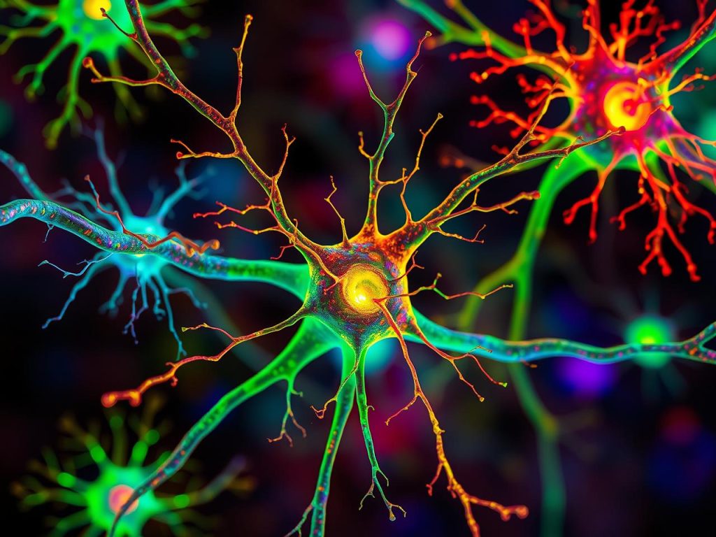
New Neural Maps Challenge Traditional Descriptions of the Brain

AI image of neurons created by Gencraft

For more than a century, maps of the brain have been based on how brain tissue looks under the microscope. These anatomical maps divide the brain into regions according to structural variations in the tissue. But do these divisions really reflect how the brain works? A new study on mice from Karolinska Institutet, published in Nature Neuroscience, suggests that this is often not the case.

By describing the brain in terms of electrical activity of its neurons, the researchers have found a new way to understand the functional organisation of the prefrontal cortex, the brain region responsible for planning, decision-making, and other advanced cognitive functions. 

“Considering that deviations in prefrontal cortex function have been linked to virtually all psychiatric disorders, it is surprising how little is known about how this region actually works,” says Marie Carlén, Professor at the Department of Neuroscience at Karolinska Institutet.

Did not align with previous maps

Her research group recorded and analysed the activity of more than 24 000 neurons in awake mice and created the first activity-based maps of the prefrontal cortex. The maps of spontaneous and cognition-related neuron activity did not match the traditional, tissue-based maps.

“Our findings challenge the traditional way of defining brain regions and have major implications for understanding brain organisation overall,” says Marie Carlén.

The researchers found that the activity patterns of neurons reflected the hierarchy of information flow in the brain rather than the structure of the tissue. Neurons with slow, regular activity turned out to be characteristic of the prefrontal cortex, which sits at the top of this hierarchy. The same activity pattern also marked regions at the top of the prefrontal cortex’s own internal hierarchy. Slow, regular activity is thought to characterise the integration of information flows, a process that is central to cognitive functions such as planning and reasoning. 

Different neuronal activity patterns work together

Carlén and her colleagues discovered that neurons involved in decision-making were concentrated in regions high up in the prefrontal hierarchy. Surprisingly, these neurons were characterised by very fast activity patterns. 

“This suggests that cognitive processes rely on local collaboration between neurons whose activity patterns complement one another. Some neurons appear to specialise in integrating information streams, while others have high spontaneous activity that supports quick and flexible encoding of information, for instance, information needed to make a specific decision,” says Marie Carlén.”

Source: Karolinska Institutet

Heavy Menstruation and Iron Deficiency Common Among Teen Girls

Photo by Marta Branco

More than half of teenage girls experienced heavy bleeding and 40% had an iron deficiency, in a study carried out among girls in upper-secondary schools. The research, led from Lund University in Sweden, also shows that young teenage girls who experience heavy menstrual bleeding – and are therefore at greater risk of iron deficiency – can be identified using a simple questionnaire.

The 2023 study, published in PLOS One, was carried out in two Swedish upper-secondary schools; a total of 394 girls aged 15 and over took part. They responded to questions about menstruation and eating habits and provided blood samples for analysis of blood count (Hb) and iron stores (ferritin). 
Lund University has previously reported how the research team saw a particularly high risk of iron deficiency and anaemia among those eating a vegetarian or pescetarian diet. Now, researchers have gone further and investigated how the extent of menstruation affects iron deficiency and anaemia.

“We see a lot of young girls who are tired and distracted. Linking that to menstruation or diet is not obvious,” says Moa Wolff, researcher and associate professor at Lund University, specialist physician in general medicine at Region Skåne and the researcher in charge.

Girls with heavy menstruation were three times more likely to have an iron deficiency, with an even higher risk for those who restricted the quantity of meat in their diet.  

“Many of them only compare with their own previous experiences without knowing what counts as heavy menstrual bleeding. We also note that many are not aware of the over-the-counter medicines available that reduce the volume of blood,” says Lisa Söderman, gynaecologist and postdoc researcher at Karolinska Institutet who collated the results for the article in question. 

SAMANTA questionnaire could be a new tool for school health services

Part of the study evaluated a Spanish questionnaire which asks six questions. This is the first time it has been used in Sweden and the first time it has been tested on teenage girls anywhere, even though it is a validated screening instrument for adult women.
 
“Based on the answers to the questionnaire, it was possible to clearly identify which secondary school students were at risk of having low iron levels. It is easy to use and could be a valuable tool for school health services and youth clinics or other care healthcare settings where we meet these girls,” says Moa Wolff.
 
Previous research shows that iron deficiency in young people may affect their energy levels, schoolwork and general wellbeing. Iron stores – Ferritin – form an important component in the formation of red blood cells and is needed for oxygen transport. Therefore, iron deficiency with low ferritin levels can eventually lead to a drop in haemoglobin, which can result in anaemia.
 
“In the next few years, some of these girls will get pregnant. When that happens, we would like them to have good stores of iron to enable as complication-free a pregnancy as possible, with a successful labour,” says Lisa Söderman.

Source: Lund University

Full-fat Cheese Linked to a Reduced Dementia Risk

Photo by David Foodphototasty on Unsplash

Eating cheese and cream with a high fat content may be linked to a lower risk of developing dementia. This is shown by a new large-scale study from Lund University. The researchers analysed the dietary habits of more than 27 000 people and linked these to the occurrence of dementia over a follow-up period of up to 25 years.

The debate about low-fat diets has long shaped our health advice and influenced how we view food and health. For several decades, fear of saturated fat and its link to cardiovascular disease has dominated. The MIND diet1 is a diet developed with the aim of reducing the risk of dementia. The diet includes protective foods such as vegetables, nuts, fruits, berries, whole grains, and fish, while cheese is one of the foods that should be limited.

Emily Sonestedt, researcher in nutritional epidemiology at Lund University in Sweden, and her colleagues, therefore wanted to investigate whether there was any link between dairy products and dementia. They collected dietary data from 27,670 people using the Malmö Diet Cancer population study, in which the participants respond about their dietary and cooking habits. The average age at the start of the study was 58, and the participants were followed for an average of 25 years, during which time 3,208 people developed dementia. The dementia diagnoses were obtained from the Swedish patient registry. For cases diagnosed up to 2014, additional validation studies were conducted in which dementia specialists reviewed medical records, brain scans, and cognitive test results.

After adjusting for lifestyle factors such as physical activity, diet, smoking, and alcohol consumption, the researchers found that people who ate 50 grams of cheese (with more than 20 percent fat) daily had a 13 percent lower risk of developing dementia than those who ate less than 15 grams daily. 50 grams is equivalent to about five regular slices of cheese. In total, about a quarter of the participants ate more than 50 grams or more daily.

”When we went on to look at specific types of dementia, we found that there was a 29 percent lower risk of vascular dementia in people who ate more full-fat cheese. We also saw a lower risk of Alzheimer’s disease, but only among those who did not carry the APOE e4 gene variant—a genetic risk factor for Alzheimer’s disease.”

The researchers also investigated the link between high-fat cream (30-40 percent fat) and dementia. People who consumed 20 grams or more daily had a 16 percent lower risk of dementia than those who did not consume any at all. 

The results of the cheese studies support the link between vascular health and brain health.

”The updated dietary guidelines in Sweden from this year say that we can eat dairy products every day, preferably fermented varieties such as yogurt or kefir. Both we and other researchers have found in observational studies that fermented dairy products in particular may be linked to a slightly reduced risk of cardiovascular disease 2,” says Emily Sonestedt.

In previous studies3, the research team has seen links to vascular health, with cheese and fermented dairy products in particular protecting against cardiovascular disease. 

”Although higher-fat cheese and cream were associated with a reduced risk of dementia, other dairy products and low-fat alternatives did not show the same effect. Therefore, not all dairy products are equal when it comes to brain health. The few studies that have investigated this have found a correlation with cheese, so more research is needed to confirm our results and investigate whether certain high-fat dairy products really do provide some protection for the brain.”

Source: Lund University


  1. The MIND diet stands for Mediterranean–DASH Intervention for Neurodegenerative Delay – a combination of the Mediterranean diet and the DASH diet. DASH (Dietary Approaches to Stop Hypertension) is a diet developed primarily to lower high blood pressure and improve cardiovascular health.
  2. Milk and dairy products – a scoping review for Nordic Nutrition Recommendations 2023
  3. Previous publications: 
    High-fat and low-fat fermented milk and cheese intake, proteomic signatures, and risk of all-cause and cause-specific mortality
    High consumption of dairy products and risk of major adverse coronary events and stroke in a Swedish population
    Dairy products and its association with incidence of cardiovascular disease: the Malmö diet and cancer cohort
    Dairy Consumption, Lactase Persistence, and Mortality Risk in a Cohort From Southern Sweden

Common Eye Ointment can Damage Glaucoma Implants, Study Warns

Research shows that petrolatum-based eye ointments can cause the device to swell and potentially rupture, prompting an urgent update to clinical guidance.

Photo by Tima Miroshnichenko


Widely-used eye ointments can cause glaucoma implants to swell and potentially rupture, according to new research from Nagoya University in Japan. This study is the first to show, using clinical and experimental evidence, that petrolatum-based eye ointments can compromise the PRESERFLO® MicroShunt, an implant used in over 60 countries to treat glaucoma.

Glaucoma is an eye disease that damages the optic nerve and can lead to vision loss. It often results from increased intraocular pressure caused by blocked drainage of eye fluid. A recent study estimated that 76 million people globally are affected by glaucoma.

Progression of visual field loss (from left to right) due to glaucoma
(Credit: Ryo Tomita)

MicroShunt is a small filtration device implanted in the eye to improve fluid drainage in glaucoma patients. Compared to traditional surgeries, it lowers post-operative complications and reduces reliance on additional medications.

MicroShunt is made from a styrenic thermoplastic elastomer based on a polystyrene-block-polyisobutylene-block-polystyrene (SIBS) block polymer, which is highly biocompatible, flexible, and less likely to cause inflammation or scarring. However, this material is vulnerable when it comes into contact with hydrocarbon- and oil-based materials. Due to its high oil affinity, exposure to petrolatum-based eye ointments may allow oil components to penetrate the device, causing swelling and potential changes in its shape and flexibility.

The MicroShunt manufacturer’s instructions state that “the MicroShunt should not be subjected to direct contact with petrolatum-based (ie, petrolatum jelly) materials, such as ointments and dispersions.” But this precaution is not widely recognised or consistently followed in clinical practice.

“Swollen MicroShunts can be structurally fragile,” said ophthalmologist and Assistant Professor Ryo Tomita of Nagoya University Graduate School of Medicine, the study’s first author. “During surgery, I observed a rupture in a swollen MicroShunt. If more clinicians are aware of this risk, they will be able to prevent similar problems.”

Tomita and colleagues, including Assistant Professor Taiga Inooka and Associate Professor Kenya Yuki from Nagoya University Hospital and the Graduate School of Medicine collaborated with Dr. Takato Kajita and Junior Associate Professor Atsushi Noro from the Graduate School of Engineering to examine changes in the MicroShunt after exposure to a petrolatum-based eye ointment.

The medical team reviewed clinical cases, while the engineering team conducted laboratory analyses. The findings were published in Graefe’s Archive for Clinical and Experimental Ophthalmology.

Clinical evidence

The clinical study examined seven glaucoma patients whose MicroShunt implants were later removed for different reasons. The results revealed a clear pattern. In three cases, the MicroShunt was exposed outside the conjunctiva, and patients received a petrolatum-based eye ointment. All three explanted devices showed significant swelling, and two of them ruptured.

In three other cases, the MicroShunt remained covered by the conjunctiva, and no ointment was administered. These devices retained their original structure. Crucially, in one additional case, the MicroShunt was exposed outside the conjunctiva, but no ointment was applied. The device did not swell. This indicates that direct contact with the ointment, rather than conjunctival rupture alone, is the primary cause of swelling.

Photographic comparison of MicroShunt illustrating size changes
Top: MicroShunt explanted from a patient, exhibiting diffuse swelling with fracture and loss of one fin
Middle: MicroShunt explanted from another patient, showing localized swelling around the fin
Bottom: Unused MicroShunt (control)

Scale: 1 division = 1 mm   
(Credit: Ryo Tomita)

Laboratory confirmation

Laboratory experiments confirmed the clinical findings. The team immersed unused MicroShunts in petrolatum-based eye ointment to reproduce the swelling seen in clinical cases. Microscopic measurements showed significant changes. After 24 hours in the ointment, the MicroShunt’s outer diameter increased to 1.44 times its original size, and the fin-like portion widened to 1.29 times its initial value.

Chemical analysis identified the cause of this change. After 24 hours of immersion, oil components made up approximately 45% of the MicroShunt’s total weight, rising to 73% after three months. These results confirmed the primary cause of swelling to be the absorption of oil-based ointment constituents into the material.

Clinical implications

The research team emphasises that clinicians should avoid using petrolatum-based ointments on patients with MicroShunt implants, particularly when the device is exposed outside the conjunctiva. Alternative post-operative treatments should be considered, while further research is needed to assess whether swelling impacts MicroShunt performance even when rupture does not occur.

“Our study found that commonly used medical materials can cause unexpected complications if their chemical properties and usage environments are not fully understood,” Noro stated. “From both medical and engineering perspectives, we emphasise the importance of understanding the chemical properties of medical materials and appropriately managing their usage environments.”

Paper information:

Ryo Tomita, Taiga Inooka, Takato Kajita, Hideyuki Shimizu, Ayana Suzumura, Jun Takeuchi, Tsuyoshi Matsuno, Hidekazu Inami, Koji M. Nishiguchi, Atsushi Noro, and Kenya Yuki. (2026) Petrolatum-based ointment application induces swelling of the PRESERFLO MicroShunt. Graefe’s Archive for Clinical and Experimental Ophthalmology
DOI: 10.1007/s00417-025-07075-2

Can AI Help Make Prescriptions Safer in South Africa’s Busy Clinics?

AI image created with Gencraft

By Henry Adams, Country Manager, InterSystems South Africa

Across South Africa, nurses and doctors in public clinics make hundreds of important decisions every day, often under enormous pressure. They’re short on time, juggling long queues, and sometimes working with incomplete information. In those conditions, even the most experienced professionals can make mistakes. It’s human.

The truth is, our healthcare system is stretched thin, and people can only do so much. That’s why I see real potential for AI to step in as a kind of virtual pharmacist. Not to replace anyone, but to back them up by checking prescriptions, catching errors, and helping ensure patients get the right treatment quickly and safely.

From data to decision support

I’m often asked how AI can make a real difference in healthcare right now. One area where it can have an immediate impact is in prescriptions. AI-assisted systems help doctors and nurses make safer, faster decisions by analysing medical data in real time. They can check a patient’s history, allergies, and possible drug interactions in seconds, flagging risks before they become problems.

Of course, because we’re dealing with sensitive medical information, trust and data quality are crucial. These systems only work when they’re built on accurate, connected data that healthcare professionals can rely on.

That’s where the latest health technology partnerships come in. By linking proven data platforms with smart AI tools, we’re already seeing real improvements overseas. In Europe, for example, these systems are helping clinicians catch potential drug errors early and prescribe with greater confidence.

There’s no reason South Africa can’t benefit in the same way. With clinics under pressure and resources stretched, technology that connects clean, reliable data with practical AI support could help reduce errors, save time, and make care safer for everyone.

Addressing local challenges

Medication errors can happen anywhere, but in South Africa the stakes are often higher. Our public clinics are exceptionally busy, staff are stretched, and doctors and nurses are doing their best under tough conditions. When you’re working under that kind of pressure, even a small mistake in a prescription can have serious consequences for a patient.

This is where AI can really help. Imagine a system that double-checks every prescription in real time, flagging possible drug interactions, incorrect dosages, or missing information before the medicine ever reaches the patient. It’s like having an extra set of expert eyes that never get tired. Instead of slowing things down, it speeds them up and gives clinicians peace of mind knowing they’re making the safest call for each patient.

For that to work, though, the data behind the system must be reliable and up to date. As South Africa moves toward a unified digital health record, the ability for these systems to connect to existing patient information becomes crucial. When healthcare professionals can trust the data they see on screen, AI becomes a genuine partner in care, helping them work faster, smarter, and safer.

Building confidence in AI

For AI to really work in healthcare, it must be clear and trustworthy. Doctors and nurses need to know why the system is recommending a specific drug or warning about a potential issue. If it can’t explain itself, people won’t use it, and rightly so.

That’s why transparency matters. The best AI tools don’t make decisions behind closed doors; they show their reasoning and help clinicians understand what’s happening in the background. When that’s combined with reliable, well-managed data, you start to build real confidence in the system.

It’s that trust, knowing the technology supports rather than replaces clinical judgment, that will make AI-assisted prescriptions part of everyday care, not just an interesting experiment.

A collaborative path forward

Technology on its own won’t fix South Africa’s healthcare challenges, but it can make a big difference in helping people do their jobs better. AI-assisted prescriptions are a good example of how smart tools can take some of the pressure off clinicians, reduce paperwork, and help patients get safer, faster care.

What excites me most is how practical this can be. Picture a nurse in a rural clinic who needs to prescribe medication but doesn’t have easy access to a specialist. With AI support, she can get accurate, instant guidance and know her patient is getting the right treatment. Or think about a busy hospital pharmacy, where an AI system automatically checks for drug interactions across hundreds of files in seconds, preventing errors before they happen.

This isn’t some far-off idea. The technology already exists and is being used successfully elsewhere. The goal now is to make sure it’s used in a way that supports our healthcare professionals, not replaces them. They are, and always will be, at the centre of care. If we get this right, AI can become a real partner in healthcare.

Immunometabolism Might Hold the Key to Controlling Sepsis

Image from Rawpixel

Metabolic changes that “rewire” part of the immune system can intensify sepsis, the body’s dysregulated response to infection. This discovery may lead to new ways to block metabolic changes contributing to excessive and ineffective inflammation, reset the immune system, and bring sepsis under control, researchers at Vanderbilt Health reported January 15 in the journal Nature Immunology.

“Metabolism is potentially a means by which we could intervene in immune dysfunction in ICU (intensive care unit) patients including those with sepsis,” said the paper’s first and co-corresponding author, Matthew Stier, MD, PhD, assistant professor of Medicine in the Division of Allergy, Pulmonary and Critical Care Medicine at Vanderbilt Health.

“I think we can make great progress and great strides by aligning cutting-edge basic science tools with ICU patient samples to understand these mechanisms and prioritise therapies for future interventions,” he said.

Sepsis is characterised by the massive production and release of inflammatory molecules, including cytokines, that if unchecked, can lead to tissue damage, septic shock, organ failure, and death.

Despite decades of research focused on stopping this “cytokine storm” and hyperinflammation, “we have unfortunately not been able to identify successful drug therapies in sepsis,” said Stier, a physician-scientist who focuses on immunologic and metabolic dysfunction in critical illness. Targeting the inflammatory aspect of sepsis is likely important, but by itself may not be sufficient.

“We provide antibiotics and great supportive care to weather the cytokine storm,” he said, “but that doesn’t fully resolve the problem. It keeps people alive while we wait for their bodies to fix themselves — or not.”

In critical illness, including sepsis, the body’s normal metabolic processes become impaired. This includes immunometabolism, the energy-generating processes that fuel the immune system.

At the same time, the immune system’s protective functions become exhausted, resulting in an acquired immunosuppression, which leaves patients vulnerable to secondary infections, persistent organ dysfunction, repeated hospitalisations and death.

While prior research has defined the characteristics of metabolism and immune dysfunction, this study was among the first to explore the mechanisms of immunometabolic dysfunction in sepsis and their association with immunosuppression, often called “immunoparalysis.”

Stier and his colleagues used cutting-edge technologies, including single-cell sequencing and flow cytometry, to study immune cells collected from the blood of critically ill patients.

The blood samples were collected and stored through the Sepsis Clinical Resource and Biorepository (SCARAB), a unique, highly collaborative ICU biobank developed by Julie Bastarache, MD, and Lorraine Ware, MD, professors of Medicine in the Division of Allergy, Pulmonary and Critical Care Medicine.

Two of the most important elements of the body’s immune responses are CD4+ T “helper” cells, inflammatory “foot soldiers” of the immune system that are distinguished by the CD4 surface protein they express, and regulatory T (Treg or “suppressor”) cells, which guard against over-active immune responses.

To study the impact of critical illness and sepsis on these cells, the Vanderbilt Health team used SCENITH, a flow cytometry-based method developed by French researchers that enables researchers to functionally profile energy metabolism with single-cell resolution.

“This technique allowed us to do something prior studies hadn’t done … to look at the metabolism of every single cell set on its own and identify subset-specific metabolic adaptations,” Stier said.

The key finding: Treg cells undergo metabolic “reprogramming” in patients with critical illness and sepsis, leading to altered tryptophan metabolism and response to reactive oxygen species in a way that enhances their immunosuppressive capability, at the expense of CD4+ T “helper” cells.

“The metabolic turmoil of critical illness appears to give Treg cells a survival and functional advantage, contributing to the harmful immunoparalysis seen in sepsis,” he said.

“This is very much a preclinical paper,” Stier cautioned. Yet it demonstrates the feasibility of deeply dissecting immunometabolic mechanisms using ICU patient biospecimens and highlights the importance of such insights to prioritise future therapeutic targets in critical illness and sepsis, he said.

Source: Vanderbilt University Medical Center

Switching Memories On and Off with Epigenetics

Photo by Laura Louise Grimsley on Unsplash

Our experiences leave traces in the brain, stored in small groups of cells called “engrams”. Engrams are thought to hold the information of a memory and are reactivated when we remember, which makes them very interesting to research on memory and age- or trauma-related memory loss.

At the same time, scientists know that the biology of learning is accompanied by epigenetic changes, which refers to the ways the cell regulates genes by adding chemical “post-it notes” on DNA.

But the question of whether the epigenetic state of a single gene in turn can cause a memory to change has thus far remained unanswered.

A team led by Professor Johannes Gräff at EPFL’s Laboratory of Neuroepigenetics combined CRISPR-based gene control with a technique that tags engram cells in mice. They focused on Arc, a gene that helps neurons adjust their connections to other neurons. By targeting the control region of Arc, the team asked whether flipping its epigenetic “switch” could directly change memory. They published their findings in Nature Genetics.

An “epigenetic switch”

The researchers developed specialised, CRISPR-based tools that could either dial down or boost Arc activity in memory neurons. Some, like the KRAB-MeCP2 tool, were designed to switch off gene activity by adding repressive marks that make the DNA less accessible, while others opened the DNA and turned the gene on. These tools were essentially an “epigenetic switch” for the Arc gene.

They then used harmless viruses to deliver these tools directly into the hippocampus of mice, a brain region central for storing and retrieving memory. The mice were then trained to link a specific place with a mild foot shock. By changing the epigenetic state of Arc in the neurons, the scientists could see whether the animals remembered the shock or not. They also added a “safety switch” that could undo the editing and reset the memory state.

The study showed that epigenetically silencing Arc in engram cells made the mice not learn, while boosting it made their memory stronger. These changes could be reversed in the same animal, showing that this epigenetic “switch” can dial memory expression up or down. Even memories that were already several days old, which are usually hard to change, could be modified. On the molecular level, the editing caused changes in gene activity and DNA packaging that matched the behavioural effects.

Controlling memory expression

The study is the first direct demonstration that changing the epigenetic state in memory cells is necessary and sufficient to control memory expression. It points to new ways of exploring how memories are stored and altered, which could eventually also be relevant in humans.

In the future, similar approaches could help researchers better understand conditions where memory processing goes awry, such as traumatic memories in PTSD, drug-related memories in addiction, or the memory problems that appear in neurodegenerative diseases.

Source: EPFL

Childhood Leukaemia Aggressiveness Depends on Timing of Genetic Mutation

Credit: National Cancer Institute

A team of researchers at the Icahn School of Medicine at Mount Sinai has uncovered why children with the same leukaemia-causing gene mutation can have dramatically different outcomes: it depends on when in development the mutation first occurs.  

The study, led by Elvin Wagenblast, PhD, Assistant Professor of Oncological Sciences, and Pediatrics, at the Icahn School of Medicine at Mount Sinai, was published in Cancer Discovery. It shows that leukemia beginning before birth is often more aggressive, grows faster, and is harder to treat. This adds a missing dimension to precision medicine for childhood leukaemia. 

Dr. Wagenblast and his team at the Wagenblast Lab set out to answer a central question about how a normal blood stem cell can become cancerous. They applied cutting-edge CRISPR/Cas9 genome-editing approaches in human primary blood stem cells to model different developmental stages of acute myeloid leukaemia, one of the most aggressive types of blood cancer. 

Using CRISPR technology, the team induced the NUP98::NSD1 fusion oncoprotein, a cancer-promoting protein created when two genes abnormally fuse, into human blood stem cells from multiple developmental stages, ranging from prenatal to postnatal, adolescence, and adulthood. This approach created the first humanised experimental model that tracks how the same mutation behaves differently depending on when in life it arises. 

The results were striking: stem cells produced during prenatal development transformed easily into leukaemia, creating a highly aggressive and more primitive form of leukaemia. Stem cells produced postnatally became increasingly resistant to transformation and required additional mutations to become cancerous. Prenatal-origin leukaemia stem cells, which are abnormal blood stem cells that arise before birth and can cause certain childhood leukaemias, were more dormant (quiescent) and relied heavily on certain energy sources specific to the cancer state, which were not seen in the leukaemias that originated later in life. Although these prenatal leukaemia stem cells were more dormant, this quiescent state makes them harder to eliminate with standard treatments, helping explain why prenatal-origin leukaemias behave more aggressively, despite identical genetics. 

By analysing single-cell gene expression data from their models, the investigators identified a prenatal gene signature that predicts whether a child’s leukaemia likely began before birth. In patients, this signature strongly correlated with significantly worse clinical outcomes. 

“This work tells us that age matters at the cellular level,” said Dr Wagenblast. “The same mutation behaves very differently depending on when it happens. Understanding this gives us a new way to identify the highest-risk patients and to tailor therapies that go beyond standard genetic classifications.” 

The team tested therapies against the most aggressive leukaemia stem cells and discovered that these cells were especially vulnerable to venetoclax, a Food and Drug Administration-approved drug already used in the clinic. Venetoclax-based combinations, including with standard chemotherapy, significantly reduced aggressiveness in the experimental models. 

“These findings give clinicians mechanistic support to use venetoclax combinations in NUP98-rearranged acute myeloid leukaemia, particularly in younger patients whose disease likely started before birth,” said Dr Wagenblast. 

Understanding when leukaemia begins may help doctors choose more effective therapies earlier, reducing trial-and-error approaches and preventing resistance and relapse later on. 

Conceptually, the study shifts how scientists understand childhood cancer. The developmental timing of the first mutation is not a minor detail. It fundamentally shapes disease biology, treatment resistance, and relapse risk. 

The research opens the door to new diagnostic tools that can identify prenatal-origin leukaemias, venetoclax-based combination therapies that more precisely target vulnerable leukaemia stem cells, and clinical trials that incorporate developmental timing into risk assessment. 

Next, the team plans to develop therapies that more directly target the metabolic program unique to prenatal-origin leukaemias, with the goal of selectively eliminating leukaemia stem cells while sparing healthy blood stem cells. 

Source: Mount Sinai

The Making of South Africa’s Medical Aid Crisis

As of this month, South African medical aid scheme contributions have increased by between 6–9% – nearly triple the Council for Medical Schemes’ recommended 3.3% guideline. While lower than last year’s double-digit surge, the underlying problem remains: premiums keep climbing while benefit coverage keeps shrinking, exposing cracks in private healthcare that are becoming impossible to ignore.

“We’re watching private healthcare price ordinary South Africans out of the market, one annual increase at a time,” says Lungile Kasapato, CEO of PPO Serve, a healthcare management company that has been implementing value-based care in South Africa for more than a decade. “Medical schemes are caught in an impossible position – unable to control what providers charge, they’re left managing what they cover. The result is diminishing benefits, rising co-payments, and mounting out-of-pocket costs for members.”

The root of the problem lies in how healthcare is paid for. Fee-for-service, the dominant reimbursement model, rewards volume over outcomes. More tests, more procedures, more bed days – each generates revenue regardless of whether they actually improve patient health. This narrow focus fragments care and drives costs up while keeping value low.

“No amount of funding can fix a payment model that drives the wrong incentives,” Kasapato explains. “Real change requires rethinking not just what we pay for, but how we pay for it.”

Value-based care offers a fundamentally different approach: putting patients at the centre, rewarding proactive care, and linking payment directly to health outcomes. PPO Serve’s The Value Care Team demonstrates what this looks like in practice. GP-led multidisciplinary teams receive monthly, risk-adjusted payments based on patient complexity, supporting holistic care and linking meaningful incentives to measurable results. Rather than maximising billable services, providers focus on optimising patients’ overall health.

For members, this means care is no longer limited by rigid benefit caps or pre-authorisation hurdles, but structured around what genuinely enhances the efficient delivery of their care. A dedicated care coordinator guides patients through decisions made collaboratively by their GP and allied health professionals, with each team member sharing accountability for better outcomes.

But scaling models like this requires medical schemes and public funders to step up. “The challenge isn’t proving value-based care works – it’s embedding it in an infrastructure built for an entirely different system,” says Kasapato. “Claims processing, scheme administration, provider networks – every layer of private healthcare is designed with fee-for-service in mind. Transitioning to outcome-based payment means rebuilding that system and accepting the upfront investment and friction that comes with structural change. The alternative is stark: a private healthcare market that collapses under its own cost pressures, pricing out members faster than schemes can adjust. South Africa is already on that trajectory.”

“If we’re serious about universal health coverage and the long-term sustainability of the private sector, we can’t keep treating symptoms while ignoring causes,” says Kasapato. “Value-based care models are already demonstrating what’s possible. The question isn’t whether transformation is worth the investment – it’s whether we can afford to delay it any longer. The more organisations that embrace a strategic purchasing role, the greater the potential for meaningful change, not just for medical schemes but for South Africa’s healthcare system and the millions who rely on it.”

R23.9 Billion, 72 000 lives, One Budget Speech

Why the Health Promotion Levy can no longer be delayed

Johannesburg, 19 January 2026: When Petrus Cockrell wakes up each morning, the first thing he reaches for is his wheelchair. Diabetes took both his legs before he turned 50. It robbed him of his mobility, his job and the simple joy of walking beside his dog.Petrus is one of millions of South Africans living with a disease that did not need to progress this far. Behind every statistic is someone like him, a parent, a worker, a caregiver whose life has been cut short or forever altered by a preventable illness.

With the National Budget Speech scheduled for February, the Healthy Living Alliance (HEALA) is calling on government to increase the Health Promotion Levy (HPL) on sugary drinks from 11% to 20%, a life-saving decision backed by evidence.

The HPL is part of South Africa’s broader package of health taxes, alongside tobacco and alcohol excise duties, which have long been used to protect the public from preventable harm.

“Every amputation, every blindness diagnosis, every child who loses a parent to diabetes is a reminder that we have waited too long,” says Nzama Mbalati, CEO of HEALA. “The HPL is not a standalone experiment; it is a proven health tax. Government has used health taxes successfully for decades. Strengthening the HPL simply extends that legacy to protect South Africans from excessive sugar consumption.”

The urgency of this demand is underscored by modelling from PRICELESS SA (University of the Witwatersrand).  The data indicates that increasing the HPL to 20% could prevent 619 000 new diabetes cases, save approximately 72 000 lives, prevent 85 000 strokes and save South Africa R23.9 billion in healthcare costs over 25 years.2

“We treat people every day for conditions that should never have progressed this far. The HPL is not just a tax, it is a protective shield for millions of South Africans,” says medical doctor and health advocate Dr Darren Green, featured in HEALA’s upcoming campaign. “Strengthening it means fewer amputations, fewer patients on dialysis and fewer children growing up without parents. Very few interventions deliver such measurable health benefits, especially for communities already carrying the heaviest burden.”

As tariff disputes and import pressures dominate sugar industry news, HEALA emphasises that tariffs and the HPL must not be conflated. Tariffs are trade instruments designed to stabilise industries. The HPL is a public health instrument designed to save lives.

“We cannot allow tariff debates to derail a health tax that works,” Mbalati adds. “Just as we use tobacco and alcohol taxes to protect South Africans from harm, the HPL is a critical part of our national health tax framework. Strengthening it is a public health necessity, not an industry target.”

HEALA’s documentary series continues to reveal the human cost of diabetes. Alphinah, who lost both legs and her eyesight; Mpho, who believed sugar was harmless until he lost his leg at 45 and now Petrus, each offering a powerful reminder that these outcomes were preventable. Their message is clear: if they had known sooner, their lives would look different. Government now has the power to prevent thousands more from walking the same path.

HEALA calls on the public to stand with Petrus and millions of others by demanding decisive government action. As the Budget Speech approaches and the Health Promotion Levy faces growing pressure from industry interference, South Africans are urged to sign the petition supporting the increase of the HPL to 20% before the Minister of Finance takes the podium in February. Sign the petition at www.heala.org.

References:

  1. HEALA Diabetes Documentary Series (2025).
  2. PRICELESS SA. The Cost of Not Setting the Sugar-Sweetened Beverage Tax at 20%. (2025).