Month: May 2025

Under Different Anaesthetics, Same Result: Unconsciousness by Shifting Brainwave Phase

MIT study finds that an easily measurable brain wave shift may be a universal marker of unconsciousness under anaesthesia

Photo by Anna Shvets on Pexels

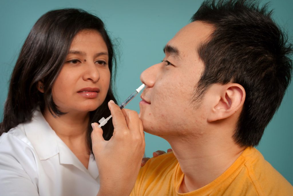
At the level of molecules and cells, ketamine and dexmedetomidine work very differently, but in the operating room, they do the same exact thing: anaesthetise the patient. By demonstrating how these distinct drugs achieve the same result, a new study in animals by neuroscientists at The Picower Institute for Learning and Memory at MIT identifies a potential signature of unconsciousness that is readily measurable to improve anaesthesiology care.

What the two drugs have in common, the researchers discovered, is the way they push around brain waves, which are produced by the collective electrical activity of neurons. When brain waves are in phase, meaning the peaks and valleys of the waves are aligned, local groups of neurons in the brain’s cortex can share information to produce conscious cognitive functions such as attention, perception and reasoning, said Picower Professor Earl K. Miller, senior author of the new study in Cell Reports. When brain waves fall out of phase, local communications, and therefore functions, fall apart, producing unconsciousness.

The finding, led by graduate student Alexandra Bardon, not only adds to scientists’ understanding of the dividing line between consciousness and unconsciousness, Miller said, but also could provide a common new measure for anesthesiologists who use a variety of different anesthetics to maintain patients on the proper side of that line during surgery.

“If you look at the way phase is shifted in our recordings, you can barely tell which drug it was,” said Miller, a faculty member in The Picower Institute and MIT’s Department of Brain and Cognitive Sciences. “That’s valuable for medical practice.  Plus if unconsciousness has a universal signature, it could also reveal the mechanisms that generate consciousness.”

A figure from the paper summarises the main findings. Under either ketamine or dexmedetomidine general anaesthesia, brain waves become shifted out of phase within a hemisphere and more into phase across hemispheres.

If more anesthetic drugs are also shown to affect phase in the same way, then anaesthesiologists might be able to use brain wave phase alignment as a reliable marker of unconsciousness as they titrate doses of anesthetic drugs, Miller said, regardless of which particular mix of drugs they are using. That insight could aid efforts to build closed-loop systems that can aid anaesthesiologists by constantly adjusting drug dose based on brain wave measurements of the patient’s unconsciousness.

Miller has been collaborating with study co-author Emery N. Brown, an anaesthesiologist and Professor of Computational Neuroscience and Medical Engineering, on building such a system. In a recent clinical trial with colleagues in Japan, Brown demonstrated that monitoring brain wave power signals using EEG enabled an anaesthesiologist to use much less sevoflurane during surgery with young children. The reduced doses proved safe and were associated with many improved clinical outcomes, including a reduced incidence of post-operative delirium.

Phase findings

Neuroscientists studying anaesthesia have rarely paid attention to phase, but in the new study, Bardon, Brown and Miller’s team made a point of it as they anaesthetised two animals.

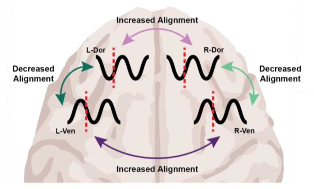
After the animals lost consciousness, the measurements indicated a substantial increase in “phase locking,” especially at low frequencies. Phase locking means that the relative differences in phase remained more stable. But what caught the researchers’ attention were the differences that became locked in: Within each hemisphere, regardless of which anesthetic they used, brain wave phase became misaligned between the dorsolateral and ventrolateral regions of the prefrontal cortex.

Surprisingly, brain wave phase across hemispheres became more aligned, not less. But Miller notes that case is still a big shift from the conscious state, in which brain hemispheres are typically not aligned well, so the finding is a further indication that major changes of phase alignment, albeit in different ways at different distances, are a correlate of unconsciousness compared to wakefulness.

“The increase in interhemispheric alignment of activity by anesthetics seems to reverse the pattern observed in the awake, cognitively engaged brain,” the Bardon and Miller team wrote in Cell Reports.

Determined by distance

Distance proved to be a major factor in determining the change in phase alignment. Even across the 2.5 millimetres of a single electrode array, low-frequency waves moved 20-30 degrees out of alignment. Across the 20 or so millimetres between arrays in the upper (dorsolateral) and lower (ventrolateral) regions within a hemisphere, that would mean a roughly 180-degree shift in phase alignment, which is a complete offset of the waves.

The dependence on distance is consistent with the idea of waves traveling across the cortex, Miller said. Indeed in a 2022 study, Miller and Brown’s labs showed that the anaesthetic propofol induced a powerful low-frequency traveling wave that swept straight across the cortex, overwhelming higher-frequency straight and rotating waves.

The new results raise many opportunities for follow-up studies, Miller said. Does propofol also produce this signature of changed phase alignment? What role do travelling waves play in the phenomenon? And given that sleep is also characterised by increased power in slow wave frequencies, but is definitely not the same state as anaesthesia-induced unconsciousness, could phase alignment explain the difference?

Source: Picower Institute

Alzheimer’s Drug Lecanemab Well Tolerated in Real-world Use

Side effects of lecanemab are manageable, study finds

Created with Gencraft. CC4.0

The Food and Drug Administration’s approval in 2023 of lecanemab – a novel Alzheimer’s therapy shown in clinical trials to modestly slow disease progression – was met with enthusiasm by many in the field as it represented the first medication of its kind able to influence the disease. But side effects of brain swelling and bleeding emerged during clinical trials that have left some patients and physicians hesitant about the treatment. [Especially considering its $26 500 per year cost – Ed.]

Medications can have somewhat different effects once they are released into the real world with broader demographics. Researchers at Washington University School of Medicine in St. Louis set out to study the adverse events associated with lecanemab treatment in their clinic patients and found that significant adverse events were rare and manageable.

Consistent with the results from carefully controlled clinical trials, researchers found that only 1% of patients experienced severe side effects that required hospitalisation. Patients in the earliest stage of Alzheimer’s with very mild symptoms experienced the lowest risk of complications, the researchers found, helping to inform patients and clinicians as they navigate discussions about the treatment’s risks.

The retrospective study, published in JAMA Neurology, focused on 234 patients with very mild or mild Alzheimer’s disease who received lecanemab infusions in the Memory Diagnostic Center at WashU Medicine, a clinic that specialises in treating patients with dementia.

“This new class of medications for early symptomatic Alzheimer’s is the only approved treatment that influences disease progression,” said Barbara Joy Snider, MD, PhD, a professor of neurology and co-senior author on the study. “But fear surrounding the drug’s potential side effects can lead to treatment delays. Our study shows that WashU Medicine’s outpatient clinic has the infrastructure and expertise to safely administer and care for patients on lecanemab, including the few who may experience severe side effects, leading the way for more clinics to safely administer the drug to patients.”

Lecanemab is an antibody therapy that clears amyloid plaque proteins, extending independent living by 10 months, according to a recent study led by WashU Medicine researchers. Because amyloid accumulation is the first step in the disease, doctors recommend the drug for people in the early stage of Alzheimer’s, with very mild or mild symptoms. The researchers found that only 1.8% of patients with very mild Alzheimer’s symptoms developed any adverse symptoms from treatment compared with 27% of patients with mild Alzheimer’s.

“Patients with the very mildest symptoms of Alzheimer’s will likely have the greatest benefit and the least risk of adverse events from treatment,” said Snider, who led clinical trials for lecanemab at WashU Medicine. “Hesitation and avoidance can lead patients to delay treatment, which in turn increase the risk of side effects. We hope the results help reframe the conversations between physicians and patients about the medication’s risks.”

Hesitation around lecanemab stems from a side effect known as amyloid-related imaging abnormalities, or ARIA. The abnormalities, which typically only affect a very small area of the brain, appear on brain scans and indicate swelling or bleeding. In clinical trials of lecanemab, 12.6% of participants experienced ARIA and most cases were asymptomatic and resolved without intervention. A small percentage (2.8%) experienced symptoms such as headaches, confusion, nausea and dizziness. Occasional deaths have been linked to lecanemab in an estimated 0.2% of patients treated.

The Memory Diagnostic Center began treating patients with lecanemab in 2023 after the drug received full FDA approval. Patients receive the medication via infusions every two weeks in infusion centers. As part of each patient’s care, WashU Medicine doctors regularly gather sophisticated imaging to monitor the brain, which can detect bleeding and swelling with great sensitivity. Lecanemab is discontinued in patients with symptoms from ARIA or significant ARIA without symptoms, and the rare patients with severe ARIA are treated with steroids in the hospital.

In looking back on their patients’ outcomes, the authors found the extent of side effects aligned with those of the trials – most of the clinic’s cases of ARIA were asymptomatic and only discovered on sensitive brain scans used to monitor brain changes. Of the 11 patients who experienced symptoms from ARIA, the effects largely resolved within a few months and no patients died.

“Most patients on lecanemab tolerate the drug well,” said Suzanne Schindler, MD, PhD, an associate professor of neurology and a co-senior author of the study. “This report may help patients and providers better understand the risks of treatment, which are lower in patients with very mild symptoms of Alzheimer’s.”

Source: WashU Medicine

Researchers Map 7000-year-old Genetic Mutation that Protects Against HIV

Modern HIV medicine is based on a common genetic mutation. Now, researchers have traced where and when the mutation arose – and how it protected our ancestors from ancient diseases.

Photo by Sangharsh Lohakare on Unsplash

What do a millennia-old human from the Black Sea region and modern HIV medicine have in common? Quite a lot, it turns out, according to new research from the University of Copenhagen.

18–25% of the Danish population carries a genetic mutation that can make them resistant or even immune to HIV. This knowledge is used to develop modern treatments for the virus.

Until now, it was unknown where, when, or why the mutation occurred. But by using advanced DNA technology, researchers have now solved this genetic mystery.

“It turns out that the variant arose in one individual who lived in an area near the Black Sea between 6700 and 9000 years ago,” says Professor Simon Rasmussen from the Novo Nordisk Foundation Center for Basic Metabolic Research (CBMR) at the University of Copenhagen, corresponding author of a new study mapping the mutation. He adds:

“HIV is a relatively new disease – less than 100 years old – so it’s almost coincidental and very fascinating that a genetic variation that arose thousands of years ago also protects against a modern virus like HIV.”

Analyzed 900 skeletons

To determine where and when the mutation arose, researchers first mapped it by analysing the genetic material of 2000 living people worldwide. They then developed a new AI-based method to identify the mutation in ancient DNA from old bones.

The researchers examined data from over 900 skeletons dating from the early Stone Age to the Viking Age.

“By looking at this large dataset, we can determine where and when the mutation arose. For a period, the mutation is completely absent, but then it suddenly appears and spreads incredibly quickly. When we combine this with our knowledge of human migration at the time, we can also pinpoint the region where the mutation originated,” explains first author Kirstine Ravn, senior researcher at CBMR.

Thus, the researchers were able to locate the mutation in a person from the Black Sea region up to 9000 years ago – an individual from whom all carriers of the mutation descend.

Immune weakening was beneficial back then

But why do so many Danes carry a millennia-old genetic mutation that protects against a disease that didn’t exist back then?

The researchers believe the mutation arose and spread rapidly because it gave our ancestors an advantage:

“People with this mutation were better at surviving, likely because it dampened the immune system during a time when humans were exposed to new pathogens,” explains Leonardo Cobuccio, co-first author and postdoc at CBMR. He and Kirstine Ravn elaborate:

“What’s fascinating is that the variation disrupts an immune gene. It sounds negative, but it was likely beneficial. An overly aggressive immune system can be deadly – think of allergic reactions or severe cases of viral infections like COVID-19, where the immune system often causes the damage that kills patients. As humans transitioned from hunter-gatherers to living closely together in agricultural societies, the pressure from infectious diseases increased, and a more balanced immune system may have been advantageous.”

Source: University of Copenhagen – The Faculty of Health and Medical Sciences

CAR-T Cell Therapy Causes ‘Brain Fog,’ Study Shows

Killer T cells about to destroy a cancer cell. Credit: NIH

After treatment with CAR-T cells, immune cells engineered to attack cancer, patients sometimes tell their doctors they feel like they have “brain fog,” or forgetfulness and difficulty concentrating.

A new Stanford Medicine-led study shows that CAR-T cell therapy causes mild cognitive impairments, independent of other cancer treatments, and that this happens via the same cellular mechanism as cognitive impairment from two other causes: chemotherapy and respiratory infections such as flu and COVID-19. The study, conducted mostly in mice, which was published in Cell, also identifies strategies for reversing the problem.

Medications that ameliorate brain fog will enable better recovery from cancer immunotherapies, the researchers said.

“CAR-T cell therapy is enormously promising,” said senior author, Michelle Monje, MD, PhD, professor in paediatric neuro-oncology. “We need to understand all its possible long-term effects, including this newly recognised syndrome of immunotherapy-related cognitive impairment, so we can develop therapeutic approaches to fix it.”

The study’s lead authors are Anna Geraghty, PhD, senior staff scientist in the Monje lab, and MD/PhD student Lehi Acosta-Alvarez.

Cognitive impairment after CAR-T cell therapy is typically mild; patients are not developing dementia, for instance. But it is frustrating and may not resolve on its own, Monje said. In mice, her team reversed the impairment using compounds similar to existing medications or medications in clinical development – meaning a treatment could be available relatively quickly, she said.

“We’re deeply interested in how cancer therapies affect cognition because it affects patients’ quality of life,” Monje said. “And this is especially important for kids because their brains are still developing.”

Investigating brain fog

CAR-T cell therapy was approved in the US for acute lymphoblastic leukaemia in 2017. The treatment involves removing some of the patient’s own immune cells, known as T cells, and engineering them to attack targets on cancer cells. The modified T cells are returned to the patient’s body, where they recognise and destroy cancer.

In addition to leukaemia, CAR-T cells are now used to treat other blood cancers, including multiple myeloma and some kinds of lymphoma, and they are being tested in clinical trials for various solid tumours. Monje and her colleagues have an ongoing trial of CAR-T cells for deadly brain stem and spinal cord tumours in children, which is beginning to show success.

Although patients report brain fog after CAR-T cell therapy, studies to measure how much cognitive impairment the therapy causes are only just emerging.

The research team wanted to get a comprehensive understanding of the situations in which CAR-T cell therapy might cause cognitive impairment. They studied mice that had tumours induced in the brain, blood, skin and bone. The researchers wanted to understand the influence on cognition of CAR-T cell treatment in combination with the tumours’ location (originating in, spreading to or staying outside the brain), as well as the degree to which the engineered cells evoked additional, accompanying immune responses. Before and after CAR-T cell treatment, the researchers used standard cognitive tests on the mice, measuring how mice responded to a novel object and navigated a simple maze.

CAR-T therapy caused mild cognitive impairment in mice with cancers originating in, metastasizing to and located completely outside the brain. The only mice tested that did not develop cognitive impairment after CAR-T treatment were those that had bone cancer that causes minimal additional inflammation beyond the cancer-fighting activity of the CAR-T cells.

“This is the first study to demonstrate that immunotherapy on its own is sufficient to cause lasting cognitive symptoms,” Monje said. “It’s also the first paper to uncover the mechanisms. We found the exact same pathophysiology we’ve seen in brain fog syndromes that occur after chemotherapy, radiation, and mild respiratory COVID-19 or influenza.”

The researchers demonstrated that the brain’s immune cells, called microglia, are key players in the problem. First, the microglia become activated by the body’s immune response. The activated, “annoyed” microglia produce inflammatory immune molecules known as cytokines and chemokines, which in turn have widespread effects throughout the brain. They are particularly harmful for oligodendrocytes, the brain cells responsible for making myelin, the fatty substance that insulates nerve fibres and helps nerves transmit signals more efficiently. Reduction in the nerves’ insulation translates into cognitive impairment.

Examining tissue samples

The scientists also analysed samples of brain tissue from human subjects who participated in the team’s ongoing clinical trial of CAR-T cells for spinal cord and brain stem tumours. Using post-mortem tissue samples, the researchers confirmed that microglia and oligodendrocytes appear dysregulated in the same way the team had observed in mice after CAR-T therapy.

In mice, the research team tested strategies to resolve the cognitive problems. They gave a compound that depleted microglia in the brains of the mice for a two-week period. After that transient depletion, the microglia  returned in the brain in a normal, non-reactive state. The mice were no longer cognitively impaired.

The researchers also gave the mice a medication that enters the brain and interferes with signals from damaging chemokines, blocking a specific receptor for these molecules.

“That alone rescued cognition,” Monje said, adding that the researchers are now exploring how to safely translate the two strategies – transiently depleting microglia or interrupting chemokine signals – in people who have had CAR-T cell therapy.

“This research further illustrates that there is a unifying principle underpinning brain fog syndromes,” said Monje, a member of the Stanford Cancer Institute. “And this particular study is so exciting because not only have we identified the cells central to this pathophysiology, we’ve found a molecular target we can investigate to treat it.”

Source: Stanford Medicine

Privacy and Safety Highest in New Survey of Parental Concerns About Screen Time

Photo by Emily Wade on Unsplash

As kids spend more time on screens, a new national survey conducted by Ipsos on behalf of The Kids Mental Health Foundation, founded by Nationwide Children’s Hospital, identifies parents’ greatest fears for their children around screen time.

The top three fears parents have around their child and screen time are: privacy and safety concerns (47%), exposure to misinformation (36%) and not socialising in person (34%). Fewer parents ranked concerns around body image and schoolwork high on their list.

“My biggest concerns with screens are making sure that my kids don’t get exposed to things before I’m ready for them to and making sure that people aren’t trying to contact them,” said Xia Chekwa, a mom of three kids in Columbus, Ohio. “They’re aware that not everywhere is a safe place, not everything is a safe thing to watch.”

Eight in 10 parents say they actively do something to manage the screen time of kids. Parents who set screen-time boundaries say setting time limits works the best (58%), followed by encouraging offline hobbies (53%) and using parental control apps (34%).

“When it comes to screen time, we can’t expect kids to set their own limits and boundaries. because this technology is made to keep us using it,” said Ariana Hoet, PhD, executive clinical director of The Kids Mental Health Foundation and a paediatric psychologist at Nationwide Children’s. “As parents, we have to pay attention to how much they are using technology – what they are consuming on it, what are they doing with it, and who are they interacting with through various platforms of games or social media.”

The Kids Mental Health Foundation offers free, evidence-informed resources to help parents understand how to set healthy screen time boundaries and understand how phones, tablets, computers and more impact the mental health and well-being of kids.

Dr Hoet says having conversations with kids about technology and screen time is key.

Sit with them, watch how they use it, ask them questions, be engaged,” said Dr. Hoet. “And not only does that help your child feel like, oh, you’re interested in me and what I’m doing, but it helps you learn as the parent or caregiver.”

Chekwa believes having a social media plan and setting healthy boundaries with technology now will help her oldest daughter in the future.

“Eventually, there’s going to come a time when we’re not there,” said Chekwa. “And we want to make sure that she knows, and she can decipher and use her intuition for herself and not just because mom and dad said so.”

Survey methodology

This survey was conducted online within the United States by Ipsos on the KnowledgePanel® from April 4 to 6, 2025. This poll is based on a nationally representative probability sample of 1085 adult parents of children under the age of 18. The margin of sampling error is plus or minus 3.2 percentage points at the 95% confidence level, for results based on the entire sample of adults. The margin of sampling error takes into account the design effect, which was 1.14.

Source: Nationwide Children’s Hospital

Lack of Sleep can Increase the Risk of Cardiovascular Disease

Photo by Andrea Piacquadio

Even a few nights with insufficient sleep promote molecular mechanisms linked to a greater risk of heart problems. This has been shown in a new study in which the researchers investigated how sleep deprivation affects biomarkers (in this case, proteins) associated with cardiovascular disease. The Uppsala University-led study is published in the journal Biomarker Research.

“Unfortunately, nearly half of all Swedes regularly experience disturbed sleep, and this is particularly common among shift workers. That is why we wanted to try to identify mechanisms that affect how lack of sleep can increase the risk of cardiovascular disease. Ultimately, the purpose was to identify opportunities to address these problems,” says Jonathan Cedernaes, physician and docent at Uppsala University, who led the study.

A chronic lack of sleep is a growing public health problem and in large population studies it has been linked to an increased risk of heart attack, stroke and atrial fibrillation. Heart health is influenced by several lifestyle factors, including sleep, diet and exercise. In order to separate out the effects of sleep, a number of conditions were controlled in the laboratory environment such as diet and physical activity.

How the study was conducted

The authors studied 16 healthy young men of normal weight. They all had healthy sleep habits. The participants spent time in a sleep laboratory where their meals and activity levels were strictly controlled in two sessions. In one session, participants got a normal amount of sleep for three consecutive nights, while during the other session, they got only about four hours of sleep each night. During both sessions, morning and evening blood samples were taken, and following high-intensity exercise lasting 30 minutes.

Inflammatory proteins increased after sleep loss

The researchers measured the levels of around 90 proteins in the blood and were able to see that the levels of many of these that are associated with increased inflammation rose when the participants were sleep-deprived. Many of these proteins have already been linked to an increased risk of cardiovascular disease such as heart failure and coronary artery disease.

“Many of the larger studies that have been done on the link between sleep deprivation and the risk of cardiovascular diseases have generally focused on slightly older individuals who already have an increased risk of such diseases. That is why it was interesting that the levels of these proteins increased in the same way in younger and previously perfectly healthy individuals after only a few nights of sleep deprivation. This means that it’s important to emphasise the importance of sleep for cardiovascular health even early in life,” says Jonathan Cedernaes.

The effects of exercise can be affected by lack of sleep

Physical exercise generated a slightly different response after lack of sleep. However, a number of key proteins increased equally, whether the person was sleep-deprived or not. Thus, proteins that can be linked to the positive effects of exercise increased, even if the person had too little sleep. The researchers have previously shown that exercise in the presence of sleep deprivation can result in a slightly increased load on the heart’s muscle cells.

“With this study, we have improved our understanding of what role the amount of sleep we get plays in cardiovascular health. It’s important to point out that studies have also shown that physical exercise can offset at least some of the negative effects that poor sleep can cause. But it’s also important to note that exercise cannot replace the essential functions of sleep,” says Jonathan Cedernaes.

Hopefully help to develop better guidelines

“Further research is needed to investigate how these effects might differ in women, older individuals, patients with heart disease, or those with different sleep patterns. Our ongoing research will hopefully help to develop better guidelines on how sleep, exercise and other lifestyle factors can be harnessed to better prevent cardiovascular diseases,” says Jonathan Cedernaes.

Source: Uppsala University

Abdominal Ascites Fuels Immune Failure in Ovarian Cancer

Human NK cells have large nuclei stained in blue and droplets of fat stores stained in red. Image: Dr Karen Slattery, Trinity College Dublin.

New research led by Irish scientists has uncovered how lipid-rich fluid in the abdomen, known as ascites, plays a central role in weakening the body’s immune response in advanced ovarian cancer. The findings offer new insights into immune suppression in ovarian cancer and open promising avenues for future immunotherapy approaches

Over 70% of patients with ovarian cancer are diagnosed at an advanced stage, often presenting with large volumes of ascites. This ascites fluid not only supports the spread of cancer throughout the abdominal cavity but also significantly impairs the body’s immune defences.

Understanding how ascites affects the immune system is important for developing better treatments that use the immune system to fight cancer. 

In this recent study, researchers from Trinity and University College Dublin explored how ascites disrupts immune cell function, with a particular focus on natural killer (NK) cells and T cells, which are key players in the body’s ability to eliminate tumours.

By analysing the contents of ascites fluid from ovarian cancer patients, the team identified a group of fat molecules called phospholipids as key drivers of this immune dysfunction. 

Dr Karen Slattery, Research Fellow in the Trinity Translational Medicine Institute, is the first author of the research article just published in the leading international journal Science Immunology.

She said: “We found that these lipids interfere with NK cell metabolism and suppress their ability to kill cancer cells. Crucially, we also discovered that blocking the uptake of these phospholipids into NK cells using a specific receptor blocker can restore their anti-tumour activity, which offers a compelling new target for therapeutic intervention.” 

“This work adds a critical piece to the puzzle of why ovarian cancer is so aggressive and has such poor outcomes. While the immune system is naturally equipped to detect and destroy cancer cells, this function is switched off in many individuals with ovarian cancer, and we now know that this is in part due to the fat-rich environment created by ascites.” 

Prof Lydia Lynch, formerly based in Trinity and now in Princeton University, is the senior author of the research article. She said: “This study marks a significant advancement in ovarian cancer research, identifying a new mechanism underpinning immune failure and laying the foundation for new therapies that could restore immune function in these patients. By targeting the fat-induced suppression of immune cells, future treatments could empower the body’s own immune defences to fight back and in doing so, improve outcomes for ovarian cancer patients.”

Source: Trinity College Dublin

HIV Drugs May Offer ‘Substantial’ Alzheimer’s Protection

Neurons in the brain of an Alzheimer’s patient, with plaques caused by tau proteins. Credit: NIH

UVA Health scientists are calling for clinical trials testing the potential of HIV drugs called NRTIs to prevent Alzheimer’s disease after discovering that patients taking the drugs are substantially less likely to develop the memory-robbing condition.

The researchers, led by UVA’s Jayakrishna Ambati, MD, previously identified a possible mechanism by which the drugs could prevent Alzheimer’s. That promising finding prompted them to analyse two of the nation’s largest health insurance databases to evaluate Alzheimer’s risk among patients prescribed the medications. In one, the risk of developing Alzheimer’s decreased 6% every year the patients were taking the drugs. In the other, the annual decrease was 13%.

“It’s estimated that over 10 million people around the world develop Alzheimer’s disease annually,” said Ambati, founding director of UVA’s Center for Advanced Vision Science and the DuPont Guerry III Professor in the School of Medicine’s Department of Ophthalmology. “Our results suggest that taking these drugs could prevent approximately 1 million new cases of Alzheimer’s disease every year.”

NRTIs restrain inflammasomes

NRTIs, or nucleoside reverse transcriptase inhibitors, are used to prevent the HIV virus from replicating inside the body. But Ambati and his team previously determined that the drugs can also prevent the activation of inflammasomes, important agents of our immune system. These proteins have been implicated in the development of Alzheimer’s disease, so Ambati and his colleagues wanted to see if patients taking the inflammasome-blocking drugs were less likely to develop Alzheimer’s.

To do that, they reviewed 24 years of patient data contained in the U.S. Veterans Health Administration Database – made up heavily of men – and 14 years of data in the MarketScan database of commercially insured patients, which offers a broader representation of the population. They looked for patients who were at least 50 years old and were taking medications for either HIV or hepatitis B, another disease treated with NRTIs. They excluded patients with a previous Alzheimer’s diagnosis.

In total, the researchers identified more than 270 000 patients who met the study criteria and then analysed how many went on to develop Alzheimer’s. Even after adjusting for factors that might cloud the results, such as whether patients had pre-existing medical conditions, the researchers determined that the reduction in risk among patients on NRTIs was “significant and substantial,” they report in a new scientific paper.

The researchers note that patients taking other types of HIV medications did not show the same reduction in Alzheimer’s risk as those on NRTIs. Based on that, they say that NRTIs warrant clinical testing to determine their ability to ward off Alzheimer’s. 

If successful, the benefits could be tremendous, as Alzheimer’s rates are climbing dramatically. Nearly 7 million Americans are living with the disease today, but that number is expected to climb to 13 million by 2050. Further, the estimated annual cost of care for Alzheimer’s and other dementias could rise from $360 billion to almost $1 trillion, the Alzheimer’s Association reports.

“We have also developed a new inflammasome-blocking drug called K9, which is a safer and more effective version of NRTIs,” Ambati said. “This drug is already in clinical trials for other diseases, and we plan to also test K9 in Alzheimer’s disease.”

Source: University of Virginia Health

Girl Effect in the Youth Drive at VUT to Close the HIV Treatment Gap

Image caption, left to right: Dr Lisa Mulenga, Country Director of Girl Effect and Gauteng MEC for Health, Ms. Nomantu Nkomo-Ralehoko, engaging with a student at VUT.

Johannesburg, 13 May25: Girl Effect South Africa, a non-profit organisation, joined the Department of Health, South African National Aids Council, Higher Health, and other partners at the ‘Close the Gap Higher Education’ event which took place on Friday, 9 May, at the Vaal University of Technology (VUT). The campaign aims to connect young people with essential health services, encourage HIV testing and treatment, and help close the country’s significant treatment gap.

With young people making up a large proportion of the estimated 5.7 million South Africans living with HIV but not on antiretroviral therapy (ART), the campaign focuses on improving access to youth-friendly healthcare on campuses and in communities. The VUT activation is part of a national strategy to achieve the UNAIDS 95-95-95 targets, which aims to ensure that 95% of people living with HIV know their status, 95% of those are on treatment, and 95% of those on treatment achieve viral suppression.

Girl Effect brings its experience in youth-centred communication, behaviour change, and media to help break down stigma and promote informed, confident decision-making among young people. Its focus is especially on adolescent girls and young women, who remain at higher risk of HIV infection and are often underserved by the health system.

Through its flagship programme, Jik’iZinto, Girl Effect connects young women with transformative health education and digital engagement, empowering them to make informed decisions about their health and wellbeing.

Over 1 687 students accessed HIV counselling, testing, ART initiation and contraceptive services at the event and 38 700 female and male condoms where distributed amongst the young people. The young people were educated on oral Pre-Exposure Prophylaxis (PrEP), a daily pill that significantly reduces the risk of contracting HIV. Young people were also encouraged to conduct screenings for STIs, TB, and chronic diseases. Additionally, students actively participated in youth dialogues and peer-to-peer health education.

“Too often, young people face barriers, whether social, structural or emotional, that prevent them from seeking the healthcare they need,” said Dr Lisa Mulenga, Country Director of Girl Effect South Africa. “At Girl Effect, we work to remove those barriers by creating platforms where young people can access accurate information, engage with relatable content, and feel supported in making decisions about their health.”

Dr Mulenga, a public health and health systems expert with over two decades of experience, leads Girl Effect’s national strategy and programme delivery. The organisation collaborates with government departments, civil society and local partners to strengthen health messaging and improve access to services for underserved groups.

Gauteng MEC for Health, Ms. Nomantu Nkomo-Ralehoko, engaged with stakeholders during the event, culminating in a pledge signing ceremony. This event demonstrated the health department’s commitment to collaborative efforts in addressing health challenges and promoting meaningful partnerships.

The ‘Close the Gap Higher Education ’ campaign aligns with the National Strategic Plan on HIV, TB and STIs (2023 – 2028), which calls for improved service integration, greater community outreach, and targeted youth interventions. In addition to healthcare services, the VUT activation featured student-led dialogues, physical wellness activities, live performances, and keynote addresses from key decision-makers.

The campaign is being delivered in partnership with Shout It Now, Soul City, LoveLife, Aids Healthcare Foundation, the South African Police Service (SAPS), and various youth organisations. The goal is not only to increase uptake of services but to change the way young people experience healthcare, making it accessible, welcoming, and relevant to their lives.

Needle-free Influenza Vaccines with Broad Protection

Photo by CDC on Unsplash

A research team led by the University of Hong Kong have achieved an award-winning breakthrough in developing broadly protective, live-attenuated influenza vaccines (LAIV). These innovative LAIV platforms offer the potential to develop universal influenza vaccines that induce a more robust immune response against various virus subtypes, including both human and avian strains. Since they induce mucosal immune responses, they can also be administered nasally.

Current seasonal influenza vaccines protect primarily against three selected strains and require annual updates. Their efficacy can significantly decline if the circulating viruses do not match the strains chosen each year for each hemisphere. Moreover, these vaccines fail to guard against human infections caused by animal influenza viruses, such as avian strains, which pose a potential global pandemic threat. The World Health Organization (WHO) has underscored the urgent need for a new generation of universal influenza vaccines.

The research team developed two innovative approaches to create next-generation LAIVs. The first strategy involved engineering a human influenza virus with then gene for α-Gal, which is already targeted by human antibodies. Vaccine-infected cells then express α-Gal on their surfaces and boost vaccine-induced immune responses, including antibody-mediated cytotoxicity, opsonisation and phagocytosis.

The research data showed that the vaccine is attenuated in mouse models. Vaccinated mice showed strong innate and adaptive immune responses, including antibody and T-cell responses. These immune responses conferred broad protection against various influenza A virus subtypes, including human H1N1 and H3N2, and avian H5N1 strains.

The second approach to developing next-generation LAIVs involved introducing hundreds of silent mutations to a human influenza virus, shifting its codon usage from that of a human influenza virus to that of an avian influenza virus-like pattern. This shift resulted in the attenuation of the virus in mammalian cells, making it safe for use as an LAIV.

Additionally, the mutant virus replicated perfectly in chicken eggs, which is crucial for current effective vaccine manufacturing processes. With this approach, the viral protein expression of the LAIV remained identical to the original wild-type virus, providing a robust immune response against the viruses. The research team successfully generated several attenuated viruses with different human influenza virus backbones, including H1N1 and H3N2.

The development of these two award-winning LAIVs represents a significant advancement in the quest for broadly protective and efficient influenza vaccines. This new generation of LAIVs can both protect humans from seasonal influenza viruses and address the threat posed by emerging viruses, like avian influenza viruses.

“The advantages of LAIVs lie in their intranasal administration, which has been shown to induce mucosal immune responses along the respiratory tract, providing additional protection against infection,” highlighted Professor Leo Poon Lit-man, Chair Professor of Public Health Virology and Head of the Division of Public Health Laboratory Sciences, School of Public Health, HKUMed. “This needle-free delivery method alleviates the fear of vaccination, particularly in young children, so it will help mitigate vaccine hesitancy.”

Source: The University of Hong Kong