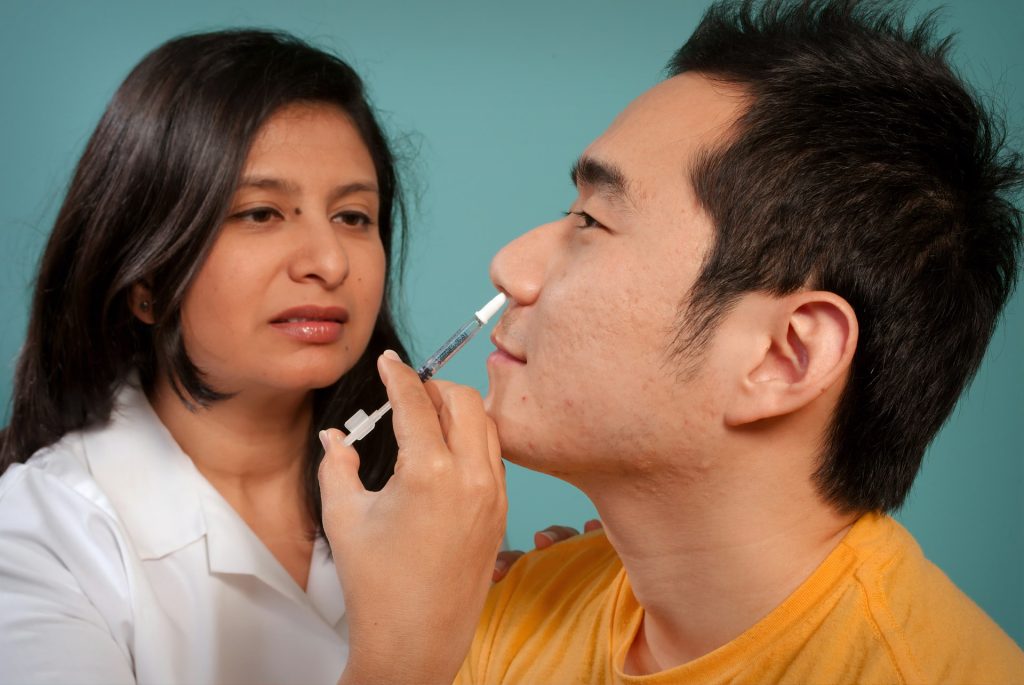
A clinical trial led by Weill Cornell Medicine investigators showed that a nasal spray that patients administer at home, without a physician, successfully and safely treated recurrent episodes of a condition that causes arrythmia. The study, published in the Journal of the American College of Cardiology, provides real-world evidence that a wide range of patients can safely and effectively use the experimental drug etripamil to treat recurrent paroxysmal supraventricular tachycardia (PSVT) episodes at home. For many, this could spare the need for repeated hospital trips for more invasive treatments.
The study is the latest in a series of studies by lead author Dr James Ip, professor of clinical medicine at Weill Cornell Medicine and a cardiologist at New York-Presbyterian/Weill Cornell Medical Center, and colleagues to demonstrate the potential of nasal spray calcium-channel blocker etripamil as an at-home treatment for PSVT.
Patients with PSVT experience sudden and recurrent rapid heart rhythms triggered by abnormal electrical activity in the upper chambers of the heart.
Though the episodes are not commonly life-threatening, they can be frightening and cause shortness of breath, chest pain, dizziness or fainting and lead to frequent emergency department visits. Treatment for PSVT often requires hospitalisation to receive intravenous medication. Some patients undergo cardiac ablation.
Dr Ip and colleagues previously showed that almost two-thirds of patients with PSVT who took one or more doses of the calcium channel blocker etripamil without a physician present experienced symptom relief on average in 17 minutes.
The latest study builds on those findings, showing that etripamil is safe and effective under more real-world circumstances in a larger patient population, and could be safely used to treat multiple episodes of PSVT.
The new study enrolled 1116 patients at 148 sites in the United States, Canada and South America. It did not require a pretest dose supervised by a physician as the previous studies did. It also included patients with a history of atrial fibrillation or atrial flutter, who were excluded from the previous studies. Patients monitored their heart for one hour with a home electrocardiogram monitor after self-administering the first dose, took an additional dose if necessary, and were allowed to self-treat up to four PSVT episodes with etripamil. Two-thirds of the patients experienced relief within an hour, and the average time needed for symptom relief was 17 minutes. Mild, temporary nasal symptoms such as runny nose, nasal congestion or discomfort, and bloody nose were common after the first use of etripamil but became less common with subsequent use.
Neurons in the brain of an Alzheimer’s patient, with plaques caused by tau proteins. Credit: NIH
In findings published in Cell Reports, researchers discovered that the biological instructions within vesicles that communicate between cells differed significantly in postmortem brain samples donated from patients suffering from Alzheimer’s disease.
Small extracellular vesicles (sEVs) are tiny containers are produced by most cells in the body to ferry a wide variety of proteins, lipids and byproducts of cellular metabolism, as well as RNA nucleic acid codes used by recipient cells to construct new proteins.
Because this biologically active cargo can easily elicit changes in other cells, scientists are interested in brain sEVs as a medium for passing along normal as well as bungled instructions for misfolded proteins that accumulate in the brain as neurodegenerative diseases such as Alzheimer’s disease progress.
To be a potential contributor to the buildup of unwanted proteins, sEVs would have to carry blueprints with sufficient information to enable other cells to produce the problematic proteins. Most previous research had indicated that the messenger RNA (mRNA) carrying plans for proteins were chopped into too many shorter fragments to allow recipient cells to change their construction patterns.
“We found quite the opposite to be true in our study,” says senior author Jerold Chun, MD, PhD, professor in the Center for Genetic Disorders and Aging Research at Sanford Burnham Prebys. “We identified more than 10 000 full-length mRNAs through the use of a relatively newer DNA sequencing technique called PacBio long-read sequencing.”
The team isolated sEVs from the prefrontal cortex of 12 postmortem brain samples donated from patients diagnosed with Alzheimer’s disease and 12 from donors without Alzheimer’s disease (or any other known neurological disease). Nearly 80% of identified mRNAs were full-length, allowing them to be transcribed by recipient cells into viable proteins.
“To corroborate the results of long-read sequencing in the human samples, we also looked at vesicles isolated from mouse cells,” says first author Linnea Ransom, PhD, postdoctoral fellow. “We found similar averages of between 78% and 86% full-length transcripts in three brain cell types: astrocytes, microglia and neurons.”
The researchers also compared the sequence of genes reflected in the sEV mRNA transcriptome. In Alzheimer’s disease samples, 700 genes showed increased expression whereas nearly 1500 were found to have reduced activity.
The scientists determined that the 700 upregulated genes are associated with inflammation and immune system activation, which fits within known patterns of brain inflammation present in neurodegenerative diseases such as Alzheimer’s disease. The researchers also found many genes associated with Alzheimer’s disease in prior genome-wide association studies also were present in Alzheimer’s disease sEVs.
“The changes in gene expression contained in these vesicles reveal an inflammatory signature that may serve as a window into disease processes occurring in the brain as Alzheimer’s disease progresses,” says Chun.
Following this study, Chun and team will dig deeper into how cells package sEVs and how the enclosed mRNA codes lead to functional changes in other brain cells affected in Alzheimer’s disease. Better understanding of sEVs and their mRNA contents may enable the discovery of biomarkers that could be used to improve early detection of Alzheimer’s disease and potentially other neurological conditions, while identifying new disease mechanisms to provide new therapeutic targets.
“Additionally, sEVs naturally occur as a vehicle for transporting biologically active cargo between cells, so it also may be possible to leverage them as a targeted delivery system for future brain therapies” says Chun.
By HualinXMN – Own work, CC BY-SA 4.0, https://commons.wikimedia.org/w/index.php?curid=133759262
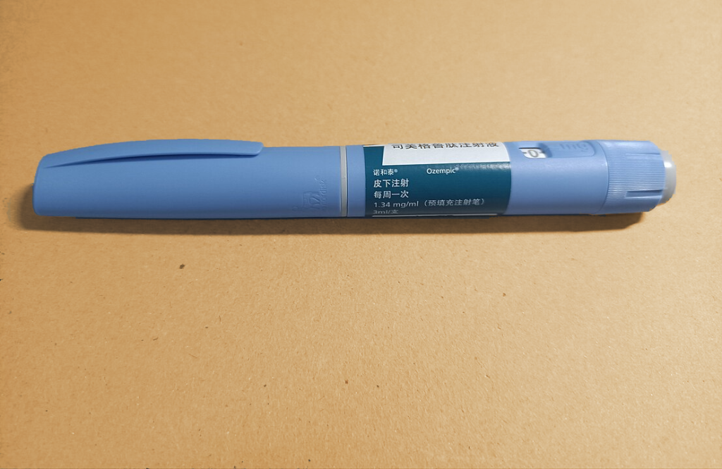
GLP-1 analogues have become increasingly popular to treat diabetes and obesity, but there have been concerns that they might increase the risk of thyroid cancer. Now an extensive Scandinavian study led by researchers at Karolinska Institutet has found no evidence of such a link. The study is published in The BMJ.
GLP-1 receptor agonists, also known as GLP-1 analogues, reduce blood sugar levels and appetite. They are widely used in the treatment of type 2 diabetes and obesity, with their clinical use steadily increasing. Earlier studies and adverse event data have suggested that these drugs could be associated with an increased risk of thyroid tumours. However, due to limitations in data and methodology, clear conclusions could not be drawn, leading to uncertainty about this potential side effect.
“Many people take these medicines, so it is important to study potential risks associated with them,” says Björn Pasternak, principal researcher at the Department of Medicine, Solna, at Karolinska Institutet in Sweden. “Our study covers a broad group of patients and provides strong support that GLP-1 analogues are not associated with an increased risk of thyroid cancer.”
The researchers analysed national register data from Denmark, Norway, and Sweden of about 145 000 patients treated with GLP-1 analogues, mainly liraglutide or semaglutide, and 290 000 patients treated with another diabetes drug (DPP4 inhibitors). The risk of thyroid cancer was compared between the groups over an average follow-up period of just under four years.
GLP-1 treatment was not associated with an increased risk of thyroid cancer. The results were consistent also when compared to a third diabetes medication group (SGLT2 inhibitors).
“We cannot rule out that the risk of certain subtypes of thyroid cancer is increased in smaller patient groups that we could not study here, for example in people with a high congenital risk of medullary thyroid cancer who are advised against using these drugs,” says Peter Ueda, assistant professor at the Department of Medicine, Solna, at Karolinska Institutet.
The ongoing research program at Karolinska Institutet investigates the effects and potential side effects of newer diabetes medications such as GLP-1 analogues and SGLT2 inhibitors. These medications are now being used to treat broader patient groups, including those with obesity, heart failure, and kidney failure.
“We know from randomised clinical trials that they have positive effects, but clinical reality is different with patients varying in disease severity, comorbidities, and adherence to treatment recommendations,” says Björn Pasternak. “It’s therefore essential to investigate how these medicines perform in everyday clinical settings.”
Hospitalised cancer patients who engaged in a 10-minute virtual reality (VR) session experienced significantly lessened pain in a trial published in CANCER, a peer-reviewed journal of the American Cancer Society. Participants still experienced sustained benefits a day later.
Most cancer patients experience pain, and treatment usually involves medications including opioids. VR sessions that immerse the user in new environments have been shown to be a noninvasive and nonpharmacologic way to lessen pain in different patient populations, but data are lacking in individuals with cancer. To investigate, Hunter Groninger, MD, of Georgetown University School of Medicine and MedStar Health and his colleagues randomized 128 adults with cancer with moderate or severe pain to a 10-minute immersive VR intervention involving calm, pleasant environments or to a 10-minute two-dimensional guided imagery experience on an iPad tablet.
The investigators found that both interventions lessened pain, but VR sessions had a greater impact. Based on patient-reported scores from 0 to 10, patients in the guided imagery group reported an average decrease of 0.7 in pain scores, whereas those in the VR group reported an average drop of 1.4. Twenty four hours after the assigned intervention, participants in the VR group reported sustained improvement in pain severity (1.7 points lower than baseline before the VR intervention) compared with participants in the guided imagery group (only 0.3 points lower than baseline before the active control intervention).
Participants assigned to the VR intervention also reported improvements related to pain “bothersomeness” (how much the pain bothered them, regardless of the severity of the pain) and general distress, and they expressed satisfaction with the intervention.
“Results from this trial suggest that immersive VR may be a useful non-medication strategy to improve the cancer pain experience,” said Dr Groninger. “While this study was conducted among hospitalized patients, future studies should also evaluate VR pain therapies in outpatient settings and explore the impact of different VR content to improve different types of cancer-related pain in different patient populations. Perhaps one day, patients living with cancer pain will be prescribed a VR therapy to use at home to improve their pain experience, in addition to usual cancer pain management strategies like pain medications.”
Brain organoids (BOs), though often referred to as “mini brains,” are not truly human brains. But the concerns over these lab-grown brain tissues, especially when they are developed from human foetal tissues, can be very human indeed.
In a paper published in EMBO Reports, researchers from Hiroshima University offer valuable insights into the complexities inherent in brain organoid research, highlighting often-overlooked ethical dilemmas for better decision-making, especially for foetal brain organoids (FeBOs).
Brain organoids are three-dimensional human brain tissues derived from stem cells. They replicate the complexity of the human brain in vitro, allowing researchers to study brain development and diseases.
Traditionally, brain organoids (BOs) are grown from pluripotent stem cells, an especially potent sub-type that is typical of early embryonic development, but new technologies now make it possible to generate these organoids from human foetal brain cells.
The research comes amid increasingly heated debates over human BOs. Central concerns are that lab-grown BOs might achieve consciousness and the ethical implications of transplanting them into animal models. The discourse includes matters of consent, commercialisation, integration with computational technologies, and legal ramifications. In addition, the public perception of BOs, often shaped by inaccurate media depictions.
Issues of consciousness arising and transplantation into animal models are particularly morally sensitive for tissue donors, and so rigorous informed consent is needed. With FeBOs, these become even more important. FeBOs, for example, can grow past the developmental stage of the initial foetal donor tissue.
“Our research seeks to illuminate previously often-overlooked ethical dilemmas and legal complexities that arise at the intersection of advanced organoid research and the use of foetal tissue, which is predominantly obtained through elective abortions,” said Tsutomu Sawai, an associate professor at Hiroshima University and lead author of the study.
The study highlights the urgent need for a sophisticated and globally harmonised regulatory framework tailored to navigate the complex ethical and legal landscape of FeBO research. One example is the 14-day rule used in embryo research, as neurogenesis does not occur in embryos prior to 14 days post-fertilisation. Using FeBOs derived from 12-15 week old foetuses therefore raises significant ethical questions, especially as there is a proposed 20-week ethical boundary.
The paper emphasises the importance of informed consent protocols, ethical considerations surrounding organoid consciousness, transplantation of organoids into animals, integration with computational systems, and broader debates related to embryo research and the ethics of abortion.
“Our plan is to vigorously advocate for the development of thorough ethical and regulatory frameworks for brain organoid research, including FeBO research, at both national and international levels,” said Masanori Kataoka, a fellow researcher at Hiroshima University.
“Rather than being limited to issues of consciousness, it’s imperative, now more than ever, to systematically advance the ethical and regulatory discussion in order to responsibly and ethically advance scientific and medical progress,” Sawai said.
Moving forward, the research duo plans to continue supporting the advancement of ethical and regulatory discussions surrounding brain organoid research. By promoting responsible and ethical progress in science and medicine, they aim to ensure that all research involving brain organoids, including FeBOs, is conducted within a framework that prioritises human dignity and ethical integrity.
Withdrawing aspirin one month after percutaneous coronary intervention (PCI) in high-risk heart patients and keeping them on ticagrelor alone safely improves outcomes and reduces major bleeding by more than half when compared to patients taking aspirin and ticagrelor combined (also known as dual antiplatelet therapy or DAPT), which is the current standard of care.
These are the results from the ULTIMATE-DAPT study announced during a late-breaking trial presentation at the American College of Cardiology Scientific Sessions on Sunday, April 7, and published in The Lancet.
This is the first and only trial to test high-risk patients with recent or threatened heart attack (acute coronary artery syndromes, or ACS) taking ticagrelor with a placebo starting one month after PCI, and compare them with ACS patients taking ticagrelor with aspirin over the same period. The significant findings could change the current guidelines for standard of care worldwide.
“Our study has demonstrated that withdrawing aspirin in patients with recent ACS one month after PCI is beneficial by reducing major and minor bleeding through one year by more than 50 percent. Moreover, there was no increase in adverse ischaemic events, meaning continuing aspirin was causing harm without providing any benefit,” says Gregg W. Stone, MD, the study co-chair of ULTIMATE-DAPT, who presented the trial results.
“It is my belief that it’s time to change the guidelines and standard clinical practice such that we no longer treat most ACS patients with dual antiplatelet therapy beyond one month after a successful PCI procedure. Treating these high-risk patients with a single potent platelet inhibitor such as ticagrelor will improve prognosis,” adds Dr Stone.
The study analysed 3400 patients with ACS at 58 centres in four countries between August 2019 and October 2022. All of the patients had undergone PCI, a non-surgical procedure in which interventional cardiologists use a catheter to place stents in the blocked coronary arteries to restore blood flow. The patients were stable one month after PCI and were on ticagrelor and aspirin. Researchers randomised the patients after one month, withdrawing aspirin in 1700 patients and putting them on ticagrelor and a placebo, while leaving the other 1700 patients on ticagrelor and aspirin. All patients were evaluated between 1 and 12 months after the procedure.
During the study period, 35 patients in the ticagrelor-placebo group had a major or minor bleeding event, compared to 78 patients in the ticagrelor-aspirin group, meaning that the incidence of overall bleeding incidents was reduced by 55 percent by withdrawing aspirin. The study also analysed major adverse cardiac and cerebrovascular events including death, heart attack, stroke, bypass graft surgery, or repeat PCI. These events occurred in 61 patients in the ticagrelor-placebo group compared to 63 patients in the ticagrelor-aspirin group, and were not statistically significant – further demonstrating that removing aspirin did no harm and improved outcomes.
“It was previously believed that discontinuing dual antiplatelet therapy within one year after PCI in patients with ACS would increase the risk of heart attack and other ischaemic complications, but the present study shows that is not the case, with contemporary drug-eluting stents now used in all PCI procedures. Discontinuing aspirin in patients with a recent or threatened heart attack who are stable one month after PCI is safe and, by decreasing serious bleeding, improves outcomes,” Dr Stone adds. “This study extends the results of prior work that showed similar results but without the quality of using a placebo, which eliminates bias from the study.”
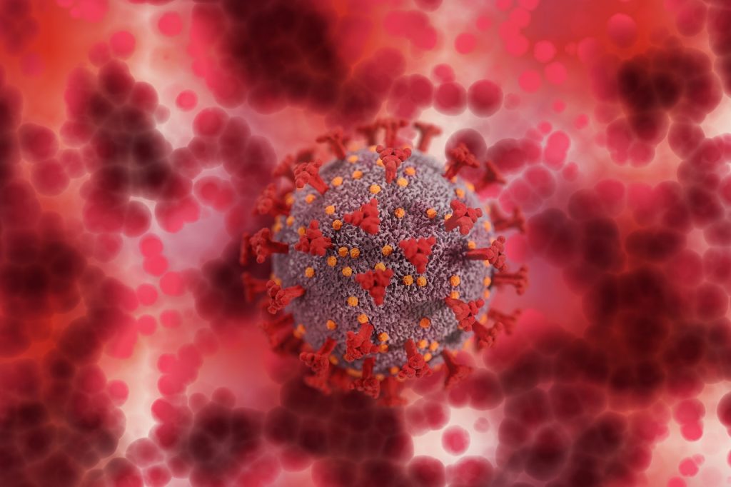
A certain variant of a key anti-inflammatory gene protects men under age 75 from severe illness and death when hospitalised from COVID, a genetic analysis of their blood shows. According to the authors of a major study published in The Journal of Infectious Diseases, the protective gene in question, an interleukin-1 receptor antagonist (IL1RN) variant, appears to tamp down inflammation, which can get out of control in severe cases SARS-CoV-2 infection.
The study showed that 124 men between the ages of 19 and 74 who possessed the IL1RN variant, called rs419598, were less likely to become severely ill after hospitalisation for COVID, and 80% less likely to die from the disease.
IL1RN is expressed naturally in the body. Different types of interleukin genes are known to dial inflammation up or down in the context of arthritis, and researchers say the results of the current study suggest that a similar dynamic influences the interleukin-1-related inflammation seen in COVID patients.
The findings, from researchers at NYU Grossman School of Medicine, stand out because historically more men than women are known to die from COVID, and the IL1RN rs419598 variant appears to selectively protect only men up to age 74, but not beyond that as age-related chronic illnesses unfold.
The research team used sequencing technologies for the study to determine the presence of specific genes or variations in the letter code that makes up genes in blood samples from 2589 men and women hospitalised for COVID at NYU Langone’s Tisch Hospital in Manhattan from March 2020 to March 2021.
More than half of the men and women in the study were older than age 60 and obese, factors that are known to increase the risk of death from the viral infection. Overall, more men than women (240 men, at 60.5%; and 157 women, at 39.5%) died from their disease, with women 20% less likely to die than men.
“Our study results show that among hospitalised patients, while women are still overall less likely than men to die from COVID-19, those men age 74 and younger who possess the IL1RN gene variant rs419598 are much less likely to suffer the severe inflammation tied to SARS-CoV-2 infection and less likely to die from the disease,” said study colead investigator and molecular biologist Mukundan Attur, PhD. Attur is an associate professor in the Department of Medicine at NYU Langone Health.
Among the study’s other findings was that average blood levels of the anti-inflammatory protein IL-1Ra, coded by IL1RN, were 14 times higher in 181 hospitalised men than in healthy male study controls from the general population, and 10 times as high in 178 hospitalised women than in healthy females. The increased levels of IL-1Ra in women did not result in any statistically significant mortality reductions.
“Our analysis offers substantial evidence of the biological link between the severe inflammation seen in SARS-CoV-2 and that which occurs in rheumatoid arthritis,” said study senior investigator Steven Abramson, MD, the Frederick H. King Professor of Internal Medicine at NYU Langone.
Abramson, a rheumatologist who also serves as chair of the Department of Medicine and chief academic officer at NYU Langone, says previous research has shown that such rheumatoid inflammation is lower in people who possessed one of the three IL1RN variants analysed in the study.
More importantly, Abramson says, the new research suggests that restraining the interleukin-1 biological pathway, which is in part tamped down by the anti-inflammatory protein IL-1Ra, could help prevent the severe inflammation seen in SARS-CoV-2 infection. Further research, he says, is warranted into whether IL-1-inhibiting therapies, such as the IL1 receptor antagonists anakinra, canakinumab, and rilonacept, are effective against Covid infection.
Abramson already has plans to investigate if the IL-1 pathway plays a role in long Covid, when people experience new or lingering symptoms, such as fatigue and ‘brain fog’, months after recuperating from their initial infection.
Abramson points out that the new study adds to the growing scientific evidence about the biological factors that contribute to gender differences seen in deaths from COVID, which are known to vary widely across the United States.
Pregnancy may carry a cost, according to a new study involving 1735 young people in the Philippines, and shows that women who reported having been pregnant looked biologically older than women who had never been pregnant, and women who had been pregnant more often looked biologically older than those who reported fewer pregnancies.
Notably, the number of pregnancies fathered was not associated with biological aging among same-aged cohort men, which implies that it is something about pregnancy or breastfeeding specifically that accelerates biological aging. The findings are published in the Proceedings of National Academy of Sciences.
This study, from the Columbia University Mailman School of Public Health, builds on epidemiological findings that high fertility can have negative side effects on women’s health and longevity. What was unknown, however, was whether the costs of reproduction were present earlier in life, before disease and age-related decline start to become apparent. Until now, one of the challenges has been quantifying biological aging among the young. This challenge was overcome by using a collection of new tools that use DNA methylation (DNAm) to study different facets of cellular aging, health, and mortality risk. These tools, called ‘epigenetic clocks’ allow researchers to study aging earlier in life, filling a key gap in the study of biological aging.
“Epigenetic clocks have revolutionised how we study biological aging across the lifecourse and open up new opportunities to study how and when long-term health costs of reproduction and other life events take hold”, said Calen Ryan, PhD, associate research scientist in the Columbia Aging Center, and lead author.
“Our findings suggest that pregnancy speeds up biological aging, and that these effects are apparent in young, high-fertility women,” said Ryan. “Our results are also the first to follow the same women through time, linking changes in each woman’s pregnancy number to changes in her biological age.”
The relationship between pregnancy history and biological age persisted even after taking into account various other factors tied to biological aging, such as socioeconomic status, smoking, and genetic variation, but were not present among men from the same sample. This finding, noted Ryan, points to some aspect of bearing children – rather than sociocultural factors associated with early fertility or sexual activity – as a driver of biological aging.
Despite the striking nature of the findings, Ryan encourages readers to remember the context: “Many of the reported pregnancies in our baseline measure occurred during late adolescence, when women are still growing. We expect this kind of pregnancy to be particularly challenging for a growing mother, especially if her access to healthcare, resources, or other forms of support is limited.”
Ryan also acknowledged that there is more work to do, “We still have a lot to learn about the role of pregnancy and other aspects of reproduction in the aging process. We also do not know the extent to which accelerated epigenetic aging in these particular individuals will manifest as poor health or mortality decades later in life.”
Ryan said that our current understanding of epigenetic clocks and how they predict health and mortality comes largely from North America and Europe, but that the aging process can take slightly different forms in the Philippines and other places around the world.
“Ultimately I think our findings highlight the potential long-term impacts of pregnancy on women’s health, and the importance of taking care of new parents, especially young mothers.”
While a hug or friendly pat can seem soothing, the question remains: can touch really help you feel better, and does it matter who it’s from or how they touch you? To explore these questions, researchers from the Social Brain Lab at the Netherlands Institute for Neuroscience and the University Hospital Essen conducted a large-scale analysis of studies exploring touch interventions. Their results, published in Nature Human Behaviour, reveal that the benefits of touch may not depend on who – or even what – does the touching.
Though individual studies provide conflicting results, this large-scale analysis shows that touch substantially improves both physical and mental wellbeing, for example via reduction of pain, anxiety, depression, and stress in adults. But in fact, those with physical or mental health problems (and therefore most in need of support) benefit even more from touch than healthy adults. “This is especially relevant considering how often touch interventions are overlooked,” notes first author Julian Packheiser.
“A key question of our study is to leverage the hundreds of individual studies out there to identify what type of touch works best,” adds professor Christian Keysers, director of the Social Brain Lab. “What if you don’t have a friend or partner close by to hug you? Would touch from a stranger or even a machine also help? And how often? The study clearly shows that touch can indeed be optimised, but the most important factors are not necessarily those we suspect.”
Interestingly, the person touching you, how they touch you, and the duration of their touch doesn’t make a difference in terms of impact. A long-lasting massage by a therapist could therefore be just as effective as a quick hug offered by a friend. That is, until the frequency of the intervention is considered. The more often a touch intervention is offered, the greater the impact. A quick hug could therefore be even more impactful than a massage if it is offered more frequently.
Human or non-human touch?
The next question was whether touch intervention needs to be human at all. As it turns out, object or robot interventions can be equally effective at improving physical wellbeing. “There are lots of people in need of wellbeing improvements, perhaps because they’re lonely but also because they may be inflicted by clinical conditions. These results indicate that a touch-robot, or even a simple weighted blanket has the potential to help those people,” last author Frédéric Michon explains. However, the benefits of robot and object interventions are less effective for mental wellbeing. Mental health disorders like anxiety or depression might therefore require human touch after all, “perhaps suggestive of the importance for an emotional component associated with the touch,” Michon points out.
While the researchers were equally curious about human-to-animal contact, studies exploring this question are still lacking. “It would be useful to see whether an animal’s or pet’s touch could improve wellbeing, and inversely if they also benefit from it, but unfortunately there simply aren’t enough studies, or properly controlled ones, for us to draw any general conclusions on these topics,” Michon clarifies.
Touch interventions across ages
When the team looked into the impact of touch on newborns, they found out that newborns also benefited significantly from touch. However, the person conducting the touch intervention was more important: the benefits of touch are higher when done by a parent instead of a healthcare worker. “This finding could be impactful,” Packheiser adds. “Death rates due to premature births are high in some countries and the knowledge that a baby benefits more from the touch of their own parent offers another easily implementable form of support for the baby’s health.”
Due to a lack of studies, it proved difficult to draw conclusions about children and teenagers. “Large scale studies like this help us draw more general conclusions but they also help us identify where research is lacking,” Michon explains. “We hope that our findings can steer future research to explore lesser-known questions. This includes animal touch, but also touch across ages, and in specific clinical settings like autistic patients, another category that has not been explored extensively.”
People with aphantasia – who cannot visualise an image in their mind’s eye – are less likely to remember the details of important past personal events or to recognise faces, according to a review of nearly ten years of research. People who cannot bring to mind visual imagery are also less likely to experience imagery of other kinds, like imagining music, according to new research by the academic who first discovered the phenomenon.
Professor Adam Zeman, of the University of Exeter, first coined the term aphantasia in 2015, to describe those who can’t visualise. Since then, tens of thousands of people worldwide have identified with the description. Many say they knew they processed information differently to others but were unable to describe how. Some of them expressed shock on discovering that other people can conjure up an image in their mind’s eye.
Now, Professor Zeman has conducted a review of around 50 recent studies, published in Trends in Cognitive Sciences, to summarise findings in a field that has emerged since his first publication. Research indicates that aphantasia is not a single entity but has subtypes. For example, not everyone with aphantasia has a poor autobiographical memory or difficulty in recognising faces, and in a minority of people, aphantasia appeared to be linked to autism. People who cannot visualise are more likely to have scientific occupations. Unexpectedly, although people with aphantasia can’t visualise at will, they often dream visually.
Professor Zeman’s review provides evidence that whether people have aphantasia or hyperphantasia – a particularly vivid visual imagination – is linked to variations in their physiology and neural connectivity in the brain, as well as in behaviour. For example, listening to scary stories alters skin conductance in those with imagery, meaning people sweat – but this does not occur in people with aphantasia.
Aphantasia is thought to affect around 1% of the population, while 3% are hyperphantasic. These figures rise to 5–10% with more generous criteria for inclusion. Both aphantasia and hyperphantasia often run in families, hinting at the possibility of a genetic basis.
Professor Zeman, who now holds honorary contracts at the universities of Exeter and Edinburgh, said: “Coining the term ‘aphantasia’ has unexpectedly opened a window on a neglected aspect of human experience. It is very gratifying that people who lack imagery have found the term helpful, while a substantial surge of research is shedding light on the implications of aphantasia.
“Despite the profound contrast in subjective experience between aphantasia and hyperphantasia, effects on everyday functioning are subtle – lack of imagery does not imply lack of imagination. Indeed, the consensus among researchers is that neither aphantasia nor hyperphantasia is a disorder. These are variations in human experience with roughly balanced advantages and disadvantages. Further work should help to spell these out in greater detail.”
“I struggle to fully immerse myself in role-play with my children”
Solicitor Mary Wathen’s frustration that she struggled to engage in role playing games with her two young children, when she found all other engagement with her children so fulfilling, was her sign that she had aphantasia, meaning she cannot visualise imagery.
The 43-year-old, from Newent near Cheltenham, said: “One of my friends said that he uses the images in his head to enhance role play. When I asked him to explain this in more detail it became clear that he – and everyone else in the room – could easily create an image in their head and use that as the backdrop for the role play. This was totally mind-blowing to me. I just cannot understand what they really mean – where is this image and what does it look like? To me, unless you can see something with your eyes, it’s not there.”
Mary’s shock intensified when she realised her husband, has such vivid visual imagery that he is probably hyperphantasic. “He thinks in moving pictures, like movies – sometimes to the point that he can mistake those thoughts for memories. To me, that’s unfathomable.”
Mary has come to realise that her lack of visual imagery may well account for her difficulties with memory. She said: “I can comprehend and retain concepts and principles really well but I’m unable to recall facts and figures. I can’t recreate something in my head or ‘re see’ something that is not actually there in that moment.
“I’ve found it quite saddening to learn that other people can call to mind an image of their children when they’re not there. I’d love to be able to do that, but I just can’t – but I’ve learned to compensate by taking plenty of photos, so that I can relive those memories through those images.
“Whilst I’m sure there are wonderful advantages to being able to think in pictures, I think it’s important to remind myself that there are advantages to having aphantasia too. I’m a really good written and verbal communicator – I think that’s because I’m not caught up with any pictures, so I just focus on the power of the word. I’m also a deeply emotional person and perhaps that’s my brain’s way of overcompensating; I feel things as a way of experiencing them, rather than seeing them.
“I think it’s really important to raise awareness that some people just don’t have this ability – particularly as using visual imagination is a key way that young children are taught to learn and engage. Primary teachers need to know that some children just won’t be able to visualise and that could be why they’re not engaging in those kinds of activities. We need to ensure we cater for everyone and encourage other ways of learning and engaging.”