Tag: foetal development

How One ‘Forever Chemical’ Can Disrupt Foetal Facial Development

Researchers discovered perfluorodecanoic acid is the most toxic during foetal craniofacial development

Credit: Chemical Research in Toxicology (2026). doi:10.1021/acs.chemrestox.5c00468

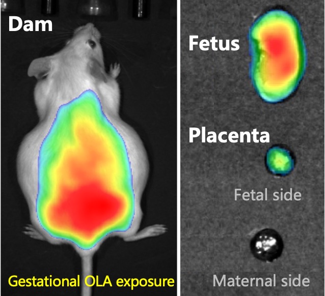
Researchers have long associated per- and polyfluoroalkyl substances (PFAS), commonly known as “forever chemicals,” to certain severe birth defects but exactly how these pollutants harm a developing foetus has remained mostly a mystery. New research now provides the first clear molecular explanation, showing how one PFAS, called perfluorodecanoic acid (PFDA), can trigger craniofacial abnormalities before birth.

The research was published in ACS Chemical Research in Toxicology.

“Most people are exposed to small amounts of PFAS in everyday life but higher exposure can occur through contaminated water, living near manufacturing sites or certain jobs like firefighting and ski waxing, which is why it’s so important to understand the chemicals better,” said the paper’s senior author Jed Lampe, PhD, associate professor at University of Colorado Anschutz Skaggs School of Pharmacy and Pharmaceutical Sciences. “We wanted to understand which PFAS compounds are truly harmful during foetus development, especially for people with higher exposure, and how they cause damage.”

There are approximately 15 000 PFAS used in consumer and industrial products but scientists are increasingly finding that only some pose serious health risks. In this study, Lampe worked with the paper’s first author Michaela Hvizdak and co-author Sylvie Kandel to test 13 commonly found PFAS and discovered PFDA as the most toxic during foetal craniofacial development.

They demonstrated a possible link between PFDA and the extensive facial changes observed in humans and laboratory animals, with some estimates indicating a 10% increased risk at extremely low exposure levels. 

“This finding moves us beyond association by providing a clear explanation for how PFDA can interfere with foetal development. It’s a critical step toward understanding a vast and complex class of environmental chemicals,” said Lampe.

The cause of underdeveloped eyes and an abnormal jaw

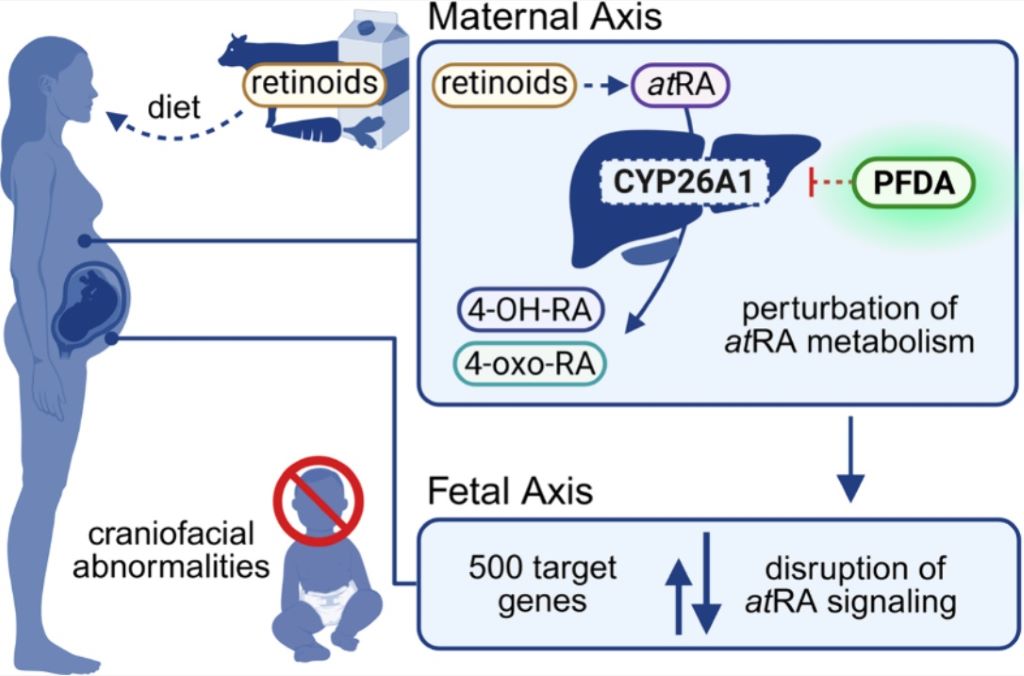
They found that PFAS disrupts retinoic acid, a molecule essential for shaping the face and head during early pregnancy. Retinoic acid regulates hundreds of genes and its levels must be controlled. Because a foetus cannot produce or safely eliminate excess retinoic acid, it relies entirely on the mother to maintain the homeostatic balance of the hormone.

The researchers discovered PFDA blocks CYP26A1, a key enzyme responsible for breaking down excess retinoic acid. When this enzyme is inhibited, retinoic acid levels can rise too high, disrupting normal facial development. PFDA also suppresses the genes that produce this enzyme through a separate biological pathway, delivering a “double hit” to the system that regulates early development.

“As a result, severe craniofacial abnormalities can develop, including underdeveloped eyes and abnormal jaw formation, which were the most common effects of PFDA exposure during foetal development,” said Lampe.

The researchers hope by providing a molecular explanation for this abnormality that the research and scientific community can work to develop targeted laboratory assays and computer‑based screening tools to quickly rank PFAS by risk. They say this could help regulators and manufacturers distinguish more dangerous compounds from safer alternatives and guide the design of less toxic chemicals in the future.

They also hope the research can open the door to practical interventions for people with higher exposure, such as firefighters and ski wax technicians. This could include looking into how to reduce exposure levels and whether strategies can be developed to lower PFDA during pregnancy to protect foetal development.

By Julia Milzer

Source: CU Anschutz Medical Campus

Breakdown Products from ‘Eco-friendly’ Plastics Impede Foetal Development in Mice

Nanoplastics from biodegradable plastics can cross the placenta and accumulate in foetal organs

PLA plastic breakdown product Oligomeric nanoplastics can penetrate the placental barrier and reach the foetus. Image credit: Dr Jia Lv (CC-BY 4.0)

When the “eco-friendly” bioplastic, polylactic acid (PLA), biodegrades, the resulting nanoplastics can accumulate in the foetuses of pregnant mice and interfere with foetal growth. Yichao Huang and De-Xiang Xu of Anhui Medical University, China, and Mingliang Fang of Fudan University, China, report these findings in a new study published March 26thin the open-access journal PLOS Biology.

PLA, which is made from corn starch and sugarcane, came onto the market as a biodegradable alternative to conventional plastics around two decades ago and has since become one of the most widely used bioplastics. Due to exponential growth in the production of PLA for packaging and medical applications, humans are increasingly exposed to its main breakdown product, oligomeric lactic acid (OLA) nanoplastics, which have recently been shown to have negative health effects.

In the new study, researchers exposed pregnant mice to OLA at doses proportional to what a human typically consumes and looked for impacts on the mouse pups. They demonstrated that OLA crosses the placenta and accumulates in various organs in the fetus. Furthermore, they showed that OLA interferes with a signaling pathway that controls the development of blood vessels in the placenta, which leads to slower growth of the fetus. This is a concern, because in humans, low birth weight is associated with an increased risk of stillbirth, as well as a higher risk of developing multiple other health problems later in life.

This work is the first animal study to evaluate the developmental health effects from the breakdown products of a supposedly eco-friendly plastic in pregnant mammals. The researchers propose that future work should focus on evaluating the exposure levels and health risks associated with eco-friendly plastics in humans, and a rethinking of our approach to plastic alternatives.

The authors add, “One of our co-authors Dr Mengjing Wang had previously discovered that the widely merchandised PLA microplastics undergo gut enzyme–mediated hydrolysis into oligomeric products that are toxic to the intestinal tract and can trigger enteritis.”

“To follow up on this work, as toxicologists, we went on to ask an additional question: do these oligomeric products, aka OLA, pose developmental threat particularly during the susceptible stage in utero?”

“What we have found was quite astonishing to us. Even under realistic exposure dose scenario during pregnancy, OLA nanoplastics can penetrate the placenta and even reach the foetus, in a mouse model. Such exposure would then cause placental vascular dysplasia and further lead to compromised foetal development.”

“While biodegradable plastics present a viable path to mitigate traditional plastic pollution, their potential health hazards necessitate a recognition in responsibility toward informed consumer intentions and conscientious usage.”

Provided by PLOS

Yawns in Healthy Foetuses Might Indicate Mild Distress

Foetuses yawn in the womb, with more yawns associated with a lower weight at birth

Photo by Mart Production on Pexels

Even in the womb, where all oxygen is provided by the parental placenta, foetuses can – and do – yawn. More yawns during observation were associated with a lower weight at birth – potentially indicating mild foetal stress in the womb, according to a study published February 25, 2026 in the open-access journal PLOS One by Damiano Menin, of the Università degli Studi di Ferrara in Italy, and colleagues.

Yawning is a behaviour found across vertebrates – and no one quite knows why. In humans, foetuses yawn in the womb from about 11 weeks. Even though there is no air to breathe, they slowly open their mouths, make motions similar to inhalation and exhalation, and close their mouths again. To understand more about foetal yawns, the authors of this study used ultrasound to observe 32 healthy foetuses (56% female, 44% male) between 23 and 31 weeks. Each foetus was observed for 22.5 minutes.

The authors found that the foetuses yawned between zero and six times during the observation period, with an average of 3.63 yawns per hour. They also showed that foetuses that yawned more during their observations were more likely to have a low weight at birth, which is considered as an indicator of mild distress – though all foetuses in the study were born healthy.

The researchers did not perform any manipulations to see if they could affect foetal yawning and also did not record measures such as foetal heart rate or maternal temperature which might potentially be associated with the behaviour. Additionally, no high-risk pregnancies were observed. Based on their research, the authors suggest that frequent foetal yawning might be a sign of mild distress in the healthy foetus.

The authors add: “We found that yawning frequencies in the womb are negatively related to birth weight, potentially indicating a stress-related response in healthy fetuses. This suggests that even before birth, yawning may serve as an indicator of a foetus’s well-being.”

Provided by PLOS

Study Explores How Prenatal Cannabis Exposure May Affect Foetal Brain Development

Source: Pixabay CC0

Researchers at UTHealth Houston are examining the biological effects of prenatal cannabis exposure and its potential impact on foetal brain development. Supported by a $3.7 million grant from the National Institutes of Health and the National Institute on Drug Abuse, the study aims to improve screening tools, public health guidance, and prenatal care strategies for pregnant people who use cannabis.

Led by Laura Goetzl, MD, MPH, a professor in the Department of Obstetrics, Gynecology, and Reproductive Sciences at McGovern Medical School at UTHealth Houston, the five-year grant will fund the study, “Foetal neuronal extracellular vesicle biomarkers of in-utero effects of maternal cannabinoid use and human foetal brain development and neurobehavioral outcomes.

“In recent years, cannabis use among pregnant women has increased, either recreationally or to help relieve nausea and vomiting during pregnancy,” Goetzl said. “Despite this rise, the effects on a baby’s brain are not well understood. Our hope through this research is that we can better identify risk factors and help health care providers give expecting mothers the best possible guidance.

The study will explore early biological signs, or biomarkers, to show how cannabis exposure influences a baby’s developing brain.

“During pregnancy, small bubbles called neuronal extracellular vesicles travel from the foetus into the mother’s bloodstream,” Goetzl said. “Through studying these small particles, we hope to gain valuable insight into foetal brain development without invasive testing.”

In collaboration with the University of Colorado, the research study will focus on how prenatal cannabis exposure may influence brain growth and neurobehavioral outcomes in children, including their potential for developing attention-deficit/hyperactivity disorder (ADHD) or autism later in life.

The project is supported by the National Institute on Drug Abuse of the National Institutes of Health under award number R01DA060319.

Source: UTHealth Houston

Music Can Influence Foetal Heart Rate in the Womb

Photo by Sergio Capuzzimati on Unsplash

Playing music has long been a way for expectant parents to connect with their children in the womb, but a group of researchers has found evidence it can calm foetal heart rates, potentially providing developmental benefits.

In the interdisciplinary journal Chaos, researchers studied the effect of classical music on a foetal heartbeat. The team used mathematical analysis tools to identify patterns in heart rate variability.

Typical measures of heart rate are an average of several beats across multiple seconds. In contrast, heart rate variability measures the time between individual beats. This measure can provide insight into the maturation of the foetal autonomic nervous system, with greater variability often indicating healthy development.

To test the effects music can have on foetal heart rate, the group of researchers recruited 36 pregnant women and played a pair of classical pieces for their foetuses. For the experiment, they chose “The Swan,” by French composer Camille Saint-Saëns, and “Arpa de Oro,” by Mexican composer Abundio Martínez.

By attaching external heart rate monitors, the researchers could measure the foetal heart rate response to both songs. And by employing nonlinear recurrence quantification analysis, they could identify changes in heart rate variability during and after the music was played.

“Overall, we discovered that exposure to music resulted in more stable and predictable foetal heart rate patterns,” said author Claudia Lerma. “We speculate that this momentary effect could stimulate the development of the foetal autonomic nervous system.”

In addition to the overall effects of playing music, the researchers looked at the differences between the two classical pieces. While both were effective, they found that the Mexican guitar melody had a stronger effect.

“When contrasting ‘The Swan’ with ‘Arpa de Oro,’ we did notice some significant differences,” said author Eric Alonso Abarca-Castro. “In particular, the second piece appeared to have a stronger impact on some measures, indicating that it produced heart rate patterns that were more predictable and regular. Factors like rhythmic characteristics, melodic structure, or cultural familiarity may be linked to this differentiation.”

For expectant parents at home, the researchers suggest that classical music could help promote fetal development.

“Our results suggest that these changes in foetal heart rate dynamics occur instantly in short-term fluctuations, so parents might want to consider exposing their foetuses to quiet music,” said Abarca-Castro. “Parents who play soothing music may stimulate and benefit the foetal autonomic system.”

The authors plan to continue to explore this effect, looking at different genres and types of music to further their understanding.

“To ascertain whether rhythmic or cultural variations elicit distinct foetal cardiac responses, we intend to increase the size of our sample and expand our investigation to include a variety of musical styles beyond classical pieces,” said author José Javier Reyes-Lagos.

Source: American Institute of Physics

Antiseizure Drugs during Pregnancy may Affect Neurodevelopment

Photo by SHVETS production

Children whose mothers have taken antiseizure drugs during pregnancy are more likely than others to receive a neuropsychiatric diagnosis. This is according to a comprehensive study by researchers at Karolinska Institutet and elsewhere, published in Nature Communications. However, the researchers emphasise that the absolute risk is low.

Antiseizure drugs are used to treat epilepsy and to stabilise mood in certain psychiatric conditions. However, some of these drugs, such as valproate, are known to affect the foetus if used during pregnancy. 

The current study included data from over three million children in the UK and Sweden, 17 495 of whom had been exposed to antiseizure drugs during pregnancy. 

As expected, children exposed to valproate were more likely to be diagnosed with autism, intellectual disability or ADHD compared to children not exposed to antiseizure drugs. Children exposed to topiramate had a 2.5-fold increased risk of intellectual disability, while those exposed to carbamazepine had a 25 per cent increased risk of being diagnosed with autism and a 30 per cent increased risk of intellectual disability. 

No increased risk with lamotrigine 

However, the researchers found no evidence that taking the antiseizure drug lamotrigine during pregnancy increases the risk of neuropsychiatric diagnoses in the child. 

“Our findings suggest that while certain medications may pose some risk, lamotrigine may be a less risky option, but active monitoring of any antiseizure medication is critical to ensure safety and effectiveness, particularly during pregnancy,” says Brian K. Lee, Professor at Drexel University Dornsife School of Public Health, USA, and affiliated researcher at the Department of Global Public Health, Karolinska Institutet, Sweden. 

The researchers emphasise that the absolute risk of the child receiving a neuropsychiatric diagnosis is low and that there may also be risks associated with not taking antiseizure medication during pregnancy. 

“If you’re pregnant or trying to become pregnant, and taking one of these medications, it may be worth talking with your physician to make sure you’re taking the best medicine for your needs, while minimising risk to future children,” says Viktor H. Ahlqvist, researcher at the Institute of Environmental Medicine, Karolinska Institutet, and joint first author with Paul Madley-Dowd at the University of Bristol, UK. 

The results support previous findings from smaller studies that found links between antiseizure drugs during pregnancy and the risk of neuropsychiatric diagnoses in the child. One difference is that the new study found no statistically significant association between topiramate or levetiracetam and ADHD in the child. 

Source: Karolinska Institutet

A Tiny Chromosomal Deletion is Linked to Spina Bifida

Photo by Mart Production on Pexels

A group of researchers at the University of California San Diego School of Medicine led an investigation that offers new insight into the causes of spina bifida, the most common structural disorder of the human nervous system.

The group’s work reveals the first link between spina bifida and a common chromosomal microdeletion in humans. The study demonstrates that individuals carrying this chromosomal deletion – present in one of 2500 live births – demonstrate a risk of spina bifida more than 10 times greater than the general public.

The study, published in Science, also underscores the potential role of folic acid (aka vitamin B-9) in reducing the risk of spina bifida.

Professor Joseph G. Gleeson at Rady Children’s Institute for Genomic Medicine, is the senior author of the study. He explained that spina bifida, also known as meningomyelocele, affects one in every 3000 newborns. Unfortunately, the causes are mostly unknown. A few mutations were reported but could only explain a tiny fraction of risk, Gleeson added.

To uncover the genetic causes of the disease, Gleeson’s UC lab joined with colleagues around the world to establish the Spina Bifida Sequencing Consortium in 2015. The consortium began focusing on a tiny deletion in chromosome 22. Chromosome microdeletions refer to a condition in which several genes in a chromosome are missing. The group’s target condition, known as 22q11.2del, has been implicated in a number of other disorders. They began looking for 22q11.2del in spinal bifida patients.

“All patients we recruited have the most severe form of spina bifida, and all underwent best-practice comprehensive genomic sequencing,” Gleeson said. “We identified 22q11.2del in 6 out of 715 patients. This may not seem a high percentage, but this is by far the most common single genetic variation that could contribute to spina bifida.”

He went on to say the group identified eight additional spina bifida patients who carried the deletion from a cohort of approximately 1500 individuals recruited because of the presence of the common 22q11.2 deletion, Gleeson said.

The researchers then narrowed the cause among the many genes in the 22q11.2 deletion to a single gene known as CRKL. Gleeson explained that there are nine other genes in this chromosomal region that could have been the cause. He said the team began a process of elimination, “knocking out” each of the mouse genes one-by-one, when they received a fortuitous email from Dolores Lamb from Weil Cornell College of Medicine. Lamb had noted some of the mice in their vivarium that were missing Crkl and showed spina bifida. (Study co-first author Keng Ioi Vong, PhD, explained that researchers use all capital letters to describe the gene in humans, and lower-case for mice.) Lamb’s group heard about the Gleeson lab project through the Spina Bifida Association.

“This finding really got us excited because it meant that CRKL disruption might be sufficient for spina bifida,” said Vong. “We removed the mouse Crkl gene ourselves and confirmed that some of the mice developed neural tube defects, including spina bifida.” Most of the other genes in 22q11.2 deletion were subsequently excluded, he added.

They next turned their attention to how folic acid may modulate CRKL-mediated spina bifida. Vong noted that prior studies in humans demonstrated that folic acid supplementation prior to conception reduces the incidence of spina bifida and other neural tube defects by up to 30-50 %, but the mechanisms are still a mystery.

“When we deprived the Crkl mutant female mice of folic acid in their chow, many more of their offspring had neural tube defects, and the severity increased dramatically,” Vong explained. “This suggests that folic acid taken by pregnant women may not only reduce the risk, but also the severity of neural tube defects in their offspring.”

“We hope our findings can help the research community to better understand causes of neural tube defects, especially the causes attributable to common genetic findings like 22q11.2 deletion,” Gleeson said. “We also hope our findings can contribute to healthy pregnancies, improved women’s health, and improved outcomes for children.”

Source: University of California – San Diego

Are Brain Organoids Derived from Foetal Tissue Ethical?

Image from Pixabay.

Brain organoids (BOs), though often referred to as “mini brains,” are not truly human brains. But the concerns over these lab-grown brain tissues, especially when they are developed from human foetal tissues, can be very human indeed.

In a paper published in EMBO Reports, researchers from Hiroshima University offer valuable insights into the complexities inherent in brain organoid research, highlighting often-overlooked ethical dilemmas for better decision-making, especially for foetal brain organoids (FeBOs).

Brain organoids are three-dimensional human brain tissues derived from stem cells. They replicate the complexity of the human brain in vitro, allowing researchers to study brain development and diseases.

Traditionally, brain organoids (BOs) are grown from pluripotent stem cells, an especially potent sub-type that is typical of early embryonic development, but new technologies now make it possible to generate these organoids from human foetal brain cells.

The research comes amid increasingly heated debates over human BOs. Central concerns are that lab-grown BOs might achieve consciousness and the ethical implications of transplanting them into animal models. The discourse includes matters of consent, commercialisation, integration with computational technologies, and legal ramifications. In addition, the public perception of BOs, often shaped by inaccurate media depictions.

Issues of consciousness arising and transplantation into animal models are particularly morally sensitive for tissue donors, and so rigorous informed consent is needed. With FeBOs, these become even more important. FeBOs, for example, can grow past the developmental stage of the initial foetal donor tissue.

“Our research seeks to illuminate previously often-overlooked ethical dilemmas and legal complexities that arise at the intersection of advanced organoid research and the use of foetal tissue, which is predominantly obtained through elective abortions,” said Tsutomu Sawai, an associate professor at Hiroshima University and lead author of the study.

The study highlights the urgent need for a sophisticated and globally harmonised regulatory framework tailored to navigate the complex ethical and legal landscape of FeBO research. One example is the 14-day rule used in embryo research, as neurogenesis does not occur in embryos prior to 14 days post-fertilisation. Using FeBOs derived from 12-15 week old foetuses therefore raises significant ethical questions, especially as there is a proposed 20-week ethical boundary.

The paper emphasises the importance of informed consent protocols, ethical considerations surrounding organoid consciousness, transplantation of organoids into animals, integration with computational systems, and broader debates related to embryo research and the ethics of abortion.

“Our plan is to vigorously advocate for the development of thorough ethical and regulatory frameworks for brain organoid research, including FeBO research, at both national and international levels,” said Masanori Kataoka, a fellow researcher at Hiroshima University.

“Rather than being limited to issues of consciousness, it’s imperative, now more than ever, to systematically advance the ethical and regulatory discussion in order to responsibly and ethically advance scientific and medical progress,” Sawai said.

Moving forward, the research duo plans to continue supporting the advancement of ethical and regulatory discussions surrounding brain organoid research. By promoting responsible and ethical progress in science and medicine, they aim to ensure that all research involving brain organoids, including FeBOs, is conducted within a framework that prioritises human dignity and ethical integrity.

Source: Hiroshima University

Foetal Exposure to THC could Lead to Long-lasting Health Impacts

Photo by Pavel Danilyuk on Pexels

Consuming THC (Delta-9-tetrahydrocannabinol) while pregnant could potentially affect development of the foetus and lead to life-long health impacts for offspring, according to a new study published in the journal Clinical Epigenetics.

THC is the main psychoactive ingredient in cannabis, which is growing in popularity and availability. The prevalence of cannabis use in pregnancy is also rapidly increasing, especially during the first trimester, when the foetus is most vulnerable to environmental exposures, to mitigate common symptoms like morning sickness. However, the potential effects of prenatal cannabis use on foetal development remain inconclusive, in part due to a lack of safety data. This study aimed to identify the potential long-term health impacts of THC use during pregnancy.

In a non-human primate model, Oregon Health & Science University researchers found that exposing a pregnant subject to THC altered placental and foetal epigenetics. Researchers also found that that these changes to gene regulation and expression are consistent with those seen with many common neurobehavioural conditions, including autism spectrum disorder.

“Cannabis is one of the most commonly used drugs and is widely available across the country, so there is a common perception that its completely safe to use,” said the study’s lead author Lyndsey Shorey-Kendrick, PhD, a computational biologist in the Division of Neurosciences at OHSU’s Oregon National Primate Research Center, or ONPRC. “The reality is that cannabis still carries many health risks for certain populations, including those who are pregnant. If we’re able to better understand the impacts, we can more effectively communicate the risks to patients and support safer habits during the vulnerable prenatal period.”

In a model using nonhuman primates, researchers administered THC in a daily edible and compared its effects to a group receiving a placebo. Specifically, researchers evaluated the epigenetic changes in several key areas that indicate healthy prenatal development: the placenta and foetal lung, brain and heart.

When looking at these areas, analyses showed that THC exposure altered the epigenome, meaning a process in which the information encoded in a gene is turned into a function or observable trait. Genes are all specifically coded to contribute to different functions of the body and brain, so any impact on epigenetic processes due to drug exposure is concerning, especially during a critical developmental window such as pregnancy.

Researchers found that significant changes involved genes associated with common neurobehavioral disorders, including autism spectrum disorder and attention deficit hyperactivity disorder. These conditions are linked to adverse health outcomes in childhood and adolescence, including poorer memory and verbal reasoning skills, and increased hyperactivity, impulsivity and inattention.

The research team hopes findings from this study will add to the limited existing literature on THC use during pregnancy, and help guide patient counselling and public health polices focused on cannabis in the future.

“It’s not common practice for providers to discuss cannabis use with patients who are pregnant or trying to conceive,” said the study’s corresponding author, Jamie Lo, MD, MCR, associate professor of obstetrics and gynaecology (maternal-foetal medicine) at OHSU. “I hope our work can help open up a broader dialogue about the risks of cannabis use in the preconception and prenatal period, so we can improve children’s health in the long run.”

Source: Oregon Health & Science University

Low Maternal Vitamin D Levels may Increase Schizophrenia Risk of Offspring

Photo by Pavel Danilyuk on Pexels

Neuroscientists published in the Journal of Neurochemistry, shows that maternal levels of vitamin D are key in the development of dopaminergic neurons, which are thought to be involved in schizophrenia.

Professor Darryl Eyles has built on past research out of his laboratory at the Queensland Brain Institute linking maternal vitamin D deficiency and brain development disorders, such as schizophrenia, to understand the functional changes taking place in the brain.

Schizophrenia is associated with many developmental risk factors, both genetic and environmental. While the precise neurological causes of the disorder are unknown, what is known is that schizophrenia is associated with a pronounced change in the way the brain uses dopamine, the neurotransmitter often referred to as the brain’s ‘reward molecule’.

Professor Eyles has followed the mechanisms that might relate to abnormal dopamine release and discovered that maternal vitamin D deficiency affects the early development and later differentiation of dopaminergic neurons.

The team at the Queensland Brain Institute developed dopamine-like cells to replicate the process of differentiation into early dopaminergic neurons that usually takes place during embryonic development.

They cultured the neurons both in the presence and absence of the active vitamin D hormone. In three different model systems they showed dopamine neurite outgrowth was markedly increased. They then showed alterations in the distribution of presynaptic proteins responsible for dopamine release within these neurites.

“What we found was the altered differentiation process in the presence of vitamin D not only makes the cells grow differently, but recruits machinery to release dopamine differently,” Professor Eyles said.

Using a new visualisation tool known as false fluorescent neurotransmitters, the team could then analyse the functional changes in presynaptic dopamine uptake and release in the presence and absence of vitamin D.

They showed that dopamine release was enhanced in cells grown in the presence of the hormone compared to a control.

“This is conclusive evidence that vitamin D affects the structural differentiation of dopaminergic neurons.”

Leveraging advances in targeting and visualising single molecules within presynaptic nerve terminals has enabled Professor Eyles and his team to further explore their long-standing belief that maternal vitamin D deficiency changes how early dopaminergic circuits are formed.

The team is now exploring whether other environmental risk factors for schizophrenia such as maternal hypoxia or infection similarly alter the trajectory of dopamine neuron differentiation.

Eyles and his team believe such early alterations to dopamine neuron differentiation and function may be the neurodevelopmental origin of dopamine dysfunction later in adults who develop schizophrenia.

Source: University of Queensland