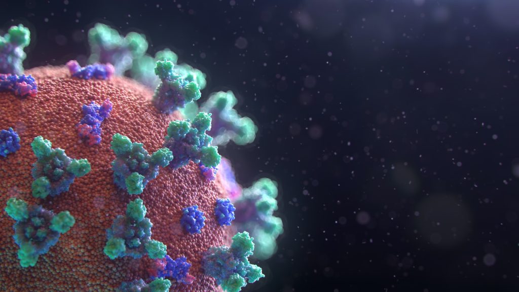
In a new entry to the growing list of lasting complications from COVID infection, a large German cohort study of over 600 000 COVIDpatients indicates that new autoimmune conditions may result from previous COVID infection. The findings, which are awaiting peer review on the MedRxiv preprint server, show that the odds of new autoimmune conditions appear to increase in line with the severity of COVID infection.
After the acute phase of infection, some people may develop long-lasting symptoms, known as post-COVID, which are consistent with COVID infection and last more than 12 weeks. Most studies to date have focused on symptoms that partly wane over time. Many studies examined a small selective sample of patients, and only a few studies included a control group or information on chronic health conditions, such as SARS-CoV-2 infection.
Compared to post-COVID emergence of cardiovascular and other diseases, autoimmune diseases are less discussed in the literature, although autoantibodies could be found in patients after SARS-CoV-2 infection. So far there is limited evidence on newly manifested autoimmune diseases after an infection based on several case reports and one recent cohort study using UK health record data. In addition, COVID itself has some similarities with systemic autoimmune rheumatic diseases, which could make diagnosis difficult.
The researchers selected a cohort from German routine health care data, identifying individuals with polymerase chain reaction (PCR)-confirmed COVID through December 31, 2020. Patients were matched 1:3 to control patients without COVID. Both groups were followed up until June 30, 2021. We used the four quarters preceding the index date until the end of follow-up to analyse the onset of autoimmune diseases during the post-acute period. The researchers calculated the incidence rates (IR) per 1000 person-years for each outcome and patient group, and estimated incidence rate ratios (IRRs) of developing an autoimmune disease conditional on a preceding COVID.
In total, 641 704 patients with COVID were included. When comparing the incidence rates in the COVID and matched control groups, the researchers found a 42.63% higher likelihood of acquiring autoimmunity for patients who had suffered from COVID. This estimate was similar for common autoimmune diseases, such as Hashimoto thyroiditis, rheumatoid arthritis, or Sjögren syndrome. The highest IRR was observed for autoimmune disease of the vasculitis group. Patients with a more severe course of COVID were at a greater risk for incident autoimmune diseases. These risk increases were as follows:
41% higher risk of Grave’s disease
42–45% higher risk of rheumatoid arthritis
25% higher risk of type 1 diabetes
27-29% higher risk of Crohn’s disease
The researchers concluded that SARS-CoV-2 infection is associated with an increased risk of developing new-onset autoimmune diseases after the acute phase of infection.
Women with preeclampsia have a higher likelihood of heart attack and stroke than their peers within just seven years of delivery, with risks remaining elevated more than 20 years later. The study in more than one million pregnant women is published today in the European Journal of Preventive Cardiology, a journal of the ESC.
“The high risk of cardiovascular disease after preeclampsia manifests at young ages and early after delivery,” said study author Dr Sara Hallum of the University of Copenhagen. “This indicates that interventions to prevent heart attacks and strokes in affected women cannot wait until middle age when they become eligible for conventional cardiovascular screening programmes.”
Preeclampsia affects up to 8% of pregnancies worldwide, and signs include hypertension and albuminuria, which develop after 20 weeks of pregnancy or soon after delivery. Symptoms include severe headache, stomach pain and nausea. “Women may mistake these for ‘normal’ pregnancy symptoms and thus not seek medical help until the condition becomes severe,” said Dr Hallum. “Most cases are mild, but preeclampsia may lead to serious complications for the mother and baby if not treated in time.”
It is well established that preeclampsia predisposes women to an elevated likelihood of cardiovascular disease later in life. This was the first study to examine how soon after pregnancy these heart attacks and strokes manifest, and the magnitude of risk in different age groups.
National registers were used to identify all pregnant women in Denmark between 1978 and 2017. Women were grouped into those with one or more pregnancies complicated by preeclampsia and those with no preeclampsia. Participants were free of cardiovascular disease before pregnancy and with follow-up for heart attack and stroke up to 39 years later. Dr Hallum said: “This allowed us to evaluate exactly when cardiovascular disease occurs in women with and without pre-eclampsia, and to estimate risk in different age groups and at various durations of follow-up.”
Up to 2% of those with pre-eclampsia in their first pregnancy had a heart attack or stroke within 20 years of delivery, compared with up to 1.2% of unaffected women. Differences in risk became apparent seven years after delivery. “A 2% incidence of acute myocardial infarction and ischaemic stroke should not be accepted as the cost of a pregnancy complicated by preeclampsia, particularly considering the young age of these women when they fall ill (below 50 years of age),” states the paper.
Overall, women with pre-eclampsia were four times more likely to have a heart attack and three times more likely to have a stroke within 10 years of delivery than those without pre-eclampsia. The risk of heart attack or stroke was still twice as high in the preeclampsia group more than 20 years after giving birth compared to unaffected women.
When the researchers examined the risk of cardiovascular disease according to age, they found that women aged 30 to 39 years with a history of preeclampsia had five- and three-fold higher rates of heart attack and stroke, respectively, than those of similar age with no history of pre-eclampsia. The raised likelihood of cardiovascular disease in those with a history of pre-eclampsia persisted throughout adulthood, with women over 50 years of age still at doubled risk compared to their peers with no history of the pregnancy complication.
Dr Hallum said: “Women are often in contact with the healthcare system during and immediately after pregnancy, providing a window of opportunity to identify those at increased risk of cardiovascular disease. The number of women with previous pre-eclampsia is large, and routine follow-up could last years or even decades. Our study suggests that the women most likely to benefit from screening are those who had pre-eclampsia after age 35 and those who had it more than once. Prevention should start within a decade of delivery, for example by treating high blood pressure and informing women about risk factors for heart disease such as smoking and inactivity.”
Scanning Electron Micrograph image of a human T cell. Credit: NIH/NIAID
New research published in the journal Immunity challenges the prevailing hypothesis for how donor stem cell grafts cause graft-versus-host disease (GVHD) and offers an alternative model that could guide development of novel therapies.
The study showed in a mouse model that GVHD, which often affects the skin, gut and liver, is maintained by donor T cells that seed those tissues soon after transplant and not by the continual recruitment of T cells from the blood as previously thought.
“This study changes the paradigm of how people think about GVHD,” said co-senior author Warren Shlomchik, MD, professor of medicine and immunology at the University of Pittsburgh School of Medicine. “It provides important mechanistic detail about what’s going on in the tissues affected by GVHD, which could ultimately inform the development of better therapeutics and lead to better outcomes for stem cell recipients.”
Allogeneic stem cell transplantation involves infusion of stem cells from a healthy donor’s blood or bone marrow to a recipient. While often lifesaving for patients with leukaemia and other blood disorders, the treatment also comes with a risk of developing GVHD, a life-threatening disease that occurs when donor alloreactive T cells attack the recipient’s healthy tissues.
According to a widely held theory, GVHD is maintained by T cells that continually migrate from secondary lymphoid organs throughout the body, including the spleen and lymph nodes, to affected tissues via the blood.
However, a different model posits that the disease is maintained locally by T cells in the tissues with little input from the blood. In the new study, Shlomchik, lead author Faruk Sacirbegovic, PhD, research assistant professor of surgery at Pitt, and their team investigated the two hypotheses for how GVHD is sustained in tissues.
The researchers developed a system to track alloreactive T cells in a mouse model of GVHD by labelling individual cells with unique tags to create different T cell “flavours.” By measuring the tags over time, they monitored where the T cells travelled and replicated.
The analysis showed that each tissue affected by GVHD had unique T cell populations with varying frequencies of each T cell flavour.
“This finding is strong evidence that the disease is locally maintained by T cells in each of the tissues,” explained Shlomchik. “If tissues were constantly getting T cells from circulating blood, then the frequencies of T cell flavors in each tissue should become more and more alike over time — but we didn’t see that.”
The team used mathematical models to predict that progenitor T cells seed out into recipient tissues early after transplant, differentiating there into disease-causing cells.
Next a series of experiments was conducted to confirm this prediction and identified these progenitors as T cells expressing a gene called Tcf7.
“We think that progenitor T cells are long-lived in target tissues and are critical for maintaining GVHD,” said co-senior author Thomas Höfer, PhD, professor of theoretical systems biology at the University of Heidelberg. “After the initial seeding phase, the disease is mostly sustained within the tissue itself without a lot of input from new T cells in the blood.”
Stem cell recipients are typically treated with immunosuppressants to prevent and treat GVHD. As these powerful drugs act systemically to suppress the immune system, they also lower immunity to infections and have other side effects.
According to the researchers, the study’s insights could eventually lead to new, targeted therapies for GVHD.
“Now that we know the identity of progenitor cells, we might be able to prevent them forming early post-transplant or target them directly after they’ve formed,” said Shlomchik. “The findings also suggest that treating GVHD in the tissues themselves would be effective – although targeting tissues beyond the skin remains a challenge.”
With better ways to minimise the risk of GVHD after stem cell transplantation, the procedure could become more widely used to treat a broader range of diseases, including blood disorders such as sickle cell anaemia and autoimmune diseases such as lupus and multiple sclerosis.
The Hive ransomware group that has targeted more than 1500 victims in over 80 countries around the world, including hospitals, has been disrupted in a months-long campaign against, the US Justice Department has announced.
Hive ransomware attacks have caused major disruptions in victim daily operations around the world and hindered responses to the COVID pandemic. In one case, a hospital attacked by Hive ransomware had to fall back to pen and paper to treat existing patients and could not take new admissions shortly after the attack.
The Justice Department revealed that the FBI had penetrated Hive’s computer network and captured its decryption keys, which were then offered to victims around the world. This saved them $130 million in ransom they would have had to otherwise pay to get their networks back.
Finally, the department announced that, in coordination with German and Dutch law enforcement, it has seized control of the servers and websites that Hive uses to communicate with its members, disrupting Hive’s ability to attack and extort victims.
Since June 2021, the Hive ransomware group has targeted more than 1500 victims around the world and received over $100 million in ransom payments.
Hive used a ransomware-as-a-service (RaaS) model featuring administrators, and affiliates. RaaS is a subscription-based model where the administrators develop an easy-to-use ransomware strain and then recruit affiliates to deploy the ransomware against victims. Affiliates identified targets and deployed this readymade malicious software to attack victims and then earned a percentage of each successful ransom payment.
Hive actors used a double-extortion model of attack: before encrypting the victim’s system, the affiliate would steal sensitive data. The affiliate then sought a ransom for both the decryption key necessary to decrypt the victim’s system and a promise to not publish the stolen data – usually the most sensitive, such as hospital patient data. After a victim pays, the affiliates and administrators split the ransom 80/20. Victims who do not pay on the Hive Leak Site. After Consulate Health Care was unable to pay the ransom, since its insurance did not cover such cyber crimes, Hive posted 550GB of personally identifiable information on its patients and employees online.
For more information about the malware, including technical information for organisations about how to mitigate its effects, is available from CISA, visit https://www.cisa.gov/uscert/ncas/alerts/aa22-321a.
SARS-CoV-2 virus. Source: Fusion Medical Animation on Unsplash
In an interview about new Omicron subvariants, leading vaccinologist Prof Shabir Madhi said that “we don’t need to be concerned” about any current threat they may pose to South Africa. However, he stressed that it can still be lethal, particularly in those without underlying T cell immunity. He also noted that boosters are also important for high-risk populations, while some sort of seasonality needs to be observed for COVID for it to make boosters worthwhile for those at low risk due to the way vaccination protection wanes.
The XBB 1.5 SARS-CoV-2 subvariant, nicknamed ‘Kraken’ by researchers, is now accounting for more than half of cases in the United States, and appears much more transmissible and antibody-evasive than the original Omicron variant which evolved in Southern Africa. Prof Pravin Manga, editor of the Wits Journal of Clicnical Medicine interviewed Prof Madhi and asked him what the emergence of Omicron subvariants meant for South Africa.
Prof Madhi, who is the Dean of the Faculty of Health Sciences at Wits University, noted that before this new XBB.1.5 variant, there were BA4 and BA5, which created a “mini surge” in the middle of last year when they arrived in SA. There were concerns that these strains seemed more antibody-resistant than previous ones, stoking fears that they would result in increased hospitalisations and deaths.
In light of the current situation, he says that “the short answer is that we don’t need to be concerned.”
One important aspect of immunity which was becoming apparent was that, although neutralising antibodies were important in protecting against contracting and transmitting the virus, “what seems to be playing a greater role in protecting against severe disease is the T cell immunity, the Natural Killer cell immunity.” This immunity is much more diverse than that from antibodies, instead of merely targeting the Spike protein is rather “multi-epitopic”, targeting the N-protein as well.
“Now this T cell immunity appears to be holding strong. It appears to be less affected by all these mutations. In fact, close to 75 to 80% of vaccine-induced T cell immunity is conserved despite the multiple mutations have arisen in Omicron and its subvariants.”
Differing impacts across countries
With regard to the impact of the virus, Prof Madhi noted that China had pursued its ‘zero COVID’ policy, along with “suboptimal” coverage of vaccines (especially among ages 60+) that were “probably not the best”, meaning that large portions of the population were essentially naïve to the virus.
SA meanwhile, had 90% of the population infected at least once with COVID, and coupled with vaccination, meant that many will have highly robust immunity, which appears to last for 9–12 months compared to vaccine-only immunity where protection starts wanes after 4–6 months.
“What is unlikely to materialise in a country such as South Africa is large numbers of hospitalisations,” he says.
Protecting at-risk populations and the need for new vaccines
At present, he says there is not a strong case for boosters, but people at greater risk, such as those over 60, people with underlying medical conditions, and compromised immune systems, hybrid immunity is likely not enough protection. In these cases probably at least four doses of vaccination. From a public health standpoint, the population under 45 without underlying conditions would require a huge effort for only a nominal benefit as they are no longer at high risk of severe disease.
Timing is also important, due to the waning of vaccine protection, as the best time to get a booster is “probably around two or three weeks before the start of the next resurgence.” Otherwise, it’s useless to get a booster now if the next resurgence is in six months and antibodies will have waned – an obvious logistical challenge for little benefit. Therefore, in order for boosters to be useful, the virus will have to settle into some sort of predictable seasonality such as with influenza.
As for people who are at risk, at least four doses are probably required, though the case for a fifth is thin. Annual boosters are a likely option, and there is a need for a second generation of vaccines. These vaccines would need to be resilient against further mutations that may arise.
Novavax, monoclonal antibodies and Paxlovid
Regarding Novavax, Prof Madhi said that it had been licensed for use in South Africa, but their bivalent vaccine was not yet available. It would not be procured by government but rather by a private company – a situation which needs to change in terms of who is allowed to bring in vaccines. Another issue is whether the no fault compensation used by the government for public sector vaccinations would be used in the private sector as well.
Prof Manga also asked about whether there had been any success with monoclonal antibody treatment, to which Prof Madhi answered that there had been some limited use in the country but overall, monoclonal antibodies were “spectacularly unsuccessful” as they were highly specific and generally unable to keep up with mutations.
In general, antivirals hold much better promise, particularly Paxlovid which is unfortunately not available in South Africa. It was disappointing that it was not available in the country,
Benefits to both pregnant mothers and babies
Regarding pregnant women and children, Prof Madhi said that their own study shows that a substantial amount of transmission takes place between mothers and children. Infants with COVID under six months are often hospitalised, especially in the first month of life. Vaccination reduces the risk of hospitalisation and protects the baby as well, with research showing that babies born to vaccinated mothers were 80% less likely to develop COVID, “which is really a huge benefit,” he noted. This is likely a little reduced with Omicron because the only thing that babies get from the mother is antibodies, not T cell immunity.
Vaccination also reduces the risk of adverse pregnancy outcomes such as stillbirth, and safety “is simply not an issue” as supported by the data. He says there is case for vaccinating pregnant women, even under 45, in the second trimester of the pregnancy so that more antibodies are transferred to the foetus.
Researchers have developed a handheld terahertz (THz) wave imaging device to assess burns faster and more accurately than current methods. The new device uses neural network model that uses terahertz time-domain spectroscopy (THz-TDS) data for non-invasive burn assessment.
“It is important for healthcare professionals to accurately assess the depth of a burn to provide the most appropriate treatment,” said research team leader M. Hassan Arbab from Stony Brook University. “However, current methods of burn depth evaluation, which rely on visual and tactile examination, have been shown to be unreliable, with accuracy rates hovering around 60–75%. Our new approach could potentially improve the accuracy of burn severity assessments and aid in treatment planning.”
THz-TDS uses short pulses of terahertz radiation, which lies between infrared and microwave wavelengths, to probe a sample. It is being examined for assessing burn injuries because physical changes caused by a burn will produce alterations in the skin’s terahertz reflectivity.
In the journal Biomedical Optics Express, the researchers reported that their artificial neural network classification algorithm was able to accurately predict the ultimate healing outcome of in vivo burns in an animal study with 93% accuracy. Their method needs much less training data, potentially making it more practical to process big data sets obtained over large clinical trials.
“In 2018, approximately 416 000 patients were treated for burn injuries in emergency departments in the United States alone,” said Arbab. “Our research has the potential to significantly improve burn healing outcomes by guiding surgical treatment plans, which could have a major impact on reducing the length of hospital stays and number of surgical procedures for skin grafting while also improving rehabilitation after injury.”
Better burn assessment
Various technologies have been developed to improve burn assessment, but they haven’t been widely adopted in the clinic due to drawbacks such as long acquisition times, high costs and limited penetration depth and field of view. Although THz-TDS looks promising for burn assessment, early demonstrations were limited to point spectroscopy measurements, which don’t account for burn heterogeneity and spatial variations. THz spectroscopy setups also tend to be bulky and difficult to set up.
“To address these challenges, we developed the portable handheld spectral reflection (PHASR) scanner, a user-friendly device for fast hyperspectral imaging of in vivo burn injuries using THz-TDS,” said Arbab. The device allows for “rapid imaging of a 37 x 27 mm2 field of view in just a few seconds.”
Previously, the researchers used numerical methods to extract features from the THz-TDS images and machine learning techniques to estimate the severity grade of in vivo burn injuries using measurements from the PHASR scanner. However, this approach did not consider the physical dynamics and macroscopic changes of the dielectric permittivity of burned skin tissue. Dielectric permittivity describes how a material responds to an electric field, and the researchers used Debye theory to explain how biological material interacts with THz waves.
The researchers tested their method by using the PHASR scanner to obtain spectroscopic images of skin burns and measure the permittivity of the burns. The researchers used this data to create a neural network model based on labelled biopsies. The model estimated the severity of the burns with an average accuracy rate of 84.5% and predicted the outcome of the wound healing process with an accuracy rate of 93%.
The researchers note that clinical testing of both the technique and the handheld imaging device are needed before this technique could be integrated into the existing workflow of clinical burn assessment.
Potassium is essential to normal cellular function, helping the cardiac muscle work correctly and aids in the transmission of electrical signals within cells. A new mathematical model published in PLOS Computational Biology sheds light on the often mysterious process of potassium homeostasis.
Using existing biological data, researchers at the University of Waterloo built a mathematical model that simulates how an average person’s body regulates potassium, both in times of potassium depletion and during potassium intake. Because so many foods contain abundant potassium, the body is continually storing, deploying, and disposing of potassium to keep it in a healthy range, ie the process of potassium homeostasis. Understanding potassium homeostasis is essential in helping diagnose the source of the problem when something goes wrong, for example, when kidney disease or medication leads to dysregulation.
“Too much potassium in the body, or hyperkalaemia, can be just as dangerous as hypokalaemia, or too little,” said study lead author Melissa M. Stadt, a PhD student in applied mathematics. “Dysregulation of potassium can lead to dangerous and potentially fatal consequences.”
The model could be used for a virtual patient trial, allowing researchers to generate dozens of patients and then predict which ones would have hyper- or hypokalaemia based on different controls.
“A lot of our models are pieces of a bigger picture,” said Anita Layton, professor of applied mathematics and Canada 150 Research Chair in mathematical biology and medicine. “This model is one new and exciting piece in helping us understand how our incredibly complex internal systems work.”
The model is especially exciting because it allows scientists to test the muscle-kidney cross-talk signal hypothesis. Scientists have hypothesised that skeletal muscles, which store most of the body’s potassium, can directly signal to the kidneys to dump potassium when there’s too much stores, and vice versa. When the mathematical researchers tested the hypothesis in their model, it more accurately reflected existing biological data regarding potassium homeostasis, suggesting that muscle-kidney cross talk might be an essential piece in the puzzle of potassium regulation.
A real-world effectiveness study of updated bivalent mRNA vaccines has shown that bivalent boosters are more effective than original monovalent boosters at preventing hospitalisation and death from the Omicron variant. The study was published today in The New England Journal of Medicine.
“While original COVID vaccines had been demonstrated to be safe and effective prior to the FDA’s authorisation, the Pfizer and Moderna bivalent vaccines that have been deployed in the United States since last fall were approved by the FDA for emergency use on the basis of non-clinical data for those two new vaccines,” explains Dr Danyu Lin, lead author on the study. “We were able to evaluate not only the effectiveness of the two bivalent boosters but also compare their effectiveness to that of monovalent boosters.”
Researchers at the at the University of North Carolina’s Gillings School of Global Public Health compared the incidence of severe Omicron infection resulting in hospitalisation or death for individuals aged 12 and up who received a monovalent or bivalent booster dose to those who did not. The study analysed vaccination and infection data of more than six million North Carolina residents from May to December of 2022, during which the Omicron variant’s BA.4.6/BA.5 and BQ.1/BQ.1.1 strains were predominant in the United States. Both the Pfizer and Moderna bivalent vaccines were included in the study, which also considered different age groups, previous infection status, and the number of booster doses already received.
The effectiveness of the booster was highest at roughly four weeks after administration and decreased afterward. Average effectiveness against severe infection resulting in hospitalisation or death over a three-month period was 25% for one monovalent booster dose and 62% for one bivalent booster dose.
“The increased effectiveness found in this study demonstrates why it’s important for people to protect themselves with the updated booster even if they had already gotten the original booster dose,” says Dr Zack Moore, State Epidemiologist with the North Carolina Department of Health and Human Services.
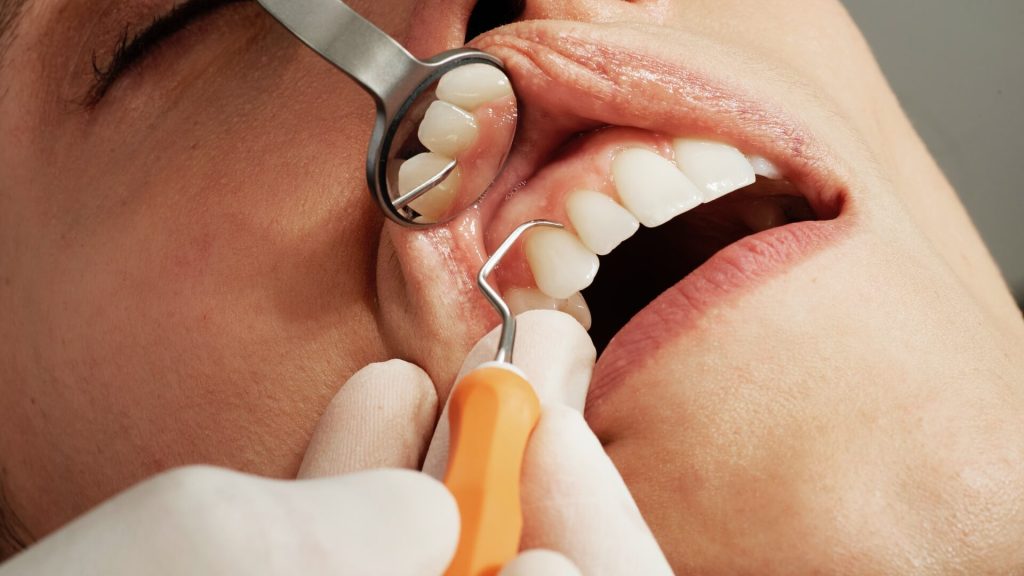
Periodontitis can lead to a litany of dental issues from bad breath to bleeding and tooth loss, and has long been suspected to be connected to other negative health outcomes in the body. Researchers at Hiroshima University have now found evidence that periodontitis could be connected to atrial fibrosis and arrythmias.
In a study published in JACC: Clinical Electrophysiology, the team found a significant correlation between periodontitis and fibrosis (which is scarring to an appendage of the heart’s left atrium that can lead to an irregular heartbeat called atrial fibrillation) in a sample of 76 patients with cardiac disease.
“Periodontitis is associated with a long-standing inflammation, and inflammation plays a key role in atrial fibrosis progression and atrial fibrillation pathogenesis,” said first author Shunsuke Miyauchi, assistant professor with the Hiroshima University’s Health Service Center. He is also affiliated with the university’s Graduate School of Biomedical and Health Sciences. “We hypothesised that periodontitis exacerbates atrial fibrosis. This histological study of left atrial appendages aimed to clarify the relationship between clinical periodontitis status and degree of atrial fibrosis.”
The left atrial appendages were surgically removed from the patients, and the researchers analysed the tissue to establish the correlation between severity of the atrial fibrosis and severity of the gum disease. They found that the worse the periodontitis, the worse the fibrosis, suggesting that the inflammation of gums may intensify inflammation and disease in the heart.
“This study provides basic evidence that periodontitis can aggravate atrial fibrosis and can be a novel modifiable risk factor for atrial fibrillation,” said corresponding author Yukiko Nakano, professor of cardiovascular medicine in Hiroshima University’s Graduate School of Biomedical and Health Sciences.
According to Nakano, in addition to improving other risk factors such as weight, activity levels, tobacco and alcohol use, periodontal care could aid in comprehensive atrial fibrillation management. However, she cautioned that this study did not establish a causal relationship, meaning that while gum disease and atrial fibrosis degrees of severity appear connected, researchers have not found that one definitively leads to the other.
“Further evidence is required for establishing that periodontitis contributes to the atrial fibrosis in a causal manner and that periodontal care can alter fibrosis,” Nakano said. “One of our goals is to confirm that periodontitis is a modifiable risk factor for atrial fibrillation and to promote dental specialists’ participation in comprehensive atrial fibrillation management. Periodontitis is an easy modifiable target with lower cost among known atrial fibrillation risk factors. Thus, the achievement of this study series may bring benefits for many people worldwide.”
Next, the researchers said they hope to conduct future clinical trials to clarify if periodontal intervention reduces atrial fibrillation occurrence and improves patient outcomes.
Experts led by researchers from the Murdoch Children’s Research Institute have created the world’s first international clinical guidelines to help prevent and treat heart complications in children undergoing cancer treatment.
Published in JACC:Advances, the guidelines cover cardiovascular disease assessment, screening and follow-up, for paediatric patients receiving cancer treatment with new molecular therapies, immunotherapy, chemotherapy and radiotherapy.
The expert consensus has defined the high-risk group of cancer patients who should undergo a heart check-up, standardised an approach to screening and surveillance during treatment and provided recommendations to protect vulnerable young hearts.
Murdoch Children’s Associate Professor Rachel Conyers said while international guidelines to monitor poor heart side effects during therapy exist for adult patients, none were specific to children.
Associate Professor Conyers said the success of new cancer drugs had increased the chances of cardiac side effects that occur early on during therapy, sometimes within days, which warranted closer heart health surveillance and earlier monitoring.
“Recent advances in treating childhood cancer have resulted in survival rates of more than 80 percent. However, improving serious health outcomes in survivors remains an important and essential focus and prevention is key,” she said.
“Heart complications are a leading cause of death for childhood cancer survivors, second only to cancer relapse. Modern treatments including precision medicine have broadened the agents that can cause heart problems.”
Childhood cancer survivors are 15 times more likely to have heart failure and eight times more likely to have heart disease than the general population.
Associate Professor Conyers said the guidelines would be an indispensable tool for clinicians to significantly reduce the harmful impact of cancer drugs on children’s hearts.
“The guidelines are a major advance for the cardio-oncology field as before this there was no defined approach for surveillance or follow up of pediatric patients during treatment despite new therapeutics having early heart complications such as high blood pressure, abnormal heart beats and heart failure,” she said.
The Australian and New Zealand expert group consisted of pediatric and adult cardiologists and pediatric oncologists who undertook a Delphi consensus approach across 11 areas of cardio-oncology care. The Australian New Zealand Children’s Oncology Group endorsed the study with the guidelines useful for any tertiary institutes treating pediatric oncology patients or initiating cardio-oncology clinics.