Vaccines that provide long-lasting protection against influenza, coronaviruses and respiratory syncytial virus (RSV) have proved exceptionally difficult to develop. In a new review article in Cell Host & Microbe, NIH researchers explore the challenges and outline approaches to improved vaccines and describe a promising path forward: mucosal vaccines.
Unlike the respiratory viruses that cause measles, mumps and rubella — for which vaccination or recovery from illness provides decades-long protection against future infection – flu, RSV, SARS-CoV-2 and “common cold” coronaviruses share several characteristics that enable them to cause repeated re-infections. These include very short incubation periods, rapid host-to-host transmission and replication in the nasal mucosa rather than throughout the body. This last feature – non-systemic replication – means these viruses do not stimulate the full force of the adaptive immune response, which typically takes a week or more to mount.
A next generation of improved vaccines for mucosa-replicating viruses will require advances in understanding on several fronts, the authors say. For instance, more must be learned about interactions between flu viruses, coronaviruses and RSV and the components of the immune response that operate largely or exclusively in the upper respiratory system. Over time, these interactions have evolved and led to “immune tolerance,” wherein the human host tolerates transient, limited infections by viruses that are generally non-lethal to avoid the destructive consequences of an all-out immune system attack.
The authors note that mucosal immunisation appears to be an optimal route of vaccination for the viruses of interest, when feasible. However, to develop useful mucosal vaccines, significant knowledge gaps must be filled including finding ideal vaccine formulations; determining dosage size, frequency and timing; and developing techniques for overcoming immune tolerance.
Cambridge scientists have successfully trialled an artificial pancreas for use by patients living with type 2 diabetes. They report in Nature Medicine that the device doubled the amount of time patients were in the target range for glucose compared to standard treatment and halved the time spent experiencing high glucose levels.
The artificial pancreas developed by University of Cambridge researchers combines an off-the-shelf glucose monitor and insulin pump with an app developed by the team, known as CamAPS HX. This app is run by an algorithm that predicts how much insulin is required to maintain glucose levels in the target range.
The researchers have previously shown that an artificial pancreas run by a similar algorithm is effective for patients living with type 1 diabetes, from adults through to very young children. They have also successfully trialled the device in patients with type 2 diabetes who require kidney dialysis.
Today, in Nature Medicine, the team report the first trial of the device in a wider population living with type 2 diabetes (not requiring kidney dialysis). Unlike the artificial pancreas used for type 1 diabetes, this new version is a fully closed loop system, whereas patients with type 1 diabetes need to tell their artificial pancreas that they are about to eat to allow adjustment of insulin, for example, with this version they can leave the device to function entirely automatically.
The researchers recruited 26 patients who were randomised to one of two groups – the first group would trial the artificial pancreas for eight weeks and then switch to the standard therapy of multiple daily insulin injections; the second group would take this control therapy first and then switch to the artificial pancreas after eight weeks.
The team used several measures to assess how effectively the artificial pancreas worked. The first was the proportion of time that patients spent with their glucose levels within a target range of between 3.9 and 10.0mmol/L. On average, patients using the artificial pancreas spent two-thirds (66%) of their time within the target range, compared to control (32%).
A second measure was the proportion of time spent with glucose levels above 10.0mmol/L. Over time, high glucose levels raise the risk of potentially serious complications. Patients taking the control therapy spent two-thirds (67%) of their time with high glucose levels — this was halved to 33% when using the artificial pancreas.
Average glucose levels fell from 12.6mmol/L when taking the control therapy to 9.2mmol/L while using the artificial pancreas.
The app also reduced levels of a molecule known as glycated haemoglobin, or HbA1c. Glycated haemoglobin develops when haemoglobin, a protein within red blood cells that carries oxygen throughout the body, joins with glucose in the blood, becoming ‘glycated’. By measuring HbA1c, clinicians are able to get an overall picture of what a person’s average blood sugar levels have been over a period of weeks or months. For people with diabetes, the higher the HbA1c, the greater the risk of developing diabetes-related complications. After the control therapy, average HbA1c levels were 8.7%, while after using the artificial pancreas they were 7.3%.
No patients experienced dangerously-low blood sugar levels (hypoglycaemia) during the study. One patient was admitted to hospital while using the artificial pancreas, due to an abscess at the site of the pump cannula.
Dr Charlotte Boughton from the Wellcome-MRC Institute of Metabolic Science at the University of Cambridge, who co-led the study, said: “Many people with type 2 diabetes struggle to manage their blood sugar levels using the currently available treatments, such as insulin injections. The artificial pancreas can provide a safe and effective approach to help them, and the technology is simple to use and can be implemented safely at home.”
Dr Aideen Daly, also from the Wellcome-MRC Institute of Metabolic Science, said: “One of the barriers to widespread use of insulin therapy has been concern over the risk of severe ‘hypos’ — dangerously low blood sugar levels. But we found that no patients on our trial experienced these and patients spent very little time with blood sugar levels lower than the target levels.”
Feedback from participants suggested that participants were happy to have their glucose levels controlled automatically by the system, and nine out of ten (89%) reported spending less time managing their diabetes overall. Users highlighted the elimination of the need for injections or fingerprick testing, and increased confidence in managing blood glucose as key benefits. Downsides included increased anxiety about the risk of hypoglycaemia, which the researchers say may reflect increased awareness and monitoring of glucose levels, and practical annoyances with wearing of devices.
The team now plan to carry out a much larger multicentre study to build on their findings and have submitted the device for regulatory approval with a view to making it commercially available for outpatients with type 2 diabetes.
Using new microscopic methods in combination with machine learning-based image analysis, researchers fromAlbert Ludwigs University of Freiburghave discovered new structures on the surface of living B cells that affect the distribution and possibly the function of their antigen receptors. The researchers’ study has been published in The EMBO Journal.
B cells recognise pathogens through specialised receptors on their surface. For the first time, scientists were able to observe how these receptors are distributed on the surface of living and moving cells. They found that the B cell surface is shaped into a characteristic landscape of interconnected ridges and protrusions. On this landscape, the IgM-class B cell antigen receptors (IgM-BCR) accumulate in specific areas. The precision of the receptors’ localisation and their clustering into larger units likely constitute a mechanism that controls receptor signalling and facilitates antigen sensing and thereby the activation of B cells.
In most immunological textbooks, lymphocytes are depicted as round, ball-like cells whose smooth surface carries randomly distributed receptors. The notion of a smooth unstructured B cell surface has already been challenged by electron micrographs of fixed and frozen lymphocytes, revealing thin membrane protrusions called microvilli on the cells’ surface. These tentacle-like structures help immune cells to search for molecular markers of pathogens, so-called antigens. B lymphocytes recognise such antigens through different classes of their B cell antigen receptors (BCR). These antigen receptors are complex molecular machines that, when activated, interact with other molecules to initiate a signalling cascade, leading to the differentiation of B cells into plasma cells and the production of protective antibodies.
The research group of Prof. Dr. Michael Reth collaborated with other imaging specialists to analyse how the IgM-BCR is distributed across the 3D surface of living B cells. For this, they used a technique called lattice light sheet microscopy, LLSM for short. “This method can capture volumetric images of living cells at a very high speed,” explains Dr. Deniz Saltukoglu from Freiburg University, the first author of the study. “In other types of high-resolution microscopy, cells need to be attached to a flat surface, which completely alters the B cells’ outer structures. LLSM allowed us to observe the cells in an environment that mimics biological tissues, meaning that the structures and movements that we saw were largely undisturbed,” she says.
The researchers then developed custom image analysis tools to quantify and objectively characterise the microscopic data. “We needed to segment the images and isolate morphological features,” says Saltukoglu. “So far this had only been done with two-dimensional data, so we had to develop new computational tools for volumetric, time course data.” For this, the researchers drew inspiration from algorithms that are used to map geographical data for archaeological surveys. With this approach, they found that the B cell surface carries a network of elevated ridges, with microvilli growing from the intersections of the network. Within this “cellular landscape”, the IgM-BCRs form clusters that concentrate along the ridges, in close proximity to the bases of the microvilli. The position of these clusters was linked with the dynamic movement of the ridges on the cells’ surface.
“We think that the 3-D location of the antigen receptors controls their activity,” says Reth. “Localisation at the microvilli base may prevent their unwanted activation. Once B cells receive a danger signal, they extent their microvilli and we assume that the IgM-BCR clusters then get recruited to the tip where they are localized in an optimal position for antigen sensing.”
This hypothesis is in line with other findings from Reth’s group, which suggest that the IgM-BCRs are regulated via lateral interactions with regulatory coreceptors. This means that the position and distribution of antigen receptors likely represent additional control mechanisms that affect signalling and activation of cells of the immune system.
Despite stringent infection-control efforts around the world, hospital-acquired infections (HAIs)keep on popping up from new strains of bacteria. In Science Translational Medicine, researchers report evidence pointing to an unexpected source of such bacteria: the hospitalised patients themselves.
From experiments with mice, researchers at Washington University School of Medicine in St. Louis discovered that urinary tract infections (UTIs) can arise after sterile tubes, called catheters, are inserted into the urinary tract, even when no bacteria are detectable in the bladder beforehand. Such tubes are commonly used in hospitals to empty the bladders of people undergoing surgery. In the mice, inserting the tubes activated dormant Acinetobacter baumannii bacteria hidden in bladder cells, triggering them to emerge, multiply and cause UTIs, the researchers said.
The findings suggest that screening patients for hidden reservoirs of dangerous bacteria could supplement infection-control efforts and help prevent deadly HAIs.
“You could sterilise the whole hospital, and you would still have new strains of A. baumannii popping up,” said co-senior author Mario Feldman, PhD, a professor of molecular microbiology. “Cleaning is just not enough, and nobody really knows why. This study shows that patients may be unwittingly carrying the bacteria into the hospital themselves, and that has implications for infection control. If someone has a planned surgery and is going to be catheterised, we could try to determine whether the patient is carrying the bacteria and cure that person of it before the surgery. Ideally, that would reduce the chances of developing one of these life-threatening infections.”
The notoriously multidrug-resistant A. baumannii is a major threat to patients, causing many cases of UTIs in people with urinary catheters, pneumonia in people on ventilators, and bloodstream infections in people with central-line catheters into their veins.
The researchers set out to investigate why so many A. baumannii UTIs develop after people receive catheters.
Most UTIs among otherwise healthy people are caused by the bacterium Escherichia coli. Research has shown that E. coli can hide out in bladder cells for months after a UTI seems to have been cured, and then re-emerge to cause another infection.
The researchers investigated whether A. baumannii can hide inside cells like E. coli can. They studied mice with UTIs caused by A. baumannii. They used mice with weakened immune systems because, like people, healthy mice can fight off A. baumannii.
Once the infections had resolved and no bacteria were detected in the mice’s urine for two months, the researchers inserted catheters into the mice’s urinary tracts with a sterile technique. Within 24 hours, about half of the mice developed UTIs caused by the same strain of A. baumannii as the initial infection.
“The bacteria must have been there all along, hiding inside bladder cells until the catheter was introduced,” said co-senior author Scott J. Hultgren, PhD, a professor and expert on UTIs. “Catheterisation induces inflammation, and inflammation causes the reservoir to activate, and the infection blooms.”
Since A. baumannii rarely causes symptoms in otherwise healthy people, many people who carry the bacteria may never know they’re infected, the researchers said. According to the researchers’ literature search, 2% of healthy people carry A. baumannii in their urine.
“I wouldn’t put much weight on the precise percentage, but I think we can say with certainty that some percentage of the population is walking around with A. baumannii,” Feldman said. “As long as they’re basically healthy, it doesn’t cause any problems, but once they’re hospitalised, it’s a different matter. This changes how we think about infection control. We can start considering how to check if patients already have Acinetobacter before they receive certain types of treatment; how we can get rid of it; and if other bacteria that cause deadly outbreaks in hospitals, such as Klebsiella, hide in the body in the same way. That’s what we’re working on figuring out now.”
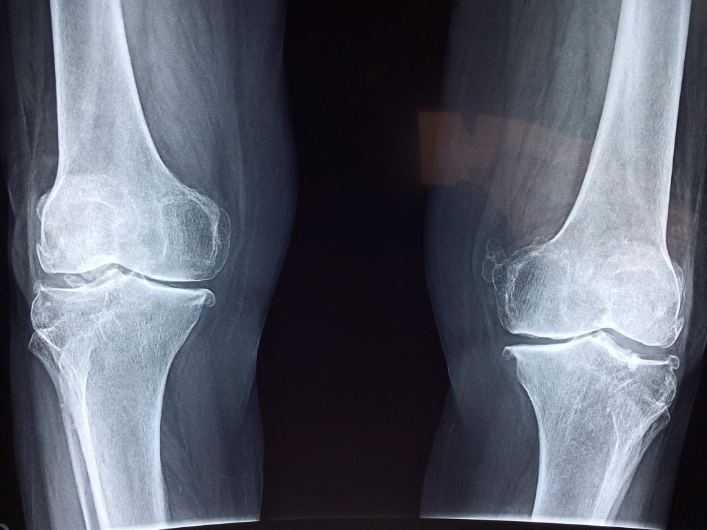
In APL Bioengineering, researchers report on an injectable hydrogel that treats infections around hip and knee replacement prosthetics without the problems caused by current treatments. Testing showed that the gel inhibits common bacteria and promotes tissue regrowth.
After hip and knee replacement surgeries, pathogenic bacteria can adhere to the surface of the joint prosthesis and form a dangerous biofilm. Gold standard clinical methods use potent antibiotics and further surgery, including removal of infected tissue and transplantation of new tissue, to treat these infections. However, these strategies run into problems with hyper-resistant bacteria caused by the abuse of antibiotics, persistent damage caused by tissue removal, difficulties in obtaining tissue donors, and toxicity and immune system complications.
A team from Shanghai Jiao Tong University School of Medicine created ablack phosphorus-enhanced antibacterial injectable hydrogel to re-establish biological barriers in soft tissue and suppress persistent infections. The gel has a porous structure, excellent injectability, and rapid self-healing properties.
“It is important to explore a new strategy for treatment of infected soft tissue wounds because it is directly related to prognosis,” said author Ruixin Lin. “We aspire to develop a simpler, safer method to help more patients avoid suffering and help more doctors make the right choices.”
In vitro tests showed the hydrogel had good stability and low toxicity to tissue cells. Irradiating the gel with near infrared light causes it to release silver ions. This process was highly efficient at inhibiting the common bacteria S. aureus.
“Furthermore, an in vivo infected wound model showed that the hydrogel could not only inhibit the persistent infection of the wound, but also accelerate the deposition of collagen fibres and angiogenesis, thereby realizing the repair of the natural barrier of soft tissue,” said Lin.
The novel hydrogel provides a safe and feasible synergistic antibacterial strategy for infected soft tissue healing. The team believes that it solves current clinical problems, such as stubborn infections caused by antibiotic resistance, and provides new ideas for minimally invasive treatment. They hope to see it used in the clinic after conducting sufficient studies on its underlying mechanisms.
A nationally representative study published in JAMA found that older adults with greater severity of hearing loss were more likely to have dementia, but the likelihood of dementia was lower among hearing aid users compared to non-users.
The findings are consistent with prior studies showing that hearing loss might be a contributing factor to dementia risk over time, and that treating hearing loss may lower dementia risk.
“This study refines what we’ve observed about the link between hearing loss and dementia, and builds support for public health action to improve hearing care access,” says lead author Alison Huang, PhD, MPH, a senior research associate in the Bloomberg School’s Department of Epidemiology and at the Cochlear Center for Hearing and Public Health, also at the Bloomberg School.
Hearing loss is a critical public health issue affecting two-thirds of Americans over 70. The growing understanding that hearing loss might be linked to the risk of dementia, which impacts millions, and other adverse outcomes has called attention to implementing possible strategies to treat hearing loss.
For the new study, Huang and colleagues analysed a nationally representative dataset from the National Health and Aging Trends Study (NHATS). Funded by the National Institute on Aging, the NHATS has been ongoing since 2011, and uses a nationwide sample of Medicare beneficiaries over age 65, with a focus on the 90-and-over group as well as Black individuals.
The analysis covered 2413 individuals, about half of whom were over 80 and showed a clear association between severity of hearing loss and dementia. Prevalence of dementia among the participants with moderate/severe hearing loss was 61% higher than prevalence among participants who had normal hearing. Hearing aid use was associated with a 32% lower prevalence of dementia in the 853 participants who had moderate/severe hearing loss.
The authors note that many past studies were limited in that they relied on in-clinic data collection, leaving out vulnerable populations that did not have the means or capacity to get to a clinic. For their study, the researchers collected data from participants through in-home testing and interviews.
How hearing loss is linked to dementia isn’t yet clear, and studies point to several possible mechanisms. Huang’s research adds to a body of work by the Cochlear Center for Hearing and Public Health examining the relationship between hearing loss and dementia.
Sixty-three percent of participants who had severe alcohol use disorder who went through a detoxification programme saw improvement within the first ten days, according to the results of a French study. The findings were published in Alcohol and Alcoholism.
Impaired cognition is known to have an impact on the efficacy of substance use rehabilitation programmes, but substance use, such as alcohol, is known to have in impact on cognition. Therefore, in order to better tailor treatment programmes, it is important to know how quickly normal cognitive levels can be restored.
To assess recovery of alcohol-related neuropsychological deficits in a group of patients with pure severe alcohol use disorder (AUD) during a detoxification program using the Brief Evaluation of Alcohol-Related Neuropsychological Impairment (BEARNI) test.
Thirty-two patients admitted to French hospitals with severe AUD using DSM-IV criteria (24 men, mean age = 45.5 ± 6.8 years old) were assessed using the BEARNI 8 ± 2 days after alcohol cessation (T1) and then were reassessed within 18 ± 2 days after alcohol cessation (T2). The primary study endpoint was the number of patients initially impaired at T1 who recovered cognitive functions at T2 assessment.
At T1, 59% (n = 19) patients with pure severe AUD had at least one impaired cognitive function assessed by the BEARNI. At T2, 63% of the patients with AUD with deficits at T1 had normal BEARNI cognitive scores. Among the subtests, the highest percentage of participants with normal subtest scores were 100% in the verbal fluency category, 67% in visuospatial, 63% in the memory category and 60% in alphabetical span.
The researchers also noted that those participants in the present study who recovered within 18 days of abstinence, did so earlier than reported in previous studies.
“Additional studies assessing cognitive improvements during abstinence, and especially earlier in abstinence, are needed,” the authors concluded. “Further studies should also assess the early course of social cognition, attentional bias and inhibition deficits in patient with alcohol use disorder early in abstinence, given their clinical impact.”
Premature birth is the main cause of brain injury and cerebral palsy in babies. Evidence shows that babies can be protected from brain injury by giving magnesium sulfate to women who are at risk of premature birth, reducing the risk of cerebral palsy by a third. From a societal and lifetime perspective, the health gains and cost savings associated with the preventative treatment generated a net monetary benefit of £866 per preterm baby, according to an evaluation published in Archives of Disease in Childhood.
The prevention of cerebral palsy in pre-term labour (PReCePT) programme was developed in 2014 and aimed to support all maternity units in England to increase the use of magnesium sulfate in premature births. It was then piloted in five NHS trusts in the West of England, and this pilot was evaluated by the NIHR Applied Research Collaboration West (NIHR ARC West). It has since been rolled out across England via the AHSN Network as a national programme.
The evaluation of the national programme, also led by NIHR ARC West, found that PReCePT was both effective and cost-effective. The researchers looked at data from the UK National Neonatal Research Database for the year before and year after PReCePT was implemented in maternity units in England.
While use of magnesium sulfate had been increasing before, the study showed that PReCePT was able to accelerate uptake. It increased by 6.3 percentage points on average across all maternity units in England during the first year, over and above the increase that would be expected over time as the practice spread organically. After also adjusting for variations in when maternity units started the programme, the increase in use of magnesium sulfate was 9.5 percentage points. By May 2020, on average 86.4% of eligible mothers were receiving magnesium sulfate.
The researchers also estimated that the programme’s first year could be associated with a lifetime saving to society of £3 million. This accounts for the costs of the programme, administering the treatment and of cerebral palsy to society over a lifetime, and the associated health gains of avoiding cases. This is across all the extra babies the programme helped get access to the treatment during the first year.
In the five pilot sites, the improved use of magnesium sulfate has been sustained over the years since PReCePT was implemented. As the programme costs were mostly in the first year of implementation, longer-term national analysis may show that PReCePT is even more cost-effective over a longer period.
John Macleod, NIHR ARC West Director, Professor in Clinical Epidemiology and Primary Care at the University of Bristol and principal investigator of the evaluation, said: “Our in-depth analysis has been able to demonstrate that the PReCePT programme is both effective and cost-effective. The programme has increased uptake of magnesium sulfate, which we know is a cost-effective medicine to prevent cerebral palsy, much more quickly than we could have otherwise expected.
Professor Lucy Chappell, Chief Executive Officer of the National Institute for Health and Care Research, said: “This important study shows the impact of taking a promising intervention that had been shown to work in a research setting and scaling it up across the country. Giving magnesium sulfate to prevent cerebral palsy in premature babies is a simple, inexpensive intervention that can make such a difference to families and the health service. We look forward to seeing ongoing use of magnesium sulfate across our maternity units so that these benefits continue.”
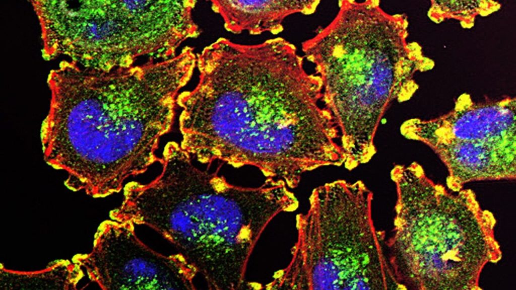
Melanoma cells. Source: National Cancer Institute.
Fewer cases of melanoma were observed among regular users of vitamin D supplements than among non-users, researchers in Finland found. People taking vitamin D supplements regularly also had a considerably lower risk of skin cancer, according to a study of nearly 500 people with increased skin cancer risk, which was published in Melanoma Research.
A key micronutrient, vitamin D may play a role in many diseases. Previous studies investigating the link between vitamin D and skin cancers, have been inconclusive or contradictory, but they mainly focussed on serum levels of calcidiol, which is a metabolite of vitamin D. Serum calcidiol levels have been associated with both a slightly higher or lower risk of different skin cancers. This may be partly due to the fact that serum calcidiol analyses do not provide information on vitamin D metabolism in the human skin, which can express enzymes that generate biologically active vitamin D metabolites or inactivate them.
The new study took a different approach: 498 adult patients with an increased risk of a skin cancer, such as basal cell carcinoma, squamous cell carcinoma or melanoma, were recruited and classified into low risk, moderate risk and high risk. Based on their use of oral vitamin D supplements, the patients were divided into three groups: non-users, occasional users and regular users. Serum calcidiol levels were analysed in half of the patients and found to correspond to their self-reported use of vitamin D.
A key finding of the study is that there were considerably fewer cases of melanoma among regular users of vitamin D than among non-users, and that the skin cancer risk classification of regular users was considerably better than non-users’. Logistic regression analysis showed that melanoma risk among regular users was more than halved compared to non-users.
The findings suggest that even occasional users of vitamin D may have a lower risk for melanoma than non-users. However, there was no statistically significant association between the use of vitamin D and the severity of photoaging, facial photoaging, actinic keratoses, nevus count, basal cell carcinoma and squamous cell carcinoma. Serum calcidiol levels were not significantly associated with these skin changes, either. Since the research design was cross-sectional, the researchers were unable to demonstrate a causal relationship.
Other relatively recent studies, too, have provided evidence of the benefits of vitamin D in melanoma, such as of the association of vitamin D with a less aggressive melanoma.
“These earlier studies back our new findings from the North Savo region here in Finland. However, the question about the optimal dose of oral vitamin D in order to for it to have beneficial effects remains to be answered. Until we know more, national intake recommendations should be followed,” Professor of Dermatology and Allergology Ilkka Harvima of the University of Eastern Finland notes.
A newly published study in iScience sheds light on the biological underpinnings in sex differences in obesity-related disease, with researchers observing “striking” differences in the cells that build blood vessels in the fatty tissue of male versus female mice.
Men are more likely than women to develop conditions associated with obesity such as cardiovascular disease, insulin resistance and diabetes, says study leader Professor Tara Haas at York University.
“People have used rodent models to study obesity, and the diseases that are associated with obesity – like diabetes – but they’ve typically always studied male rodents, because females are resistant to developing the same kinds of diseases,” says Haas. “We were really interested in exploring that difference because, to us, it spoke of something really fascinating happening in females that protects them.”
In earlier work, Haas and her team saw that when mice become obese, females grow a lot of new blood vessels to supply the expanding fat tissue with oxygen and nutrients, whereas males grow a lot less. For this study, Haas and her co-authors focused on differences in the endothelial cells that make up the building blocks of these blood vessels in fat tissue.
The team used software to help sift through thousands of genes to zero in on the ones that would be associated with blood vessel growth. They discovered that processes associated with the proliferation of new blood vessels were high in the female mice, whereas the males had a high level of processes associated with inflammation.
“It was very striking the extent of inflammation-associated processes that were prevalent in the males,” Haas recalls. “Other studies have shown that when endothelial cells have that kind of inflammatory response, they’re very dysfunctional, and they don’t respond to stimuli properly.”
York PhD student Alexandra Pislaru, who works in Haas’ lab and is a co-first author of the study, participated in this project as part of her dissertation.
“It is exciting to observe the continuing resilience that female endothelial cells display even when stressed by a long-term high-fat diet,” Pislaru says. “The findings from our study can help researchers to get a better understanding of why obesity manifests differently in men and women.”
The researchers also examined the behaviour of the endothelial cells when they were taken out of the body and studied in petri dishes.
“Even when we take them out of the body where they don’t have the circulating sex hormones or other kinds of factors, male and female endothelial cells still behave very differently from each other,” Haas explains.
Female endothelial cells replicated faster, while male endothelial cells displayed greater sensitivity to an inflammatory stimulus. By comparing with previously published data sets, the researchers found endothelial cells from aged male mice also displayed a more inflammatory profile compared to female cells.
“You can’t make the assumption that both sexes are going to respond to the same series of events the same way,” says Haas. “This isn’t just an obesity related issue – I think it’s a much broader conceptual problem that also encompasses healthy aging. One implication of our findings is that there will be situations where the treatment that is ideal for men is not going to be ideal for women and vice-versa.”
While humans and mice have different genes that may be turned up or down, Haas believes the general findings would likely apply and is interested studying the same cells in humans in future research.