SARS-CoV-2 virus. Source: Fusion Medical Animation on Unsplash
In a study published in JAMA Network Open, researchers reported that the blood pressure medication losartan is not effective in reducing lung injury in patients with COVID.
This drug was investigated based on early reports suggesting benefit in preclinical models of the 2003 SARS virus, a close family member to the current SARS-CoV-2 virus.
The research team sought to determine if a common blood pressure medication might decrease lung injury in patients hospitalised with COVID. Their results found that losartan treatment did not reduce lung injury in patients admitted with COVID, and had no effect on mortality.
In addition, critically-ill patients treated with losartan needed additional, temporary blood pressure support. However, this did not result in worse outcomes overall.
“Even though this particular drug was not effective for the treatment of COVID-19, repurposing inexpensive and relatively safe medications remains an important approach to contain healthcare costs,” said study co-author Michael Puskarich, MD, an associate professor in emergency medicine.
“Finding effective treatments for COVID that can be widely used across both the developed and developing world remains an important ongoing area of investigation,” Dr Puskarich added.
The researchers noted that more studies of protein and cellular signalling from ALPS-COVID trial participants are ongoing.
“We hope that future study findings of these proteins may show insights into why the body responds the way it does to COVID,” said co-author Christopher Tignanelli, MD, MS, FACS, FAMIA, an assistant professor in surgery. “Critically, this will help us understand why some people develop severe disease following COVID infection and others are asymptomatic.”
Researchers have reported that they have successfully fused human retinal cells with adult stem cells, in a novel potential regenerative therapy to treat retinal damage and visual impairment.
The resulting hybrid cells stimulate the regenerative potential of human retinal tissue, something previously only thought to be found in cold-blood vertebrates.
Cell fusion events, where two different cells combine into one single entity, are known to be a possible mechanism contributing to tissue regeneration. These cell fusions result in four sets of chromosomes instead of the usual two. Though a rare phenomenon in humans, it has been reliably detected in the liver, brain, and gastrointestinal tract. Now, cell fusion events have been found also take place in the human retina, as reported in eBioMedicine.
Seeking to see if cell fusion events could differentiate into neurons, the researchers fused Müller glia, cells that play a secondary but important role in maintaining the structure and function of the retina, with adult stem cells.
“We were able to carry out cell fusion in vitro, creating hybrid cells. Importantly, the process was more efficient in the presence of a chemical signal transmitted from the retina in response to damage, resulting in rates of hybridisation increasing twofold. This gave us an important clue for the role of cell fusion in the retina,” said first author Sergi Bonilla.
The hybrid cells were injected into a growing retinal organoid, a model that closely resembles the function of the human retina. The researchers found that the hybrid cells successfully engrafted into the tissue and differentiated into cells that closely resemble ganglion cells, a type of neuron essential for vision.
“Our findings are important because they show that the Müller Glia in the human retina have the potential to regenerate neurons,” said lead researcher Professor Pia Cosma. “Salamanders and fish can repair damage caused to the retina thanks to their Müller glia, which differentiate into neurons that rescue or replace damaged neurons. Mammalian Müller glia have lost this regenerative capacity, which means retinal damage or degradation can lead to visual impairment for life. Our findings bring us one step closer to recovering this ability.”
Further work will be to understand why these hybrid cells, which have four complete sets of chromosomes, don’t result in chromosomal instability and cancer development. The authors of the study believe the retina may have a mechanism regulating chromosome segregation similar to the liver, which contains tetraploid cells that act as a genetic reservoir, undergoing mitosis in response to stress and injury.
Scanning Electron Micrograph of Pseudomonas aeruginosa. Credit: CDC/Janice Carr
While pathogens usually work against drug treatments, sometimes, they can actually strengthen them, according to a new University of Maine study published in the journal Infection and Immunity.
Polymicrobial infections, which are a combination of bacteria, viruses, fungi and parasites, are challenging to treat because it is not well understood how pathogens interact during infection and how these interactions affect the drugs treating them.
In a study published in Infection and Immunity, University of Maine researchers examined two common pathogens that often occur at similar sites, particularly in cystic fibrosis and mechanically ventilated patients: Candida albicans andPseudomonas aeruginosa.
Candida is the fourth most common hospital-acquired pathogen, and many antifungal agents only slow it rather than kill it outright. Meanwhile, P. aeruginosa infects 90% of all adult cystic fibrosis patients. Combined, C. albicans and P. aeruginosa cause more serious disease in cystic fibrosis and ventilated patients.
The researchers investigated the effectiveness of the antifungal drug fluconazole in vitro and then during infection of the zebrafish with both pathogens. Fluconazole slows fungal growth, but Candida can become tolerant to the drug and not only survive, but also evolve tolerance that leads to therapy failure and, potentially, death.
The results showed that P. aeruginosa in fact works with fluconazole to eliminate drug tolerance and clear the C. albicans infection in the culture and the zebrafish.
“Polymicrobial infections are challenging to treat not only because of the lack of understanding of how invading microorganisms interact but also because we don’t know how these interactions affect treatment efficacy. Our work demonstrates that polymicrobial interactions can indeed affect treatment efficacy and, most importantly, it highlights the importance of nutrient availability in the environment -; such as iron in our study -; and how it modulates treatment efficacy,” explained Siham Hattab, lead author of the study.
What’s more, the bacteria also enhance the drug’s ability against a second pathogenic Candida species that tends to be more resistant to the drug.
The increased effectiveness of the drug suggests to the researchers that there is still much more to learn about how current drugs work when targeting these dangerous and complex polymicrobial infections.
Senior study author, Robert Wheeler, associate professor of microbiology said: “We are really excited to have revealed that sometimes drugs against fungal infection can work even better in a more ‘real-world’ situation than in the test tube. There is still a lot to learn about how pathogens interact during infection, and it will be interesting to see how the bacteria manage to work with the drugs to target Candida.”
The World Health Organization is alerting health care professionals about the risk of lethal administration errors that can potentially occur with tranexamic acid (TXA) injection. There have been reports of TXA being mistaken for obstetric spinal anaesthesia used for caesarean deliveries resulting in inadvertent intrathecal administration.
Intrathecal TXA is a potent neurotoxin and neurological sequelae are manifested, with refractory seizures and 50% mortality. The profound toxicity of intrathecal TXA was described in 1980. In a 2019 review, Patel et al. identified 21 reported cases of inadvertent intrathecal injection of TXA since 1988, of which 20 were life-threatening and 10 fatal. It appears that mortality risk is greater after caesarean delivery. Sixteen were reported between 2009 and 2018.
WHO recommends early use of intravenous TXA within three hours of birth in addition to standard care for women with clinically diagnosed postpartum haemorrhage (PPH) following vaginal births or caesarean section. TXA should be administered at a fixed dose of 1g in 10 ml (100 mg/ml) IV at 1 ml per minute, with a second dose of 1g IV if bleeding continues after 30 minutes. In South Africa, the incidence of maternal bleeding after caesarean delivery has been characterised as a national emergency, and obstetric haemorrhage remains the third most common cause of maternal mortality at 17%.
However, problems can arise as TXA is frequently stored in close proximity with other medicines, including injectable local anaesthetics indicated for spinal analgesia (eg, for caesarean section). The presentation of some of the local anaesthetics is similar to the TXA presentation (transparent ampoule containing transparent solution), which can be administered in error instead of the intended intrathecal anaesthetic, and resulting in serious undesirable adverse effects.
Obstetricians from several countries have recently reported inadvertent intrathecal TXA administration and related serious neurological injuries. In a South African clinical alert, Bishop et al. highlighted the different appearances of TXA used in state and private hospitals, with one example in private hospitals appearing very similar (white label, red text) at first glance to spinal bupivacaine and stored in the same container. Applicable recommendations were provided by the authors.
TXA is a lifesaving medicine, however, this potential clinical risk should be considered and addressed by all operating theatre staff. Reviewing of existing operating theatre drug handling practice is required in order to decrease this risk, such as storage of TXA away from the anaesthetic drug trolley, preferably outside the theatre.
A clinical trial found that cardiac computed tomography (CT) offers similar diagnostic accuracy to catheterisation – the current standard diagnostic test for intermediate-risk patients – in people with suspected coronary artery disease, as well as being associated with a lower risk of complications. The trial’s findings were published in the New England Journal of Medicine.
The current standard diagnostic test for coronary artery disease (CAD) is coronary angiography (often along with cardiac catheterisation). This minimally invasive procedure uses dye marker visible on X-ray imaging to detect arterial narrowing. Any narrowing detected in this manner can be treated during the procedure itself using stents, which prop open the newly widened blood vessels. More than 3.5 million of these procedures are carried out in European catheterisation laboratories every year, and more are carried out every year. Approximately two million of these do not involve immediate treatment in the cath lab. In these cases, the procedure is able to rule out narrowed or blocked coronary arteries.
The main question addressed by the DISCHARGE Trial Group was whether the low-risk, non-invasive coronary CT method can provide a safe alternative to catheterization in certain patients with suspected CAD. In order to test the effectiveness of both of these diagnostic imaging techniques in patients with stable chest pain, the project followed more than 3500 patients for a duration of four years. Patients were randomised to either computed tomography or cardiac catheterisation. If their initial evaluation ruled out obstructive coronary artery disease, participants were discharged back to their referring physician for further treatment – a step which gave the trial its name: DISCHARGE. Patients who were diagnosed as having the disease were managed in accordance with European guidelines at the time of the study.
Discussing the long-term results, trial leader Professor Dr Marc Dewey said: “The trial confirmed that a CT-based management is safe in patients with stable (ie, non-acute) chest pain and suspected coronary artery disease.”
Evaluation of safety was based on the incidence of major cardiovascular events over a period of up to four years. He added: “Among the patients referred for cardiac catheterisation and included in this trial, the risk of major adverse cardiovascular events was found to be similar in both the CT and catheterisation groups, occurring in 2.1% and 3.0% of patients, respectively. The incidence of major procedure-related complications was found to be four-times lower in patients managed with an initial CT strategy.”
Other outcome measures were included in the DISCHARGE trial, such as improvements in chest pain and quality of life over the course of the trial. This new strategy could help relieve pressure on health care systems by helping to reduce the volume of catheterisation procedures. Prof Dewey said: “Now that CT has been standardised and quality-tested as part of the DISCHARGE trial, this method could be made more widely available as part of the routine clinical care of people with intermediate CAD risk.”
As a next step, the trial’s method for estimating a person’s clinical risk of having coronary artery disease will need to be further evaluated to determine whether it can improve referral and indication for CT in routine clinical care. Health economics are an important component in making decisions about reimbursement in health care systems. As mentioned in the discussion of the publication, further methodologically very rigorous cost-effectiveness analyses of CT and cardiac catheterisation are necessary and will be conducted by the DISCHARGE Trial Group.
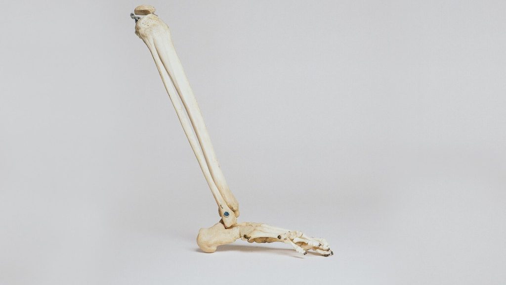
The study supports the use of widely available CEAA supplements to promote recovery and preserve function in patients undergoing surgery for repair of major fractures. “Our results suggest that this inexpensive, low-risk intervention has considerable potential to improve outcomes after fracture fixation,” according to the report by Michael Willey, MD, and colleagues of University of Iowa Hospital and Clinics, Iowa City.
The study included 400 patients undergoing operative fixation of fractures in the limbs and/or pelvis at the researchers’ trauma centre. In equal numbers and stratified by fracture severity, patients were randomises to either standard postoperative nutrition or standard nutrition plus CEAA supplementation.
CEAAs are termed “conditionally essential” because the body doesn’t usually require them. However, during times of illness or stress, the need for these conditional amino acids increases dramatically. Previous studies have reported that CEAA supplementation can improve wound-healing and other outcomes in patients with a variety of conditions, including postoperative recovery. In the new trial, patients assigned to the CEAA group received a standard supplement that included arginine, leucine, and glutamine.
At follow-up, the overall complication rate was significantly lower for patients who received CEAA supplementation (30.5%) compared with those who did not receive CEAA (43.8%). The CEAA group also had a lower rate of nonunion (5.1 vs 13.2%, respectively). Some other types of complications, including surgical-site infections, were similar between groups.
Patients who undergo operative fracture fixation are at risk of skeletal muscle wasting, which often results in weight loss as a result of reduced muscle mass. In the new study, patients receiving CEAA supplements had little or no change in fat-free body mass. In contrast, patients receiving standard nutrition had a 1kg reduction in fat-free mass at 6 weeks postoperatively, which took until 12 weeks to return to normal.
An unexpected finding was a sharply reduced mortality rate in the CEAA group (0.5% compared to 4.1 % for the control). Although the authors could not explain the lower risk of death in patients receiving CEAA, they suggest it might result from “unidentified confounding factors.”
Despite advances in surgical techniques, trauma patients undergoing operative fixation of extremity and pelvic fractures remain at risk of complications and prolonged loss of function. “Malnutrition is a potentially modifiable risk factor for mortality, fracture nonunion, wound complications, and increased length of stay,” the authors wrote.
CEAA supplementation therefore appears to be a simple, risk-free, and inexpensive means of promoting good nutrition after fracture fixation surgery. Controlling for other factors, the relative risk of complications is about 40% lower in patients receiving CEAA, with no reduction in fat-free mass during the early weeks of recovery. The researchers concluded: “This study will serve as the foundation for multicentre [randomised controlled trials] that are designed to assess the impact of CEAA nutrition supplementation in reducing complications and loss of functional muscle mass in high-risk populations.”
With the spread of omicron infections in young children, doctors have observed the rise of a previously unrecognised COVID complication: croup. Published in Pediatrics, physicians at Boston Children’s Hospital reported on 75 children admitted to the emergency department (ED) with croup and COVID.
The children appeared at the ED from from March 1, 2020 through January 15, 2022. Some cases were surprisingly severe, requiring hospitalisation and more medication doses compared to croup caused by other viruses. Just over 80% occurred during the omicron period. The report was published March 8 in a pre-publication in.
“There was a very clear delineation from when omicron became the dominant variant to when we started seeing a rise in the number of croup patients,” said Ryan Brewster, MD, first author of the report.
Laryngotracheitis, commonly known as croup, is a common respiratory illness in babies and young children. It is marked by a distinctive barking cough and sometimes stridor. It happens when viral infections cause swelling around the upper respiratory tract. In severe cases, including some seen at Boston Children’s, it can dangerously constrict breathing.
COVID studies in animals have found that the omicron strain ‘prefers’ the upper airway more than earlier variants, which mainly targeted the lower respiratory tract. This may account for the sudden appearance of croup during the omicron surge, said Dr Brewster.
In keeping with the general pattern of croup, most of the children with COVID and croup were under two years old, and 72% were boys. Except for one child with a common cold virus, none had a viral infection other than SARS-CoV-2.
Although all the children survived, nine of the 75 children with COVID-associated croup (12%) required hospitalisation and four of them (44%, or 5%of the total) required intensive care. (By comparison, before COVID, fewer than 5% of children with croup were hospitalised, and of those, only 1 to 3% required intubation.)
Overall, 97% of the children were treated with dexamethasone, a steroid. All of those who were hospitalised received racemic epinephrine via nebuliser, which is reserved for moderate or severe cases, as did 29% of children treated in the ED. Those who were hospitalised needed a median of six doses of dexamethasone and 8 nebulised epinephrine treatments to control their symptoms.
“Most cases of croup can be managed in the outpatient setting with dexamethasone and supportive care,” said Dr Brewster. “The relatively high hospitalisation rate and the large number of medication doses our COVID croup patients required suggests that COVID might cause more severe croup compared to other viruses. Further research is needed to determine the best treatment options for these children.”
Researchers at the National Robotarium in the UK, are developing an artificial intelligence (AI) ‘storytelling’ companion that will aid memory recollection, boost confidence and combat depression in patients suffering from Alzheimer’s disease and other types of dementia.
The idea for the ground-breaking ‘Agent-based Memory Prosthesis to Encourage Reminiscing’ (AMPER) project came from Dr Mei Yii Lim, a co-investigator of the project and an experienced memory modelling researcher.
In Alzheimer’s patients, memory loss occurs in reverse chronological order, with pockets of long-term memory remaining accessible even as the disease progresses. Rehabilitative care methods currently focus on physical aids and repetitive reminding techniques, but AMPER’s AI-driven user-centred approach will instead focus on personalised storytelling to help bring a patient’s memories back to the surface.
Dr Lim explained the project: “AMPER will explore the potential for AI to help access an individual’s personal memories residing in the still viable regions of the brain by creating natural, relatable stories. These will be tailored to their unique life experiences, age, social context and changing needs to encourage reminiscing.”
Having communication difficulties and decreased confidence are commonly experienced by people living with dementia and can often lead to individuals becoming withdrawn or depressed. By using AI to aid memory recollection, researchers at the National Robotarium hope that an individual’s sense of value, importance and belonging can be restored and quality of life improved.
The project’s long-term vision is to show that AI companions can become more widely used and integrated into domestic, educational, health and assistive-needs settings.
Professor Ruth Aylett from the National Robotarium is leading the research. She said: “One of the most difficult aspects of living with dementia can be changes in behavior caused by confusion or distress. We know that people can experience very different symptoms that require a range of support responses. Current intervention platforms used to aid memory recollection often take a one-size-fits-all approach that isn’t always suitable to an individual’s unique needs.”
“AI technology has the potential to play a pivotal role in improving the lives of people living with cognitive diseases. Our ambition is to develop an AI-driven companion that offers patients and their caregivers a flexible solution to help give an individual a sustained sense of self-worth, social acceptance and independence.
“Through projects like AMPER, we’re able to highlight the many ways AI and robotics can both help and improve life for people now and in the future. At the National Robotarium, we’re working on research that will benefit people in adult care settings as well as across a wide range of other sectors that will make life easier, safer and more supported for people.”
Once developed, the AI technology will be accessed through a tablet-based interface to make it more widely accessible and low-cost. The National Robotarium team will also investigate a using the AI in a desktop robot to see if a physical presence has any benefit.
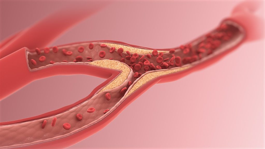
New research has shown that the link between low density lipoprotein cholesterol (LDL-C) and cardiovascular disease may not be as strong as previously thought.
The study, published in JAMA Internal Medicine, provides evidence that calls into question the efficacy of statins when prescribed with the goal of lowering LDL-C and consequently cardiovascular disease (CVD) risk.
Numerous prior studies have suggested that using statins to lower LDL-C positively affects cardiovascular health outcomes, findings which are reflected in the various iterations of expert guidelines for the prevention of CVD. Several large clinical trials have indicated that for every 1-mmol/l reduction in LDL-C levels there is a 23% reduction in CVD risk.
The new findings contradict this theory, finding that this relationship was weaker than previously thought. Lowering LDL-C with statins in fact was found to have an inconsistent and inconclusive impact on CVD outcomes such as myocardial infarction (MI), stoke, and all-cause mortality.
Additionally, it indicates that the overall benefit of taking statins may be small and will vary depending on an individual’s personal risk factors.
Commenting on the findings, the paper’s lead author Dr Paula Byrne said: “The message has long been that lowering your cholesterol will reduce your risk of heart disease, and that statins help to achieve this. However, our research indicates that, in reality, the benefits of taking statins are varied and can be quite modest.”
The researchers go on to suggest that this updated information should be communicated to patients through informed clinical decision-making and updated clinical guidelines and policy.
Even exposure to moderate light levels during nighttime sleep, compared to sleeping in a dimly lit room, harms cardiovascular function during sleep and increases insulin resistance the following morning, according to a new study published in PNAS.
“The results from this study demonstrate that just a single night of exposure to moderate room lighting during sleep can impair glucose and cardiovascular regulation, which are risk factors for heart disease, diabetes and metabolic syndrome,” said senior study author Dr Phyllis Zee at Northwestern University. “It’s important for people to avoid or minimise the amount of light exposure during sleep.”
There is evidence that daytime light exposure increases heart rate via sympathetic nervous system activation, increasing heart rate and alertness to meet the day’s challenges.
“Our results indicate that a similar effect is also present when exposure to light occurs during nighttime sleep,” Dr Zee said.
“We showed your heart rate increases when you sleep in a moderately lit room,” said Dr Daniela Grimaldi, a co-first author and research assistant professor of neurology at Northwestern. “Even though you are asleep, your autonomic nervous system is activated. That’s bad. Usually, your heart rate together with other cardiovascular parameters are lower at night and higher during the day.”
The sympathetic and parasympathetic nervous systems regulate the body physiology during the day and night. Sympathetic takes charge during the day and parasympathetic is supposed to at night, when it conveys restoration to the entire body.
The researchers found signs of insulin resistance the morning after people slept in a light room. An earlier study examined a large population of healthy people who had exposure to light during sleep, and found they were more overweight and obese, Dr Zee said.
“Now we are showing a mechanism that might be fundamental to explain why this happens. We show it’s affecting your ability to regulate glucose,” Dr Zee said.
The study participants were unaware of the biological shift in their bodies at night.
“But the brain senses it,” A/Prof Grimaldi said. “It acts like the brain of somebody whose sleep is light and fragmented. The sleep physiology is not resting the way it’s supposed to.”
Night-time exposure to artificial light is widespread in the modern world, either from light-emitting devices indoors, or from outdoor sources such as street lights. Up to 40% of people sleep with a bedside lamp on or with a light on in the bedroom and/or keep the television on.
“In addition to sleep, nutrition and exercise, light exposure during the daytime is an important factor for health, but during the night we show that even modest intensity of light can impair measures of heart and endocrine health,” said co-first author Dr Ivy Mason.
The study tested the effect of sleeping with 100 lux (moderate light) compared to 3 lux (dim light) in participants over a single night. Moderate light exposure caused the body to go into sympathetic activation. In blood vessels, sympathetic activation constricts arteries and arterioles which increases vascular resistance and decreases distal blood flow. When this occurs throughout the body, the increased vascular resistance causes arterial pressure to increase.
“These findings are important particularly for those living in modern societies where exposure to indoor and outdoor nighttime light is increasingly widespread,” Dr Zee said.
Zee’s top tips for reducing light during sleep
1) Keep lights off. If a light is necessary (eg for older people’s safety), keep it dim and close to the floor.
2) Colour is important: amber or red/orange light stimulates the brain. Avoid white or blue light.
3) Blackout shades or eye masks are good if outdoor light can’t be controlled. Move your bed so the outdoor light isn’t shining on your face
As a rule of thumb, Dr Zee said that being able to see things really well means it’s too light.