Scientists investigating why people who have had shingles have an increased stroke risk now believe the answer lies within, exosomes, lipid vesicles called that shuttle proteins and genetic information between cells. Their study, published The Journal of Infectious Diseases, details the mechanisms behind the link between shingles and strokes.
“Most people know about the painful rash associated with shingles, but they may not know that the risk of stroke is elevated for a year after infection,” said the study’s lead author Andrew Bubak, PhD, assistant research professor in the Department of Neurology at the University of Colorado School of Medicine. “Importantly, the rash is often completely healed and individuals feel normal but nonetheless are walking around with this significant elevation in stroke risk.”
Herpes zoster (HZ) or shingles is caused by the varicella zoster virus which causes chicken pox. The virus lingers in the ganglionic neurons and can reactivate, causing excruciating pain. But researchers have found that shingles can also increase the risk of stroke especially for those under age 40 where the shingles vaccine is not typically recommended.
The risk is greatest in people with the rashes on their faces, perhaps due to the proximity to the brain.
To better understand how this works, Bubak and his team began looking more closely at exosomes.
“Exosomes carry pathogenic cargo that can cause thrombosis and inflammation distant from site of actual infection,” Bubak said. “That could ultimately lead to a stroke in patients.”
Researchers collected plasma samples from 13 patients with shingles and 10 without. The samples were taken at time of infection and at 3-month follow-ups for a subset of patients and exosomes were extracted from the plasma.
The researchers found prothrombotic exosomes which could cause blood clots in those with the infection. They also discovered proinflammatory exosomes that also pose risks for stroke at the 3-month follow-up.
Bubak said the findings suggest that in a subset of people with shingles, the virus may not return to latency or the circulating exosomes that induce a prolonged prothrombotic state may persist even after therapy is done and the rash is gone. He said using antiviral agents longer with the addition of antiplatelet and anti-inflammatory agents could help.
“As well as initiatives to increase HZ vaccine uptake to decrease stroke risk, particularly in individuals with known preexisting stroke risk factors,” said Bubak. “If these findings are confirmed with a larger longitudinal study, then this could change clinical practice.”
Most physicians are unaware of the connection between shingles (which has an effective vaccine) and stroke.
“But it’s really important and so easily mitigated,” Bubak said. “Send them home with antiplatelet agents.”
Red mitochondria in airway cells become coated with green SARS-COV-2 proteins after viral infection: Researchers discovered that the virus that causes COVID-19 damages lungs by attacking mitochondria. Credit: Stephen Archer
Viruses and bacteria have a very long history. Because viruses can’t reproduce without a host, they’ve been attacking bacteria for millions of years. Some of those bacteria eventually became mitochondria, synergistically adapting to life within eukaryotic cells (cells that have a nucleus containing chromosomes).
Ultimately, mitochondria became the powerhouses within all human cells.
This is the story of how a team, assembled during the pandemic, recognized the mechanism by which these viruses were causing lung injury and lowering oxygen levels in patients: It is a throwback to the primitive war between viruses and bacteria – more specifically, between this novel virus and the evolutionary offspring of bacteria, our mitochondria.
SARS-CoV-2 is the third novel coronavirus to cause human outbreaks in the 21st century, following SARS-CoV in 2003 and MERS-CoV in 2012. We need to better understand how coronaviruses cause lung injury to prepare for the next pandemic.
How COVID-19 affects lungs
People with severe COVID-19 pneumonia often arrive at the hospital with unusually low oxygen levels. They have two unusual features distinct from patients with other types of pneumonia:
First, they suffer widespread injury to their lower airway (the alveoli, which is where oxygen is taken up).
Second, they shunt blood to unventilated areas of the lung, which is called ventilation-perfusion mismatch. This means blood is going to parts of the lung where it won’t get sufficiently oxygenated.
We already knew that mitochondria are not just the powerhouse of the cell, but also its main consumers and sensors of oxygen. Mitochondria control the process of programmed cell death (called apoptosis), and they regulate the distribution of blood flow in the lung by a mechanism called hypoxic pulmonary vasoconstriction.
This mechanism has an important function. It directs blood away from areas of pneumonia to better ventilated lobes of the lung, which optimizes oxygen-uptake. By damaging the mitochondria in the smooth muscle cells of the pulmonary artery, the virus allows blood flow to continue into areas of pneumonia, which also lowers oxygen levels.
It appeared plausible that SARS-CoV-2 was damaging mitochondria. The results of this damage – an increase in apoptosis in airway epithelial cells, and loss of hypoxic pulmonary vasoconstriction – were making lung injury and hypoxaemia (low blood oxygen) worse.
Our discovery, published in Redox Biology, explains how SARS-CoV-2, the coronavirus that causes COVID-19 pneumonia, reduces blood oxygen levels.
We show that SARS-CoV-2 kills airway epithelial cells by damaging their mitochondria. This results in fluid accumulation in the lower airways, interfering with oxygen uptake. We also show that SARS-CoV-2 damages mitochondria in the pulmonary artery smooth muscle cells, which inhibits hypoxic pulmonary vasoconstriction and lowers oxygen levels.
Attacking mitochondria
Coronaviruses damage mitochondria in two ways: by regulating mitochondria-related gene expression, and by direct protein-protein interactions. When SARS-CoV-2 infects a cell, it hijacks the host’s protein synthesis machinery to make new virus copies. However, these viral proteins also target host proteins, causing them to malfunction. We soon learned that many of the host cellular proteins targeted by SARS-CoV-2 were in the mitochondria.
How SARS-CoV-2 targets mitochondria to kill lung cells and prevent oxygen sensing. Credit: Brooke Ring, provided by Stephen Archer
Viral proteins fragment the mitochondria, depriving cells of energy and interfering with their oxygen-sensing capability. The viral attack on mitochondria starts within hours of infection, turning on genes that break the mitochondria into pieces (called mitochondrial fission) and make their membranes leaky (an early step in apoptosis called mitochondrial depolarization).
In our experiments, we didn’t need to use a replicating virus to damage the mitochondria – simply introducing single SARS-CoV-2 proteins was enough to cause these adverse effects. This mitochondrial damage also occurred with other coronaviruses that we studied.
We are now developing drugs that may one day counteract COVID-19 by blocking mitochondrial fission and apoptosis, or by preserving hypoxic pulmonary vasoconstriction. Our drug discovery efforts have already enabled us to identify a promising mitochondrial fission inhibitor, called Drpitor1a.
Our team’s infectious diseases expert, Gerald Evans, notes that this discovery also has the potential to help us understand Long COVID. “The predominant features of that condition – fatigue and neurologic dysfunction – could be due to the lingering effects of mitochondrial damage caused by SARS-CoV-2 infection,” he explains.
Bacteria are regularly attacked by viruses, called bacteriophages, that need a host to replicate in. The bacteria in turn fight back, using an ancient form of immune system called the CRISPR-cas system, that chops up the viruses’ genetic material. Humans have recently exploited this CRISPR-cas system for a Nobel Prize-winning gene editing discovery.
The ongoing competition between bacteria and viruses is a very old one; and recall that our mitochondria were once bacteria. So perhaps it’s not surprising at all that SARS-CoV-2 attacks our mitochondria as part of the COVID-19 syndrome.
Pandemic pivot
The original team members on this project are heart and lung researchers with expertise in mitochondrial biology. In early 2020 we pivoted to apply that in another field – virology – in an effort to make a small contribution to the COVID-19 puzzle.
Our discovery owes a lot to our virology collaborators. Early in the pandemic, University of Toronto virologist Gary Levy offered us a mouse coronavirus (MHV-1) to work with, which we used to make a model of COVID-19 pneumonia. Che Colpitts, a virologist at Queen’s University, helped us study the mitochondrial injury caused by another human beta coronavirus, HCoV-OC43.
Finally, Arinjay Banerjee and his expert SARS-CoV-2 virology team at Vaccine and Infectious Disease Organization (VIDO) in Saskatoon performed key studies of human SARS-CoV-2 in airway epithelial cells. VIDO is one of the few Canadian centres equipped to handle the highly infectious SARS-CoV-2 virus.
Our team’s super-resolution microscopy expert, Jeff Mewburn, notes the specific challenges the team had to contend with.
“Having to follow numerous and extensive COVID-19 protocols, they were still able to exhibit incredible flexibility to retool and refocus our laboratory specifically on the study of coronavirus infection and its effects on cellular/mitochondrial functions, so very relevant to our global situation,” he said.
Our discovery will hopefully be translated into new medicines to counter future pandemics.
Rheumatoid arthritis (RA) is a chronic autoimmune disease that is associated with aberrant immune responses. In a recent analysis published in Arthritis & Rheumatology, people with RA and those at risk for the disease had higher blood levels of antibodies against a protein expressed by Prevotella copri, a common gut bacteria.
The study compared 98 participants with established RA who were compared with 98 controls without the condition, as well as 67 participants at high risk for RA who were compared with 67 controls. The researchers measured levels of antibodies against Pc-p27, a protein expressed by P. copri.
Participants with RA had significantly higher levels of IgA anti-Pc-p27 antibodies and trends towards higher levels of IgG anti-Pc-p27 antibodies when compared to their matched controls. When stratified by early versus established RA, early RA participants had median values of IgG anti-Pc-p27 antibodies that were overall higher, whereas median values of IgA anti-Pc-p27 were statistically significantly higher in participants with established RA, compared with their matched controls.
The authors noted that additional research into the roles of this and other microorganisms in rheumatoid arthritis is warranted.
“Our hope is that these findings can help to further elucidate the complex etiologic role of bacterial commensals in people who are at-risk of developing RA and in those with RA so that targeted therapies can be developed with the goals of providing better treatment and ultimately, prevention of the disease,” said corresponding author Jennifer A. Seifert, MPH, of the University of Colorado Denver.
When dealing with risks, the often-used metric of “deaths per 100 000 population” can be a bit unwieldy, especially when explaining risk to patients. During the COVID pandemic, this became something of a problem when trying to convey the risks of vaccination to the general public. Many felt that the risk of adverse effects outweighed the protection that the vaccination conferred against the coronavirus.
But there is a different, more approachable metric – in 1979, Ronald A. Howard introduced the ‘micromort’ – defined as one chance of death in a million. One in a million is about the same odds as flipping a coin 20 times a row and getting the same result.
Though its name is sure to make some people giggle, the micromort is commonly used in actuarial sciences and decision analyses to quantify the risk of death per unit of exposure to a risk factor or event (Howard, 1980). micromorts have the advantage of being easier to interpret and comprehend than abstract numerical figures, and have been used to convey risks associated with surgical operations and medical treatments.
For example, the mortality risk of general anaesthesia is 10 micromorts per procedure (ie, a 0.001% chance of dying), which is comparable to the mortality risk of an experienced skydiver takes when doing a parachute jump. Chronic cigarette smoking reduces adult life expectancy by 6.5 years on average; said differently, averaged over a lifetime, smoking reduces life expectancy by 7 micromorts per day (3.5 hours per day) (Shaw et al., 2000).
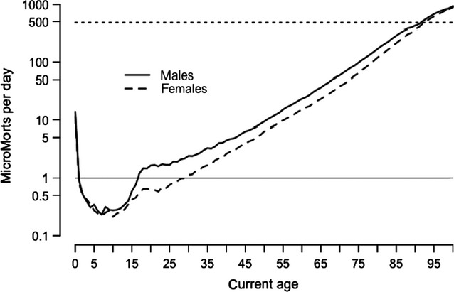
Simply being being alive carries a risk in itself: it is highest on the first day of lift at around 500 micromorts, drops rapidly in childhood and then increases steadily throughout adult life until the 90s, when each day is as risky as that person’s first day.
MicroMorts per day for average person in England and Wales—dashed line shows rate for first day of life. (Spiegelhalter, 2014)
Perhaps unsurprisingly, one UK estimate showed that travelling by motorbike carries the highest risk of death at 9.6km for each MicroMort. Travelling by car is far safer at 400km per MicroMort. But somewhat counterintuitively, walking is not much safer than travelling by motorbike: hitting the pavement results in one MicroMort per 27.2km walked. This can be partly explained by the greater amount of time spent walking and exposed to environmental hazards such as traffic, air pollution, crime and so on.
The concept of micromorts can also be applied to pregnancies and risks to the foetus. The risk per unit due to stillbirth is only about 19 or 3710 micromorts when drinking 5 units/week throughout pregnancy. For low risk women in the UK, planned first birth at home carries an additional 843 (-200 to 2620) micromorts compared with in hospital, and planned vaginal breech birth an additional 5870 (-4400 to 18 500), compared with planned caesarean. By contrast, the risk from the mother eating a serving of unpasteurised cheese, is negligible at 0.00026 micromorts (Hickson et al., 2020).
In a recent analysis published in Acta Obstetricia et Gynecologica Scandinavica, researchers found no link between women’s caffeine consumption and pregnancy or live birth rate after fertility treatments – but alcohol consumption was linked to decreased pregnancy rate after treatments with more than 84g of alcohol a week (approximately 7 standard drinks).
The link held true for their spouses as well: men’s alcohol consumption was associated with decreased live birth rate after fertility treatments in women when weekly consumption was greater than 84g.
The researchers searched the available literature and found a total of 7 studies on caffeine consumption and 9 studies on alcohol consumption were included, with a total of 26 922 women and/or their spouse who underwent fertility treatment.
Compared with those abstaining from alcohol, the chance of achieving a pregnancy after fertility treatment decreased by 7% for women who consumed 84g of alcohol per week, and the chance of partners achieving a live birth decreased by 9% for men who consumed 84g of alcohol per week.
“Couples should be aware that some modifiable lifestyle factors such as drinking habits may affect their fertility treatment outcomes. But how these factors impact the reproductive system still needs more research to elucidate,” said corresponding author Yufeng Li, MD, of Tongji Hospital, in China.
In recent years, researchers have made strides in promoting tissue regeneration in spinal cord injuries (SCI) through implanted neural stem cells or grafts in animal models. Separate efforts have shown that intensive physical rehabilitation can improve function after SCI by promoting greater or new roles for undamaged cells and neural circuits.
University of California San Diego researchers tested whether rehabilitation can pair with pro-regenerative therapies, such as stem cell grafting. They published their findings in in JCI Insight,
The researchers induced a cervical lesion in rats that impaired the animals’ ability to grasp with its forelimbs. The animals were divided into four groups: animals who underwent the lesion alone; animals who received a subsequent grafting of neural stem cells designed to grow and connect with existing nerves; animals who received rehabilitation only; and animals who received both stem cell therapy and rehabilitation.
Rehabilitation therapy for some animals began one month after initial injury, a time point that approximates when most human patients are admitted to SCI rehabilitation centers. Rehabilitation consisted of daily activities that rewarded them with food pellets if they performed grasping skills.
The researchers found that rehabilitation enhanced regeneration of injured corticospinal axons at the lesion site in rats, and that a combination of rehabilitation and grafting produced significant recovery in forelimb grasping when both treatments occurred one month after injury.
“These new findings indicate that rehabilitation plays a critically important role in amplifying functional recovery when combined with a pro-regenerative therapy, such as a neural stem cell transplant,” said first author Paul Lu, PhD, associate adjunct professor of neuroscience at UC San Diego School of Medicine and research health science specialist at the Veterans Administration San Diego Healthcare System.
“Indeed, we found a surprisingly potent benefit of intensive physical rehabilitation when administered as a daily regimen that substantially exceeds what humans are now provided after SCI.”
Senior author Mark H. Tuszynski, MD, PhD, professor of neurosciences and director of the Translational Neuroscience Institute at UC San Diego School of Medicine, and colleagues have long worked to address the complex challenges of repairing SCIs and restoring function.
In 2020, for example, they reported on the observed benefits of neural stem cell grafts in mice and in 2019, described 3D-printed implantable scaffolding that would promote nerve cell growth.
“There is a great unmet need to improve regenerative therapies after SCI,” said Tuszynski. “We hope that our findings point the way to a new potential combination treatment consisting of neural stem cell grafts plus rehabilitation, a strategy that we hope to move to human clinical trials over the next two years.”
The largest ever randomised controlled trial of intensive blood pressure lowering after thrombectomy in ischaemic stroke patients found that it led to deterioration in surrounding brain tissue and higher rates of disability, compared to less intensive treatment.
The results of the ENCHANTED2/MT trial were presented in a late-breaking session at the World Stroke Congress and simultaneously published in The Lancet. The trial was stopped early due to the significance of the findings.
Professor Craig Anderson, Director of Global Brain Health at The George Institute for Global Health, said the rapid emergence of this effect suggested the more aggressive approach was compromising the return of blood flow to the affected area.
“Our study provides a strong indication that this increasingly common treatment strategy should now be avoided in clinical practice,” he said.
Endovascular thrombectomy is an increasingly used non-surgical treatment for ischaemic stroke, in which x-ray guided microcatheters are inserted into the blood clot to dissolve it.
“A potential downside of this now widely used and effective treatment is that the rapid return of blood supply to an area that has been deprived of oxygen for a while can cause tissue damage known as reperfusion injury,” said Professor Anderson.
“This has resulted in a shift in medical practice towards more intensive lowering of blood pressure after clot removal to try and minimise this damage, but without evidence to support the benefits versus potential harms.”
To this end, researchers recruited 816 adults with acute ischaemic stroke who had elevated blood pressure after clot removal from 44 centres in China between July 2020 and March 2022. They had an average age of 67 and just over a third were female.
Of these, 407 were assigned to more-intensive (target < 120mmHg) and 409 to the less-intensive (target 140–180mmHg) systolic blood pressure control, with the target to be achieved within one hour of entering the study and sustained for 72 hours.
Researchers looked at how well the patients in both groups recovered according to a standard measure of disability, ranging from 0–1 for a good outcome without or with symptoms but no disability, scores of 2–5 reflecting increasing disability levels, and 6 being death.
Patients in the more-intensively treated group had significantly worse scores on the scale compared to those allocated to those treated less intensively.
Compared to the less-intensive group, they had more early brain tissue deterioration and major disability at 90 days but there were no significant differences in brain bleeds, mortality, or serious adverse events.
Patients who had their blood pressure more intensively controlled also rated their quality of life as significantly worse due to limitations on their physical abilities resulting from their stroke.
Prof Anderson said that after scouring the medical literature the research team had been unable to find strong enough evidence to recommend the ideal target for blood pressure control after blood clot removal in patients with acute ischaemic stroke.
“While our study has now shown intensive blood pressure control to a systolic target of less than 120mmHg to be harmful, the optimal level of control is yet to be defined,” he said.
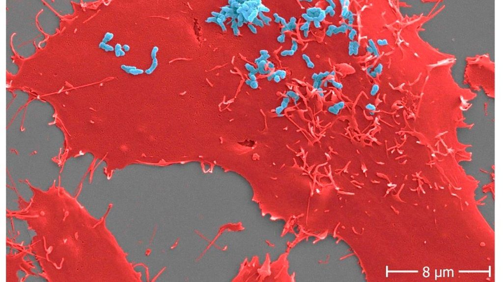
Adhesion of Bartonella henselae to human cells. B. henselae (strain Marseille) bacteria (light blue) in an early stage infection process (30 min) to human HeLa-229 cells (red). Adhesion to host cells is mediated by specific interactions between B. henselae surface proteins and components of the host extracellular matrix including molecules such as fibronectin or collagen. Scale bar: 8 μm.
Using Bartonella henselae bacteria, the cause of cat scratch disease, researchers have demonstrated for the first time that antibodies can prevent certain surface proteins of bacterial pathogens from entering host cells. The findings are important for the development of new drugs against highly resistant infectious agents.
Infections pose a significant threat to human health, especially when pathogens manage to colonise the organism and subsequently cause severe infections. The first step in such an infection always consists of the pathogens attaching themselves to the host cells’ surface. From here, the infections spread, resulting, for example, in infections of deeper tissue layers and organs.
A group of scientists surrounding Prof. Volkhard Kempf from Frankfurt University Hospital’s Institute of Microbiology and Hospital Hygiene has now succeeded in blocking this adhesion mechanism in a bacterium, thereby preventing the infection of host cells. For this purpose, the researchers examined the pathogen Bartonella henselae, usually causing cat scratch disease. Transmitted by cats, the disease mainly affects young children, whose symptoms include swollen and hardened lymph nodes around the site of infection, usually after a scratch or bite injury caused by infected cats.
Bartonella bacteria infect so-called endothelial cells, which line the blood vessels. Via their surface protein Bartonella adhesin A (BadA), they attach themselves to a protein (fibronectin) of the so-called “extracellular matrix,” a network of protein fibers that lie on top of the endothelial cells.
Breaking BadA
To determine which parts of the BadA protein are important in the bacterial adhesion process, the researchers equipped Bartonella bacteria with various genetically modified BadA variants, among others, and then analysed the extent to which these variants were still able to bind fibronectin. Once it was clear which BadA segments were responsible for the binding, the team produced antibodies against them, demonstrating for the first time that such antibodies can prevent infection by such bacteria.
Prof. Volkhard Kempf explains: “Bartonella henselae is not a very dangerous pathogen, and in most cases, cat scratch disease does not require any specific medical treatment. However, for us Bartonella henselae is a very important model organism for far more dangerous pathogens such as Acinetobacter baumannii, a serious pathogen that usually causes wound infection or pneumonia and often shows resistance to several last-choice antibiotics. The BadA protein of Bartonella henselae belongs to the so-called ‘trimeric autotransporter adhesins’, which are also responsible for adhesion to human cells in Acinetobacter and a number of other pathogens. A drug-induced blocking of these adhesins is therefore a promising novel and future approach to combat dangerous bacterial infections.”
The researchers published their findings in Diagnostics.
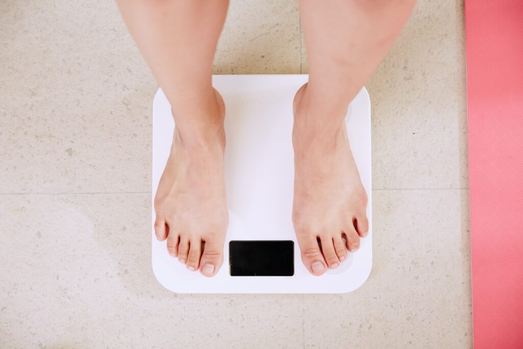
Intermittent fasting has been shown to be an effective way to lose weight, but critics have worried that the practice may have a negative impact on women’s reproductive hormones. Now, researchers bring new evidence to the tablein a study published in Obesity.
The researchers, led by Krista Varady, University of Illinois Chicago professor of nutrition, followed a group of pre- and post-menopausal obese women for a period of eight weeks on the ‘warrior diet’ method of intermittent fasting.
The warrior diet prescribes a time-restricted feeding window of four hours per day, during which dieters can eat without counting calories before resuming a water fast until the next day.
They measured the differences in hormone levels, obtained by analysing blood sample data, in groups of dieters who stuck to four- and six-hour feeding windows against a control group that followed no diet restrictions.
Varady and her team found that levels of sex-binding globulin hormone, a protein that carries reproductive hormones throughout the body, was unchanged in the dieters after eight weeks. The same held true for both testosterone and androstenedione, a steroid hormone that the body uses to produce both testosterone and oestrogen.
However, dehydroepiandrosterone or DHEA, a hormone that fertility clinics prescribe to improve ovarian function and egg quality, was significantly lower in both pre-menopausal and post-menopausal women at the end of the trial, dropping by about 14%.
While the drop in DHEA levels was the most significant finding of the study, in both pre- and post-menopausal women, DHEA levels remained within the normal range by the end of the eight-week period.
“This suggests that in pre-menopausal women, the minor drop in DHEA levels has to be weighed against the proven fertility benefits of lower body mass,” Varady said. “The drop in DHEA levels in post-menopausal women could be concerning because menopause already causes a dramatic drop in estrogen, and DHEA is a primary component of estrogen. However, a survey of the participants reported no negative side effects associated with low estrogen post-menopause, such as sexual dysfunction or skin changes.”
As an added benefit, since high DHEA has been linked to breast cancer risk, Varady said a moderate drop in levels might be helpful in reducing that risk for both pre- and post-menopausal women.
The study measured levels of oestradiol, oestrone and progesterone as well, but only in post-menopausal women, due to the changing levels of these hormones throughout pre-menopausal women’s menstrual cycles. Among post-menopausal women, there was no change in these hormones at the end of eight weeks.
Women in both the four-hour and six-hour dieting groups experienced weight loss of 3% to 4% of their baseline weight throughout the course of the study, compared with the control group, which had almost no weight loss. The dieters also saw a drop in insulin resistance and in biomarkers of oxidative stress.
Perimenopausal women, who are typically in their 40s, were excluded from the study.
Still, Varady said, “I think this is a great first step. We’ve observed thousands of pre- and post-menopausal women through different alternate-day fasting and time-restricted eating strategies. All it’s doing is making people eat less. By shortening that eating window, you’re just naturally cutting calories. Much of the negative information on intermittent fasting reported has come from studies on mice or rats. We need more studies to look at the effects of intermittent fasting on humans.”
The award was based on strides made by ASASA towards improving the quality of life of people living with AxSpa, as well as training done to build awareness in the medical fraternity around AxSpA in the country. With 36 posters entered into the awards by organisations across the globe, ASASA came out tops.
When asked about the award, van Dam said, “This was a real honour to represent South Africa at PARE. 2022 is also the first year that an African country was invited to attend PARE. Winning this award sheds light on our country and our unique problems. The delay to diagnosis of 10.8 years is just unacceptable. The access to the correct medication in both the private and public sector is also not sufficient for a debilitating, progressive disease that can lead to disability if left untreated.”
ASASA estimates that there are approximately 160 000 people suffering from the AxSpA in South Africa, with many of these sufferers undiagnosed. ASASA has made significant strides this year in the training of over 100 General Practitioners and over 250 optometrists around AxSpA diagnosis and the effects it can have on other parts of the body, like the eyes. In addition, ASASA, along with other partners, assisted in gathering data from South African respondents in the first ever live patient survey, called the International Map of Axial Spondyloarthritis (IMAS) survey, which is run by the Axial Spondyloarthritis International Federation that surveys people diagnosed with AxSpA and assesses the impact and burden that AxSpA has on the lives of patients, from their perspective.
Van Dam concluded, “There is still a lot we can do in South Africa and ASASA is busy growing its team of volunteers to help to build awareness around AxSpA in the country. We aim to continue to build support structures for patients in the country, as well as continually working with the medical fraternity, assisting with early diagnosis and access to treatment.”