The trace metal selenium could help reverse the cognitive impact of stroke and boost learning and memory in ageing brains, according to a study published in Cell Metabolism.
Previous studies on the impact of exercise on the ageing brain found levels of a protein key to transporting selenium in the blood were elevated by physical activity.
Lead researcher Dr Tara Walker said: “We’ve known for the last 20 years that exercise can create new neurons in the brain, but we didn’t really understand how,” Dr Walker said.
The research team sought to find out whether dietary selenium supplements could replicate the effects of exercise.
“Our models showed that selenium supplementation could increase neuron generation and improve cognition in elderly mice,” Dr Walker said. “The levels of new neuron generation decrease rapidly in aged mice, as they do in humans. When selenium supplements were given to the mice, the production of neurons increased, reversing the cognitive deficits observed in ageing.”
Selenium is an essential trace metal which can play an important role in human health. It is absorbed from soil and water and is found in foods such as grains, meat and nuts, with the highest levels found in Brazil nuts. The researchers also investigated whether selenium would have an impact on post-stroke cognitive decline.
“Young mice are really good at the learning and memory tasks, but after a stroke, they could no longer perform these tasks,” Dr Walker said. “We found that learning and memory deficits of stroke affected mice returned to normal when they were given selenium supplements.”
Dr Walker said the results opened a new therapeutic avenue to boost cognitive function in people who were unable to exercise due to poor health or old age.
“However, selenium supplements shouldn’t be seen as a complete substitute for exercise, and too much can be bad for you,” she said. “A person who is getting a balanced diet of fruits, nuts, veggies and meat usually has good selenium levels. But in older people, particularly those with neurological conditions, selenium supplements could be beneficial.”
Having a spinal cord injury increases risk of developing mental health conditions such as depression and anxiety by nearly 80% compared to those without the traumatic injury, a new study shows. However, chronic pain may have an equally large, negative effect on mental health.
The study, published in Spinal Cord, compared private insurance claims from more than 9000 adults with a traumatic spinal cord injury with those of more than 1 million without. Researchers accounted for a range of psychological conditions, from anxiety and mood disorders to insomnia and dementia.
People living with a spinal cord injury had a diagnosis of a mental health condition more often than those without – 59.1% versus 30.9%. While depression and adverse mental health effects are not inevitable consequences of every traumatic spinal injury, previous studies have consistently echoed higher levels of psychological morbidity among this group than the general population without spinal cord injuries.
However, this study found that chronic centralised and neuropathic pain among adults living with a spinal cord injury were robustly associated with post-traumatic stress disorder, substance use disorders and other mental health conditions. In most cases, chronic pain was an even greater influence on these conditions than exposure to living with the injury itself.
The study authors said the findings should prompt physicians to identify mental health conditions when seeing patients with spinal cord injuries and refer them for treatment.
“Improved clinical efforts are needed to facilitate screening of, and early treatment for, both chronic pain and psychological health in this higher-risk population,” said lead author Dr Mark Peterson, associate professor of physical medicine and rehabilitation at Michigan Medicine.
However, researchers note a lack of insurance coverage and limited available services will likely cause the issue to remain largely unaddressed.
“Stakeholders need to work together to lobby for more federal research funding and special policy amendments to ensure adequate and long-term insurance coverage for both physical and mental health to meet the needs of folks living with spinal cord injuries,” Dr Peterson said.
Getting adequate sleep could be key to fighting growing rates of obesity around the world, according to a study published inJAMA Internal Medicine, which focused solely on improving sleep duration in overweight individuals.
Understanding the underlying causes of obesity and how to prevent it is the best way to fight obesity, according to first author Dr Esra Tasali. “The current obesity epidemic, according to experts, is mostly explained by an increase in caloric intake, rather than lack of exercise,” she said.
In a randomised clinical trial with 80 adults, published in JAMA Internal Medicine, researchers found that young, overweight adults who habitually slept fewer than 6.5 hours a night were able to sleep for 1.2 hours longer after a personalised sleep hygiene counselling session. The sleep intervention was intended to extend time in bed duration to 8.5 hours and, compared to controls, the increased sleep duration also reduced participants’ overall caloric intake by an average of 270 kcal (calories) per day.
“Over the years, we and others have shown that sleep restriction has an effect on appetite regulation that leads to increased food intake, and thus puts you at risk for weight gain over time,” said Tasali. “More recently, the question that everyone was asking was, ‘Well, if this is what happens with sleep loss, can we extend sleep and reverse some of these adverse outcomes?”
The study examines the effects of sleep extension on caloric intake but also does so in a real-world setting, with no influence on participants’ diets. Participants slept in their own beds, tracked their sleep with wearable devices, and otherwise followed their normal lifestyle without any instructions on diet or exercise.
“Most other studies on this topic in labs are short-lived, for a couple of days, and food intake is measured by how much participants consume from an offered diet,” said Tasali. “In our study, we only manipulated sleep, and had the participants eat whatever they wanted, with no food logging or anything else to track their nutrition by themselves.”
Instead, to objectively track participants’ caloric intake, investigators relied on the “doubly labelled water” method to track change in energy stores, which uses isotopes of hydrogen and oxygen in drinking water. “This is considered the gold standard for objectively measuring daily energy expenditure in a non-laboratory, real-world setting and it has changed the way human obesity is studied,” said Professor Dale A. Schoeller, senior study author and pioneer of the method.
Overall, individuals who increased their sleep duration were able to reduce their caloric intake by an average of 270 kcal per day – which would translate to roughly 12 kg of weight loss over three years if the effects were maintained over a long term.
Perhaps the most surprising aspect of the study was the intervention’s simplicity. “We saw that after just a single sleep counselling session, participants could change their bedtime habits enough to lead to an increase in sleep duration,” said Dr Tasali. “We simply coached each individual on good sleep hygiene, and discussed their own personal sleep environments, providing tailored advice on changes they could make to improve their sleep duration. Importantly, to blind participants to sleep intervention, recruitment materials did not mention sleep intervention, allowing us to capture true habitual sleep patterns at baseline.”
Even though the study did not systematically assess factors that may have influenced sleep behaviour, “limiting the use of electronic devices before bedtime appeared as a key intervention,” said Dr Tasali.
Following just a single counselling session, participants increased their average sleep duration by over an hour a night. Despite prescribing no other lifestyle changes, most participants had a large decrease in how much they ate, with some participants’ intake reduced by 500kcal per day.
The subjects were only involved in the study for a total of four weeks, with two weeks for gathering baseline information about sleep and caloric intake, followed by two weeks to monitor the effects of the sleep intervention.
“This was not a weight-loss study,” said Dr Tasali. “But even within just two weeks, we have quantified evidence showing a decrease in caloric intake and a negative energy balance – caloric intake is less than calories burned. If healthy sleep habits are maintained over longer duration, this would lead to clinically important weight loss over time. Many people are working hard to find ways to decrease their caloric intake to lose weight – well, just by sleeping more, you may be able to reduce it substantially.”
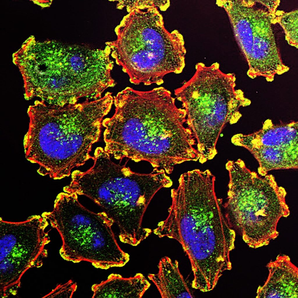
Melanoma cells. Source: National Cancer Institute.
Researchers have identified a protein which explains why some tumours metastasise, contributing to a better understanding of the process in certain types of cancer.
In a study published in Frontiers in Oncology, researchers focused on MFSD1 – the mammalian relative of a protein they had previously identified as affecting cell migration in fruit flies. created mouse cancer cells lacking the protein. Without the protein, cells travelled much faster, suggesting that MFSD1 prevents the cells from moving. The team tested their theory in living mice with breast, colon, and skin cancer. “In the absence of MFSD1, there was a strong increase in metastasis,” said lead researcher Professor Daria Siekhaus.
“We wanted to know why lower MFSD1 levels were beneficial to the tumour apart from allowing them to move more freely. As cancer cells travel through the blood for example, they experience a lot of mechanical stress,” explained first author Marko Roblek.
So the researchers performed a stress test on cancer cells with and without the protein. Using a tiny rubber scraper, Roblek tried to scrape the cells off the surface of the Petri dish in which he had grown them. While the cancer cells with MFSD1 quickly died under the mechanical stress, those without the protein tended to remain intact. Without the protein, the team concluded, certain tumour cells could more easily enter the bloodstream and find their way to other parts of the body. In another experiment, the researcher tested the cancer cell’s resistance to nutrient starvation with a similar result. Again, the cells lacking MFSD1 survived for longer.
The protein MFSD1 appeared to cause the cells’ reaction to starvation and mechanical stress by affecting specific receptors located at the cell surface. These receptors, known as integrins, ensure the cells stick to each other and the extracellular matrix. The cell produces these receptors, transports them to the cell surface and back inside the cell. If a tumour cell lacks MFSD1, they fail to recycle a certain type of integrin. “The result is that the cells stick less to the surrounding tissue and each other, which makes it easier for them to migrate,” said Prof Siekhaus.
Anonymised patient data supported this, showing a correlation between the level of MFSD1 and the patient’s prognosis. “We’ve seen that patients suffering from specific forms of breast, gastric and lung cancer who had lower levels of MFSD1 had a worse outcome. A high level of MFSD1 seems to be protective – it works like a suppressor of tumor metastasis,” said cancer researcher Roblek.
To optimise therapy for their patients, doctors are already analysing the expression of certain genes. Now, they can also look for the gene encoding the protein MFSD1. “If this marker becomes more established, doctors can use it to help classify how aggressive the cancer is and to decide between different treatment options,” suggested Prof Siekhaus.
In future studies, the team wants to focus in detail on how the protein functions on a molecular level and is curious to learn if artificially raising the amount of MFSD1 could help suppress the spread of certain tumours. The long term goal is to examine if it can be used as a therapeutic target.
Long-term paracetamol use could increase the risk of heart disease and strokes in people with high blood pressure, according to a randomised clinical trial by the University of Edinburgh.
Researchers recommend that patients with a long term prescription, usually for chronic pain, should rather choose the lowest effective dose for the shortest possible time.
The study, which appears in Circulation, is the first large randomised clinical trial to address the question of paracetamol’s effect on cardiovascular disease, and complements earlier work in observational studies.
Paracetamol was often suggested as a safer alternative to non-steroidal anti-inflammatory drugs (NSAIDs), which are known to increase blood pressure and risk of heart disease.
In the latest study, 110 patients with a history of high blood pressure were prescribed one gram of paracetamol four times a day – a routinely prescribed dose in patients with chronic pain – or a matched placebo for two weeks. All patients received both treatments, with the order randomised and blinded. The paracetamol group saw a significant increase in blood pressure, compared to the placebo group.
This rise was similar to that seen with NSAIDs, and could be expected to increase the risk of heart disease or stroke by around 20%. The findings should lead to a review of long-term paracetamol prescriptions to patients, said the researchers, especially to those with hypertension and an increased risk of heart disease or stroke.
Lead Investigator Dr. Iain MacIntyre said: “This is not about short-term use of paracetamol for headaches or fever, which is, of course, fine—but it does indicate a newly discovered risk for people who take it regularly over the longer term, usually for chronic pain.”
Principal Investigator Professor David Webb said: “We would recommend that clinicians start with a low dose of paracetamol, and increase the dose in stages, going no higher than needed to control pain. Given the substantial rises in blood pressure seen in some of our patients, there may be a benefit for clinicians to keep a closer eye on blood pressure in people with high blood pressure who newly start paracetamol for chronic pain.”
Professor Sir Nilesh Samani, Medical Director at the British Heart Foundation, who funded the study, said: “This research shows how quickly regular use of paracetamol can increase blood pressure in people with hypertension who are already at increased risk of heart attacks and strokes. It emphasises why doctors and patients should regularly review whether there is an ongoing need to take any medication, even something that may seem relatively harmless like paracetamol, and always weigh up the benefits and risks. However, if you take paracetamol occasionally to manage an isolated headache or very short bouts of pain, these research findings should not cause unnecessary concern.”
Luallen Lab members pose in their lab at SDSU. Inset: Microscope image of Bordetella atropi (pink lines) infecting roundworm intestine (green). Credit: San Diego State University
Like something out of a horror movie, a new way that one type of bacteria invades tissue within a living organism has been identified by biologists from San Diego State University.
The study, published in Nature Communications, describes how a new species of bacteria, Bordetella atropi, invades its roundworm host. The name which comes from the Greek fate Atropos responsible for cutting the threads of life, is apt because the bacteria transforms into a long thread, growing up to 100 times the usual size of one bacterium in the span of 30 hours without dividing.
By altering the genes of B. atropi, the research team discovered that this invasive threading relies on the same genes and molecules that other bacteria use when they are in a nutrient-rich environment. However, these other bacteria only use this pathway to make subtly larger cells, whereas the B. atropi bacteria grows continuously.
Other bacteria often transform into threads, called filamentation, in response to dangerous environments or DNA damage. This lets them continue to grow in size, but delay cell division until they repair the damage inflicted by the stress.
Here, however, the researchers were the first to observe filamentation as a way of spreading from cell to cell in a living organism for a purpose other than the stress response. They believe that instead the new species is invading the host cells, detecting this rich environment and triggering filamentation in order to quickly infect more cells and access additional nutrients for their growth.
“We went from finding the worm in the ground, finding the bacteria, and carrying it all the way to the molecular mechanism of how the bacteria infects the worm,” explained Robert Luallen, biology professor and principal investigator of the study. “We’re seeing things that no one’s ever seen before.”
Though B. atropi does not infect humans, it is possible that human pathogens may also make use of its spreading mechanism. Separately, the nutrient-induced filamentation process might be used by other bacteria to form biofilms, which can coat the tubing of catheters and lead to complications for patients.
In a study published in Influenza and Other Respiratory Viruses, researchers were able to detect SARS-CoV-2 viral RNA in droplets from the exhaled breaths and coughs of COVID patients.
COVID is assumed to be transmitted mainly by respiratory droplets. However, probable aerosol transmission has been reported to occur under certain conditions. The researchers sought to address the lack of information on viral load in exhaled breath samples,as well as the size and concentration of exhaled endogenously generated droplets in relation to viral load. Additionally, the relationship between the viral load in upper airway diagnostic samples and aerosol samples needed to diagnose. For the study, researchers analysed exhalations by two different methods during 20 normal breaths, 10 airway opening breaths (which involves deep inhalation followed by relaxed exhalation), and 3 coughs.
PCR detection of SARS-CoV-2 RNA in aerosols was possible in 10 out of 25 participants. Viral RNA presence in aerosol was mainly detected in cough samples (8 samples), but also in normal breaths (4 samples) and in airway opening breaths (3 samples).
“Our data confirm findings from other researchers that SARS-CoV-2 can be detected in aerosol particles < 5µm and highlight the small amount of exhaled aerosol needed for detection. Of specific interest were findings from the airway opening maneuver, which is thought to generate particles mainly from the small airways,” said lead author Emilia Viklund, PhD student at the University of Gothenburg, in Sweden. “COVID causes a lot of damage in this region, and it would be of great interest to further explore the amount of exhaled virus and the course of disease, as well as the infectious potential of exhaled virus.”
A study into the genetics of acne revealed 29 regions of the genome that underpin the condition, which could offer potential new treatment targets and may also help clinicians identify individuals at high risk of severe disease.
A common skin condition, acne is estimated to affect 80% of adolescents, with common features including spots and cysts, pigment changes and scarring. The face is the most common site, with the chest and back also frequently involved. The negative psychological consequences of acne are seen in all ages, but are of particular concern for many adolescents.
The research, published in Nature Communications, analysed nine genome wide association study datasets from patients around the world. These studies involved scanning the whole genomes of 20 165 people with acne and 595 231 without. The study identified 29 new genetic variants that are more common in people with acne. It also confirmed 14 of the 17 variants already known to be associated with the condition, which brings the total number of known variants to 46.
Professor Catherine Smith at St John’s Institute of Dermatology at Guy’s and St Thomas’ said: “Despite major treatment advances in other skin conditions, progress in acne has been limited. As well as suffering from the symptoms of acne, individuals describe consequent profound, negative impacts on their psychological and social wellbeing. It’s exciting that this work opens up potential avenues to find treatments for them.”
A number of genes associated with acne were identified, and are also linked to other skin and hair conditions. The team believe this will help to understand the causes of acne, which could be a mix of factors.
“We know that the causes of acne are complicated, with a mix of biological factors such as genetics and hormones, and environmental factors,” said Professor Michael Simpson at King’s College London. “Understanding the genetics of the condition will help us to disentangle some of these causes, and find the best way to treat the condition. This is a really promising area for further study, and opens up a lot of avenues for research.”
The study also uncovered a link between the genetic risk of acne and disease severity. Individuals with the highest genetic risk are more likely to have severe disease. While further research is required, this finding raises the potential to identify individuals at risk of severe disease for early intervention.
In a world first, Michel Roccati, a man with a completely severed spinal cord was able to walk again outside the lab with the help of a portable electrical stimulation system that causes his legs to take a step in conjunction with his intention to move and a walker to steady him.
This was a further development of a technology that in 2018 helped David M’zee, who had been left paralysed by a partial spinal cord injury suffered in a sports accident, to walk again. A research team led by Professors Grégoire Courtine, at École Polytechnique Fédérale de Lausanne (EPFL) and Jocelyne Bloch, at Lausanne University Hospital (CHUV) had developed an electrical stimulation system to help people with spinal cord injuries walk again.
They had wanted to see if electrodes could stimulate movement in the parts of the spine damaged so badly that signals no longer reach the nervous system from the brain. That pioneering study was detailed in Nature and Nature Neuroscience. Thanks to the electrodes making up for the weakness of the signals in his damaged spinal cord, M’zee was able to voluntarily move his legs and could walk several hundred metres at a time, sometimes without the aid of the rails on the treadmill.
Now a new milestone has just been reached with the technology, and the research team enhanced their system with more sophisticated implants controlled by advanced software. These implants can stimulate the region of the spinal cord that activates the trunk and leg muscles. Thanks to this new technology, three patients with complete spinal cord injury were able to walk again outside the lab. “Our stimulation algorithms are still based on imitating nature,” said Prof Courtine. “And our new, soft implanted leads are designed to be placed underneath the vertebrae, directly on the spinal cord. They can modulate the neurons regulating specific muscle groups. By controlling these implants, we can activate the spinal cord like the brain would do naturally to have the patient stand, walk, swim or ride a bike, for example.”
On a cold, snowy day last December, Michel Roccati – an Italian man who became paralysed after a motorcycle accident four years earlier – braved the icy wind to try out the system outdoors, in central Lausanne. He had recently undergone the surgical procedure in which Prof Bloch placed the new, implanted lead on his spinal cord.
Scientists attached two small remote controls to Michel’s walker and connected them wirelessly to a tablet that forwards the signals to a pacemaker in Michel’s abdomen. The pacemaker in turn relays the signals to the implanted spinal lead that stimulates specific neurons, causing Michel to move. Grasping the walker, Michel pressed a button corresponding to either the left or right leg with the firm intention of taking a step forward, and his feet rose and fell in short steps.
“The first few steps were incredible – a dream come true!” he says. “I’ve been through some pretty intense training in the past few months, and I’ve set myself a series of goals. For instance, I can now go up and down stairs, and I hope to be able to walk one kilometre by this spring.”
Two other patients have also successfully tested the new system, which is described in Nature Medicine. “Our breakthrough here is the longer, wider implanted leads with electrodes arranged in a way that corresponds exactly to the spinal nerve roots,” said Bloch. “That gives us precise control over the neurons regulating specific muscles.” Ultimately, it allows for greater selectivity and accuracy in controlling the motor sequences for a given activity.
While extensive training is necessary for patients to get comfortable using the device, the speed and scope of rehabilitation is amazing. “All three patients were able to stand, walk, pedal, swim and control their torso movements in just one day, after their implants were activated!” said Prof Courtine. “That’s thanks to the specific stimulation programs we wrote for each type of activity. Patients can select the desired activity on the tablet, and the corresponding protocols are relayed to the pacemaker in the abdomen.”
While the progress achievable in a single day is astonishing, the gains after several months are even more impressive. The three patients followed a training regimen based on the stimulation programs and were able to regain muscle mass, move around more independently, and take part in social activities like having a drink standing at a bar. What’s more, because the technology is miniaturized, the patients can perform their training exercises outdoors and not only inside a lab.
Presently there is one key limitation, Prof Bloch said: “We need at least six centimetres of healthy spinal cord under the lesion. That’s where we implant our electrodes.”
As for Roccati, after nine months of Lausanne-based rehab, he now lives independently in Italy. “I continued rehab at home, working alone, with all the devices,” he said. “And I see improvements every day.”
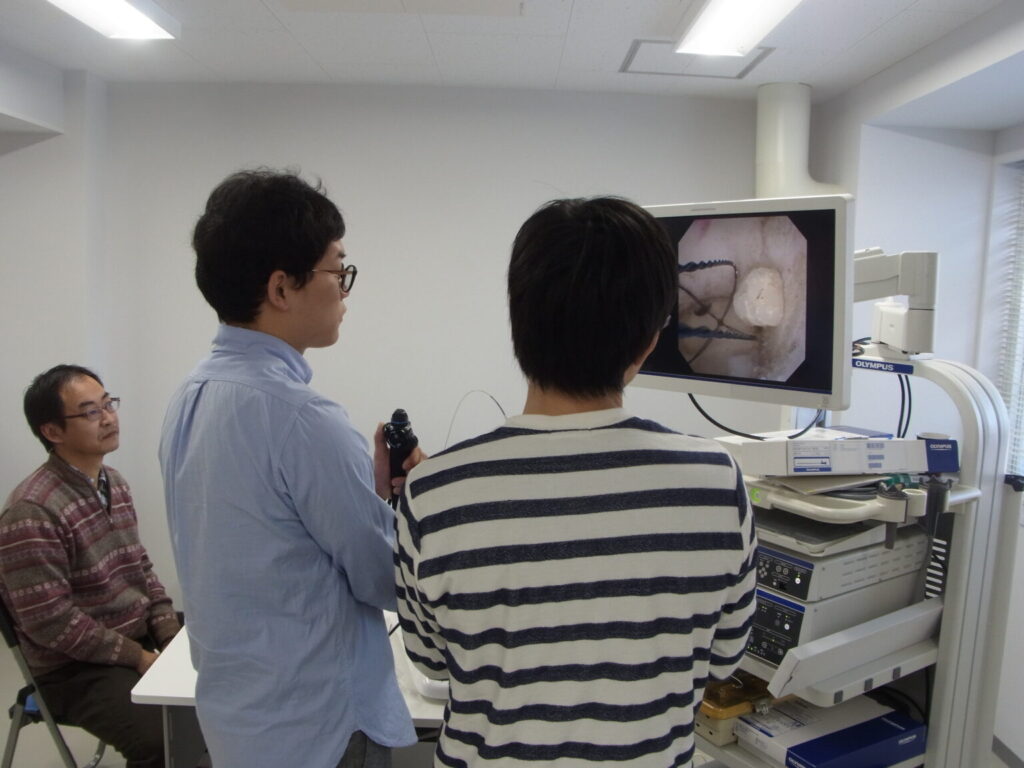
A surgical simulation unit used in the study. Credit: Takashige Abe.
An international trial found that while simulation-based training for ureteroscopy did not speed up surgeons general proficiency acquisition, it did increase skills in more complex surgeries, with fewer total complications and ureteric injuries. The results were published in the journal European Urology.
“To date, there have been limited data, mostly from small-scale studies conducted with medical students, assessing the transferability of surgical simulation,” said paper authors Takashige Abe, Associate Professor of Urology at Hokkaido University. The aim, he said, was to evaluate whether surgical residents undergoing additional simulation training can achieve proficiency sooner and with better patient outcomes, compared to usual operation room-based training.
The trial followed 65 participants in 10 countries for 18 months, or up to 25 procedures. A total of 32 participants received simulation-based training while 33 received conventional apprenticeship-style training. Both remained supervised by more experienced surgeons. Participants performed a total of 1140 surgeries, either semi-rigid or flexible ureteroscopy to remove ureteral or renal stones, respectively, demonstrating “mixed results” in proficiency.
“For our primary outcome measure, while we showed what might be deemed a clinically meaningful difference, it was not statistically significant,” Prof Abe said. “However, when stratified to each procedure type, there were higher rates of proficiency in the simulation-based training group when it came to the more technically challenging flexible ureteroscopy procedure.”
Prof Abe also noted that the simulation group scored higher on a standard assessment for each surgerythe other group.
“Simulation-based training led to higher overall proficiency scores than for conventional training, and fewer procedures were required to achieve proficiency in the complex form of the index procedure, with fewer serious complications overall,” Prof Abe said. “It is expected that the results of the trial will have a positive impact for advanced procedural training beyond the fields of surgery and urology in order to promote patients’ safety as well as better surgical outcomes.”