Month: November 2022

World First Trial of Lab-grown Red Blood Cells for Transfusion

https://www.pexels.com/photo/a-close-up-shot-of-bags-of-blood-4531306/
Photo by Charlie-Helen Robinson on Pexels

In a world first, researchers have launched a clinical trial of lab-grown red blood cells for transfusion into another person. These manufactured blood cells were grown from stem cells from donors, for transfusion into volunteers in the RESTORE randomised controlled clinical trial.

If our trial is successful, it will mean that patients who currently require regular long-term blood transfusions will need fewer transfusions in future, helping transform their care

Professor Cedric Ghevaert, chief investigator

If the technique is proven safe and effective, manufactured blood cells could in time revolutionise treatments for people with blood disorders such as sickle cell and rare blood types. It can be difficult to find enough well-matched donated blood for some people with these disorders.

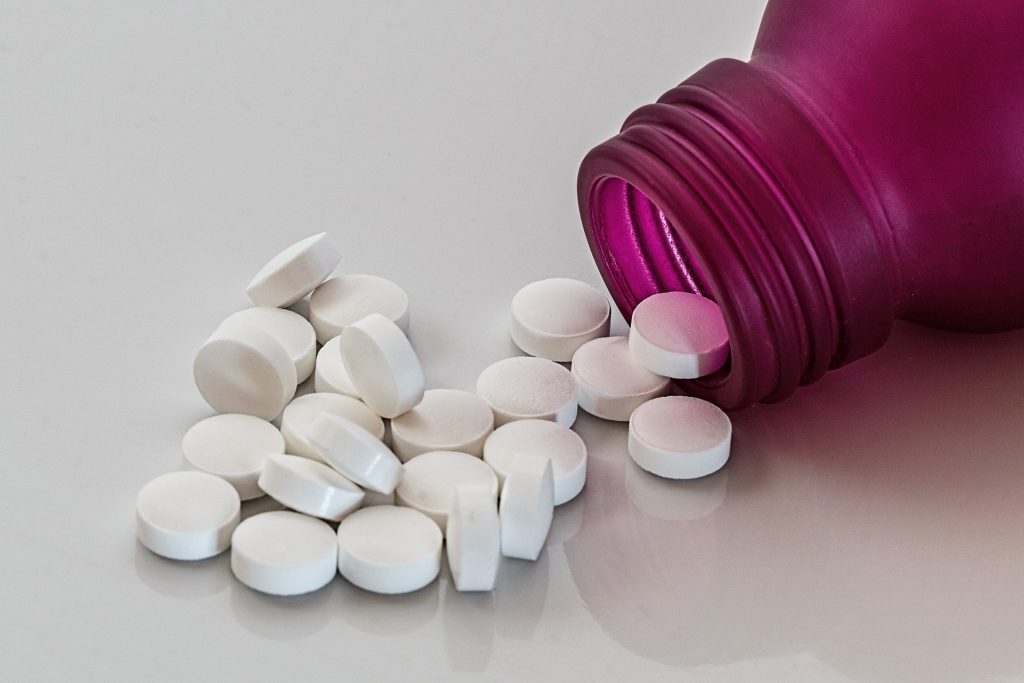
To produce the lab-grown blood cells, stem cells are first magnetically extracted from a normal 470ml blood donation. These stem cells are then coaxed into becoming red blood cells. Over the three week process, an initial pool of about half a million stem cells generates 50 billion red blood cells.

Chief Investigator Professor Cedric Ghevaert, Professor in Transfusion Medicine and Consultant Haematologist at the University of Cambridge and NHS Blood and Transplant, said: “We hope our lab grown red blood cells will last longer than those that come from blood donors. If our trial, the first such in the world, is successful, it will mean that patients who currently require regular long-term blood transfusions will need fewer transfusions in future, helping transform their care.”

Professor Ashley Toye, Professor of Cell Biology at the University of Bristol and Director of the NIHR Blood and Transplant Unit in red cell products, said: “This challenging and exciting trial is a huge stepping stone for manufacturing blood from stem cells. This is the first-time lab grown blood from an allogeneic donor has been transfused and we are excited to see how well the cells perform at the end of the clinical trial.”

The trial is studying the lifespan of the lab grown cells compared with infusions of standard red blood cells from the same donor. The lab-grown blood cells are all fresh, so the trial team expect them to perform better than a similar transfusion of standard donated red cells, which contains cells of varying ages.

Additionally, if manufactured cells last longer in the body, patients who regularly need blood may not need transfusions as often. That would reduce iron overload from frequent blood transfusions, which can lead to serious complications.

The trial is the first step towards making lab grown red blood cells available as a future clinical product. For the foreseeable future, manufactured cells could only be used for a very small number of patients with very complex transfusions needs. NHSBT continues to rely on the generosity of donors.

Co-Chief Investigator Dr Rebecca Cardigan, Head of Component Development NHS Blood and Transplant and Affiliated Lecturer at the University of Cambridge, said: “It’s really fantastic that we are now able to grow enough red cells to medical grade to allow this trial to commence. We are really looking forward to seeing the results and whether they perform better than standard red cells.”

Thus far, two people have been transfused with the lab grown red cells. They are well and healthy, and were closely monitored with no untoward side effects were reported. The amount of lab grown cells being infused varies but is around 5-10mls.

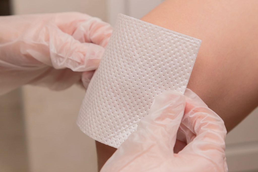
Donors were recruited from NHSBT’s blood donor base. They donated blood to the trial and stem cells were separated out from their blood. These stem cells were then grown to produce red blood cells in a laboratory at NHS Blood and Transplant’s Advanced Therapies Unit in Bristol. The recipients of the blood were recruited from healthy members of the NIHR BioResource.

A minimum of 10 participants will receive two mini transfusions at least four months apart, one of standard donated red cells and one of lab grown red cells, to see if the young lab-made red blood cells last longer than cells made in the body.

Further trials are needed before clinical use, but this research marks a significant step in using lab grown red blood cells to improve treatment for patients with rare blood types or people with complex transfusion needs.

Source: University of Cambridge

Stopping Prostate Tumours from Evading Androgen Suppression Therapy

Credit: Darryl Leja / National-Human-Genome Research Institute / National Institutes of Health

Researchers have identified an investigational therapeutic approach that could be effective against treatment-resistant prostate cancer. Results of the Phase II clinical trial performed by Cedars-Sinai Cancer investigators and published in Molecular Therapyhave led to a larger, multicentre trial that will soon be underway.

Cancer of the prostate is the second-leading cause of cancer-related death in men. Many prostate tumours are not aggressive and may require no or minimal treatment. Aggressive tumours are initially treated with surgery or radiation therapy.

In roughly a third of patients, the cancer comes back after initial treatment, said Neil Bhowmick, PhD, research scientist at Cedars-Sinai Cancer, professor of Medicine and Biomedical Sciences and senior author of the study. Those patients are usually treated with medications that suppress the actions of testosterone and other androgens, which promote prostate tumour growth.

“Patients do really well until the tumour figures a way around the androgen-suppressing therapy,” Bhowmick said. “One way that it can do this is to cause cells to make only part of the protein that the drug binds to, rendering the drug useless. The partial proteins are called splice variants.”

Through research with human cells and laboratory mice, study first author Bethany Smith, PhD, figured out that the cancer cells were using a protein called CD105 to signal to the surrounding supportive cells to make these slice variant proteins. Investigators then conducted a trial in human patients to test a drug that they hoped would keep those partial proteins from forming by inhibiting CD105.

In the trial, 9 patients whose tumours were resistant to androgen-blocking therapy continued that therapy but were also given a CD105 inhibitor called carotuximab. Forty percent of those patients experienced progression-free survival, based on radiographic imaging.

“Every single one of the patients in our trial was totally resistant to at least one androgen suppressor, and the normal course of action would be to simply try a different one or chemotherapy, which research has shown generally doesn’t stop tumor growth for more than about three months,” Bhowmick said. “Carotuximab prevented the cancer’s workaround and made the tumor sensitive to androgen-suppressing therapy.”

Importantly, Bhowmick said, carotuximab also appears to prevent androgen receptor splice variants in the supporting cells surrounding tumours, further sensitising the tumour to the androgen suppressor.

“We found that this therapy may be able to, especially in early cancers, resensitize select patients to androgen suppression. This could allow patients to avoid or delay more toxic interventions such as cytotoxic chemotherapy,” said Edwin Posadas, MD, associate professor of Medicine at Cedars-Sinai and a co-author of the study. “We also hope to find ways of predicting which patients are most likely to benefit from this approach by testing blood and tissue samples using next-generation technologies housed at Cedars-Sinai Cancer.”

Study co-author Sungyong You, PhD, director of the Urologic Oncology Bioinformatics Group, pinpointed three biomarkers that could help indicate which patients will respond to this investigational therapy, and the team will validate those markers in a new clinical trial. This will allow future studies to target patients most likely to be helped by this intervention, Bhowmick said.

Source: Cedars-Sinai Medical Center

Modern Ventilators Shown to Overstretch Lung Tissue

Source: Pixabay CC0

In pulmonary medicine, it has long been debated as to whether ventilator overstretches lung tissue, and now new research published in the American Journal of Respiratory and Critical Care Medicine has proven that they do in fact cause overstretching.

The University of California Riverside researchers showed that there were major differences between natural breathing versus the forced breathing from ventilators. These results are critical, particularly in context of the COVID pandemic and the rush to build ventilators.

“Using novel techniques, we observed that ventilators can overextend certain regions of the lungs,” said Mona Eskandari, assistant professor of mechanical engineering, who led the research. These results may explain why lung health declines for patients the longer they spend on the machines, especially in the case of disease.

Eskandari’s bMECH lab pioneered a technique to study lungs as they are made to breathe. On a custom-built ventilator designed in their lab, the researchers imitated both natural and artificial breathing. Then, they observed isolated lungs involved in both types of breathing using multiple cameras collecting fast, high-resolution images, a method called digital image correlation.

“Our setup allows us to imitate both physiological and artificial breathing on the same lung with the switch of a button,” Eskandari said. “The unique combination of our ventilator with digital image correlation gives us unprecedented insights into the way specific regions of the lungs work in concert with the whole.”

Using their innovative method to interface these two systems, UCR researchers collected evidence demonstrating that natural breathing stretches certain parts of the lung as little as 25% while those same regions stretch to as much as 60% when on a ventilator.

Scholars traditionally model the lungs like balloons, or what they refer to as thin-walled pressure vessels, where pushing air in and pulling air out are understood to be mechanically equivalent.

To explain what they observed in this study, the researchers propose moving away from thin-walled pressure vessel models and instead towards thick-walled models. Unlike thin-walled pressure vessels theory, a thick-walled model accounts for the differing levels of stress in airways resulting from ventilators pushing air in versus natural breathing, which pulls air in. This helps to explain how airways are more engaged and air is more evenly distributed in the lung during physiological breathing.

Iron lungs, the gigantic ventilators used during the late 1940s polio outbreak, acted more like a human chest cavity, expanding the lung as it naturally would. This creates a vacuum effect that pulls air into the lungs. Though this action is gentler for the lungs, these bulky systems prevented easy access to monitoring other organs in hospital care.

By contrast, modern ventilators are more portable and easier for caretakers to work with. However, they push air into the lungs that is not evenly distributed, overstretching some parts and causing a decline in lung health over time.

While it is unlikely that hospitals will return to the iron lung models, it is possible that modern machines can be altered to reduce injury.

“Now that we know about excessive strain when air is delivered to the lungs, the question for us becomes about how we can improve ventilation strategies by emulating natural breathing,” Eskandari said.

Source: University of California – Riverside

Antibiotics Reduce the Gastrointestinal Bleeding Risk of Long-term Aspirin

Bottle of pills
Source: Pixabay CC0

A major clinical trial found that the risk of gastrointestinal bleeding caused by long-term aspirin use can be reduced with a short course of antibiotics, potentially improving the safety of aspirin when used to prevent heart attacks, strokes and possibly some cancers.

The results of the HEAT (Helicobacter pylori Eradication Aspirin) trial, which was led by Professor Chris Hawkey from the University of Nottingham, are published in The Lancet.

Aspirin in low doses is a very useful preventative drug in people at high risk of strokes or heart attacks. However, on rare occasions, its blood thinning effect can provoke internal ulcer bleeding. These ulcers may be caused by Helicobacter pylori.

The STAR (Simple Trials for Academic Research) team from the University of Nottingham investigated whether a short course of antibiotics to remove these bacteria would reduce the risk of bleeding in aspirin users.

The HEAT trial, conducted in 1208 UK general practices, was a real-life study which used clinical data routinely stored in GP and hospital records, instead of bringing patients back for follow up trial visits.

The researchers recruited 30 166 who were taking aspirin. Those who tested positive for H. pylori were randomised to receive antibiotics or placebos (dummy tablets) and were followed for up to 7 years.

Over the first two and a half years, those who had antibiotic treatment were less likely to be hospitalised for ulcer bleeding than those taking placebo (6 versus 17). Protection occurred rapidly: with the placebo group, the first hospitalisation for ulcer bleeding occurred after 6 days, compared to 525 days following antibiotic treatment.

Over a longer time period, protection appeared to wane. However, the overall rate of hospitalisation for ulcer bleeding was lower than expected and this in line with other evidence that ulcer disease is on the decline. Risks for people already on aspirin are low. Risks are higher when people first start aspirin, when searching for H. pylori and treating it is probably worthwhile.

Aspirin has many benefits in terms of reducing the risk of heart attacks and strokes in people at increased risk. There is also evidence that it is able to slow down certain cancers. The HEAT trial is the largest UK-based study of its kind, and we are pleased that the findings have shown that ulcer bleeding can be significantly reduced following a one-week course of antibiotics. The long-term implications of the results are encouraging in terms of safe prescribing.

Professor Chris Hawkey, University of Nottingham’s School of Medicine and Nottingham Digestive Diseases Centre

Source: University of Nottingham

Reawakening a Foetal Gene Promotes Diabetic Wound Healing

Photo by Diana Polekhina on Unsplash

In the journal Molecular Therapy, researchers report that it may be possible to heal wounds by using a healing protein that is active in foetuses, but largely inactive in adults and absent in diabetic adults.

“We already know from previous studies at other institutions that if a foetus is wounded, it can regenerate the tissue, or repair it to be like new,” said Chandan K. Sen, PhD, at Indiana University School of Medicine. “But after birth, such regenerative wound healing ability is lost. Healing in adults is relatively inefficient often associated with undesirable scar formation.”

In the study,  the team focused on a protein called nonselenocysteine-containing phospholipid hydroperoxide glutathione peroxidase, or NPGPx. NPGPx is active in foetal tissue but becomes mostly inactive in the skin after birth.

“Nature essentially hides this foetal regenerative repair pathway in the adult body,” Sen said. “We spotted its absence, and then activated it to improve healing of diabetic wounds.”

Researchers used tissue ‘nanotransfection’ technology to deliver the NPGPx gene to the wound site. Diabetic wounds, which are complicated skin injuries in people with diabetes, are particularly difficult to treat and often lead to amputations or other complications because of how easily they can become infected.

“This is an exciting new approach to harness foetal repair mechanisms to close diabetic wounds in adults,” Sen said. “The study results show that while NPGPx has been known to be abundant in the foetal skin, but not after birth, it can be reactivated in the skin after an injury. We look forward to continued study aiming to achieve a more complete regenerative repair by improving our understanding of how NPGPx functions.”

Source: Indiana University School of Medicine

Pregnancy Permanently Alters Skeletal Composition

pregnant woman holding her belly
Source: Anna Hecker on Unsplash

Reproduction permanently alters the skeletons of females in ways not previously known, a team of anthropologists has concluded from research findings published in PLOS ONE. This discovery, based on an analysis of primates, sheds new light on how giving birth can permanently change the body.

“Our findings provide additional evidence of the profound impact that reproduction has on the female organism, further demonstrating that the skeleton is not a static organ, but a dynamic one that changes with life events,” explains Paola Cerrito, who led the research as a doctoral student in NYU’s Department of Anthropology and College of Dentistry.

Specifically, the researchers found that calcium, magnesium, and phosphorus concentrations are lower in females who have experienced reproduction. These changes are linked to giving birth itself and to lactation.

However, they caution that while other clinical studies show calcium and phosphorus are necessary for optimal bone strength, the new findings do not address overall health implications for either primates or humans. Rather, they say, the work illuminates the dynamic nature of our bones.

“A bone is not a static and dead portion of the skeleton,” notes NYU anthropologist Shara Bailey, one of the study’s authors. “It continuously adjusts and responds to physiological processes.”

It’s been long-established that menopause can have an effect on females’ bones. Less clear is how preceding life-cycle events, such as reproduction, can influence skeletal composition. To address this, the researchers studied the primary lamellar bone, the main type of bone in a mature skeleton. This aspect of the skeleton is an ideal part of the body to examine because it changes over time and leaves biological markers of these changes, allowing scientists to monitor alterations during the life span.

The researchers examined the growth rate of lamellar bone in the femora, or thigh bones, of both female and male primates who had lived at the Sabana Seca Field Station in Puerto Rico and died of natural causes. Veterinarians at the field station had monitored and recorded information on these primates’ health and reproductive history, allowing the researchers to match bone-composition changes to life events with notable precision.

Cerrito and her colleagues used electron microscopy and energy-dispersive X-ray analysis to calculate changes in concentrations of calcium, phosphorus, oxygen, magnesium, and sodium in the primates’ bones.

Their results showed different concentrations of some of these elements in females who gave birth compared males as well as females who did not give birth. Specifically, in females who gave birth, calcium and phosphorus were lower in bone formed during reproductive events. Moreover, there was a significant decline in magnesium concentration during these primates’ breastfeeding of infants.

“Our research shows that even before the cessation of fertility the skeleton responds dynamically to changes in reproductive status,” says Cerrito, now a research fellow at ETH Zurich. “Moreover, these findings reaffirm the significant impact giving birth has on a female organism – quite simply, evidence of reproduction is ‘written in the bones’ for life.”

Source: New York University

Oxygen Deficiency in Newborns may Increase Later Cardiovascular Risk

Photo by Christian Bowen on Unsplash

A population-based observational study has shown that babies suffering oxygen-deficiency complications at birth are almost twice as likely to develop cardiovascular disease in childhood or early adulthood, though such conditions are rare in youth. The findings are published in the journal The Lancet Regional Health – Europe.

According to the Karolinska Institutet researchers, the study could be the first of its kind to examine how complications related to asphyxiation at birth, which affects four million babies annually, affects the risk of cardiovascular disease later in life. Previous research has mostly concentrated on the association between asphyxia in the neonatal period and brain development.

Despite the relatively high risk, the absolute number of babies who suffer from cardiovascular disease despite asphyxiation at birth is very low. After the 30-year follow-up period, only 0.3% of those with asphyxia-related complications had a cardiovascular diagnosis, compared with 0.15% of those without complications.

Since the study was observational, the researchers are unable to establish any causality or propose any underlying mechanisms.

Largest risk increase for stroke and heart failure

The study followed over 2.8 million individuals born in Sweden between 1988 and 2018, of whom 31 419 suffered asphyxia-related complications at birth. A total of 4165 cases of cardiovascular disease were identified during the follow-up period. The increase in risk was particularly salient for stroke and heart failure, as well as for atrial fibrillation. The researchers took into account potential confounders such as birth weight and maternal lifestyle.

“Even if the absolute risk of cardiovascular disease is low at a young age, our study shows that asphyxia-related complications at birth are associated with a higher risk of cardiovascular disease later in life,” says the study’s corresponding author Neda Razaz, assistant professor at the Department of MedicineSolna, Karolinska Institutet.

Source: Karolinska Institutet

Morning Blue Light Exposure Eases PTSD Symptoms and Aids Sleep

Photo by Stormseeker on Unsplash

After being treated with a course of blue light exposure in the morning, people with post-traumatic stress disorder (PTSD) experienced better sleep, a reduction in the severity of PTSD symptoms and had more effective treatments overall, according to a new study recently published in Frontiers in Behavioral Neuroscience.

Sleep is crucial for maintaining physical and mental health, and inadequate sleep over time can impact all aspects of life with serious implications for long-term health, relationships, cognitive abilities such as learning, and healing.

The influence of sleep disruption on PTSD symptom severity is well established. Those who seek treatment to allay their PTSD symptoms often face a vicious cycle where poor sleep interferes with the effectiveness of treatments, negating any lessening of symptoms, which in turn contributes to sleep disruptions. To reduce and eliminate the emotional impact of traumatic memories, the patient needs quality sleep to integrate healing mechanisms achieved through cognitive or exposure therapy treatments.

“This research is exciting and unique because it points to an easy-to-use method for helping those with PTSD to retain the benefits of therapy long after the treatment ends,” said psychiatry professor William “Scott” Killgore, PhD, senior author on the paper, “Morning blue light treatment improves sleep complaints, symptom severity, and retention of fear extinction memory in post-traumatic stress disorder.”

Dr Killgore and the SCAN Lab team conducted a comprehensive assessment of daily morning blue-wavelength light exposure on individuals with clinically significant levels of PTSD. The goal was to ascertain if blue light therapy would help improve sleep and PTSD symptoms and sustain learned fear extinction memories, an analogue of therapeutic treatment for trauma.

Study participants committed to 30 minutes of morning light exposure daily for six weeks, with half of the participants using blue-wavelength light and half using amber light. Researchers examined the neurobiological, autonomic and behavioural outcome changes during the study.

The 43 participants who received blue light therapy not only demonstrated significant improvements in the severity of their PTSD symptoms, but also reported improvements in sleep and showed an increased retention of fear extinction memories. The control 39 controls receiving amber light did not show the same retention of the extinction memories, but rather showed a return of the original fear memories.

“While the limitations of the research include its modest sample size and difficulties monitoring compliance, the possibilities of utilising a treatment that is relatively simple, drug-free and inexpensive can offer hope for the large population of people living with the intense challenges of post-traumatic stress disorder,” Dr Killgore said.

Source: University of Arizona Health Sciences

Scientists Witness the Creation of a Hybrid Virus

In a world first, scientists have witnessed the fusion two viruses, influenza A virus (IAV) and respiratory syncytial virus (RSV), forming a single, hybrid virus particle (HVP). The discovery was published in Nature Microbiology.

Viruses often share tropism for the same system, such as respiratory viruses preferentially infecting the respiratory system. Coinfections by more than one virus represent between ~10–30% of all respiratory viral infections and are common among children. The clinical impact of viral coinfections is unclear: while some studies indicate that coinfections do not alter the outcome of disease, others report increased incidence of viral pneumonia.

Though evidence suggests virus–virus interactions play an important role in virus dynamics and transmission, viruses are typically studied in isolation. Recent work showed that interactions among respiratory viruses occur and have impacts at multiple levels, from populations, to individuals and tissues. However, studies characterising direct virus–virus interactions within cells are scarce. Here we report previously unknown interactions between IAV and RSV, two clinically important respiratory viruses that belong to different taxonomical families.

To investigate virus–virus interactions, the researchers infected human lung cells with both influenza A virus (IAV) and respiratory syncytial virus (RSV). Using super-resolution microscopy, live-cell imaging, scanning electron microscopy and cryo-electron tomography, the researchers found extracellular and membrane-associated filamentous structures consistent with hybrid viral particles (HVPs).

The researchers found that HVPs harbour surface glycoproteins and ribonucleoproteins of IAV and RSV. HVPs use the RSV fusion glycoprotein to evade anti-IAV neutralising antibodies and infect and spread among cells lacking IAV receptors. Finally, we show that IAV and RSV coinfection in primary cells of the bronchial epithelium results in viral proteins from both viruses latching on together at the apical cell surface.

“Our observations define a previously unknown interaction between respiratory viruses that might affect virus pathogenesis by expanding virus tropism and enabling immune evasion,” the researchers wrote.

“This kind of hybrid virus has never been described before,” virologist and senior author Pablo Murcia told The Guardian. “We are talking about viruses from two completely different families combining together with the genomes and the external proteins of both viruses. It is a new type of virus pathogen.”

When IAV and RSV coinfect, IAV becomes more infectious, infecting a wider array of human cells. Carrying the RSV surface proteins, IAV was able to better evade the immune system. The HVP also spread into cells lacking influenza receptors, letting it progress further down the respiratory tract.

The relationship is not mutually beneficial for the viruses as RSV loses potency. Overall though, pilfering another virus’s tools could play a role in viral pneumonia.

“RSV tends to go lower down into the lung than the seasonal flu virus, and you’re more likely to get more severe disease the further down the infection goes,” said Dr Stephen Griffin, a virologist at the University of Leeds who was not involved in the study.

“It is another reason to avoid getting infected with multiple viruses, because this [hybridisation] is likely to happen all the more if we don’t take precautions to protect our health,” he added.

The researchers also found that the combination of viruses was important; IAV did not form an effective hybrid with rhinovirus.

Iron Drives Chronic Heart Failure in Myocardial Infarction Survivors

Photo by Freestocksorg on Pexels

A multi-institution study has found that iron drives the formation of fatty tissue in the heart and leads to chronic heart failure in about 50% of myocardial infarction (MI) survivors. The discovery, recently published in Nature Communications, paves the way for treatments that have the potential to prevent heart failure.

“For the first time, we have identified a root cause of chronic heart failure following a heart attack,” said study leader Rohan Dharmakumar, PhD, of Indiana University School of Medicine.

“While advances across populations have made survival after a heart attack possible for most, too many survivors suffer long-term complications like heart failure,” said Subha Raman, MD, who is physician director of the Cardiovascular Institute. “Dr. Dharmakumar’s breakthrough science illuminates who is at risk and why and points to an effective way to prevent these complications.”

The study followed large animal models over six months. In MI with bleeding complications, scar tissue is slowly replaced by fat. Fatty tissue can’t push blood from the heart effectively, and this is what leads to heart failure and eventually to death in many survivors of haemorrhagic MI, Dharmakumar said.

“Using noninvasive imaging, histology and molecular biology techniques, and various other technologies, we have shown that iron from red blood cells is what drives this process,” he explained. “When we removed the iron, we reduced the amount of fat in the heart muscle. This finding establishes a pathway for clinical investigations to remedy or mitigate the effects associated with iron in haemorrhagic myocardial infarction patients.”

Dharmakumar’s team is currently testing iron chelation therapy to do just that in a just-launched clinical trial.

“Thanks to a clinical trial underway being led by his team at Indiana University, I’m excited to see this treatment improve the lives of millions of heart attack survivors worldwide,” said Raman.

Source: Indiana University School of Medicine