Month: August 2022

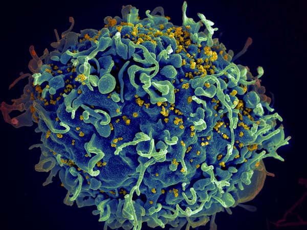
Research Throws Cold Water on COVID ‘Lab Leak’ Theory

Photo by KOBU Agency on Unsplash

The source of the COVID pandemic likely is down to live animals sold at the Huanan Seafood Wholesale Market, according to an international team of researchers.

The researchers traced the start of the pandemic to the market in Wuhan, China, where animals susceptible to the virus were sold live immediately before the pandemic began. Their findings were published in a pair of papers in the journal Science.

The publications all but rule out other explanations for the start of the pandemic, such as the ‘lab leak’ hypothesis. The authors further conclude that the first spread to humans from animals likely occurred in two separate transmission events in the Huanan market in late November 2019.

The first study looked at the locations of the first known COVID cases, as well as swab samples taken various places in the market. The second study examined genomic sequences of SARS-CoV-2 from samples collected from COVID patients during the first weeks of the pandemic in China.

The first paper, led by University of Arizona virus evolution expert Michael Worobey and Professor Kristian Andersen, was able to determine the locations of almost all of the 174 COVID cases identified by the World Health Organization in December 2019, 155 of which were in Wuhan.

A ‘bullseye’ on the market

Analyses showed that these cases were clustered tightly around the Huanan market, whereas later cases were dispersed widely throughout Wuhan. A striking percentage of early COVID patients had not visited there but turned out to live near the market. This suggests that vendors got infected first and set off a chain of infections among community members in the surrounding area, said Worobey.

“In a city covering more than 3000 square miles, the area with the highest probability of containing the home of someone who had one of the earliest COVID cases in the world was an area of a few city blocks, with the Huanan market smack dab inside it,” said Worobey.

This conclusion was supported by another finding: When the authors looked at the geographical distribution of later COVID cases, from January and February 2020, they found a “polar opposite” pattern, Worobey said. While the cases from December 2019 mapped “like a bullseye” on the market, the later cases coincided with areas of the highest population density in Wuhan.

“This tells us the virus was not circulating cryptically,” Worobey said. “It really originated at that market and spread out from there.”

Worobey and collaborators also addressed the question of whether health authorities found cases around the market simply because that is where they looked.

To rule out bias even more, from the market outwards the team removed cases ran the stats again. They found that even when two-thirds of cases were removed, the findings remained the same.

“Even in that scenario, with the majority of cases, removed, we found that the remaining ones lived closer to the market than what would be expected if there was no geographical correlation between these earliest COVID cases and the market,” Worobey said.

The study also looked at swab samples taken from market surfaces like floors and cages after Huanan market was closed. SARS-CoV-2-positive samples were significantly associated with stalls selling live wildlife.

The researchers determined that mammals now known to be susceptible to SARS-CoV-2, including red foxes, hog badgers and raccoon dogs, were sold live at the Huanan market in the weeks preceding the first recorded COVID cases. The scientists developed a detailed map of the market and showed that SARS-CoV-2-positive samples reported by Chinese researchers in early 2020 showed a clear association with the western portion of the market, where live or freshly butchered animals were sold in late 2019.

“Upstream events are still obscure, but our analyses of available evidence clearly suggest that the pandemic arose from initial human infections from animals for sale at the Huanan Seafood Wholesale Market in late November 2019,” said Prof Andersen at Scripps Research, co-senior author of both studies.

Virus likely jumped from animals to humans more than once

The second study, was an analysis of SARS-CoV-2 genomic data from early cases.

The researchers combined epidemic modeling with analyses of the virus’s early evolution based on the earliest sampled genomes. They determined that the pandemic, which initially involved two subtly distinct lineages of SARS-CoV-2, likely arose from at least two separate infections of humans from animals at the Huanan market in November 2019 and perhaps in December 2019. The analyses also suggested that, in this period, there were many other animal-to-human transmissions of the virus at the market that failed to manifest in recorded COVID-19 cases.

Using molecular clock analysis, which relies on the natural pace with which genetic mutations occur over time, researchers established a framework for the evolution of the SARS-CoV-2 virus lineages. They found that a scenario of a singular introduction of the virus into humans rather than multiple introductions would not align with molecular clock data. Earlier studies had suggested that one lineage of the virus – named A and closely related to viral relatives in bats – gave rise to a second lineage, named B. The more likely scenario in which the two lineages jumped from animals into humans on separate occasions, both at the Huanan market, Worobey said.

“Otherwise, lineage A would have had to have been evolving in slow motion compared to the lineage B virus, which just doesn’t make biological sense,” said Worobey.

The two studies provide evidence that COVID originated via jumps from animals to humans at the Huanan market, likely following transmission to those animals from coronavirus-carrying bats in the wild or on farms in China. Moving forward, the researchers say scientists and public officials should seek better understanding of the wildlife trade in China and elsewhere and promote more comprehensive testing of live animals sold in markets to lower the risk of future pandemics.

Source: University of Arizona

UK Children’s Gender Identity Clinic to Close after Controversies

Photo by Sharon McCutcheon on Unsplash

Following a highly critical independent report and accusations of inadequate and unsafe care, the UK will shut down the Tavistock gender identity clinic for children – the only one in the country. It will be replaced by a number of smaller facilities with closer links with mental health care.

The Tavistock and Portman NHS Foundation Trust clinic, named the Gender Identity Development Service (GIDS), had faced complaints of both long waiting lists for a burgeoning number of referrals, as well as rushing to assign puberty-blocking drugs and cross-sex hormones to children experiencing gender dysphoria.

Concerns had been voiced as early as 2005, when a nurse working at the clinic said that patients were being assessed too quickly and giving in to pressure from interest groups. Nevertheless, demand for its services skyrocketed in later years, from less than 100 per year in 2010 to nearly 2500 by 2018. In 2018, concerns were raised anew, with staff going on to make serious public accusations.

In July 2019, Dr Kirsy Entwhisle, a psychologist at GIDS Leeds hub, said that staff misled patients and made decisions about young people’s “bodies and lives” without “robust evidence”. Some of the children had suffered “very traumatic early experiences” which had not been addressed by the staff. The trust’s safeguarding lead, Sonia Appleby, won a claim from an employment tribunal after trust managers tried to stop her from carrying out her role when staff raised concerns.

One of the loudest critics of Tavistock Centre is Keira Bell, who at 16 was assigned puberty blockers, then cross-sex hormones at 17, and had a double mastectomy at 20 before later de-transitioning.

The former patient, who said she was suffering from anxiety and depression at the time she received treatment, said medics should have considered her mental health issues, “not just reaffirm my naïve hope that everything could be solved with hormones and surgery”.

Along with the unnamed parent of an autistic girl at the clinic, she won a ruling against the NHS assigning cross-sex hormones to children under 16 – but was overturned on appeal.

Helen, a parent of a patient at the clinic, welcomed its closure, but expressed concern for the future of her son’s treatments, according to LGBT site Pink News. While she said her son was treated quickly and received puberty blocking drugs, “From that point on, it felt like it was a little bit like they were winging it,” she said.

During therapy sessions at Tavistock, she said her son was asked a lot of questions and treated “almost like a little bit of an academic curiosity”. She criticised the fact that the same staff evaluated children for medical interventions and also offered therapy session, creating “a fear that they would stop access to medical support”. In contrast to the legal claims of Keira Bell’s and the unnamed patient, she said that GIDS refused to even discuss cross-sex hormones.

Dr David Bell (no relation to Keira Bell) welcomed the closure of Tavistock, telling the BBC: “Some children have got the double problem of living with the wrong treatment, and the original problems weren’t addressed – with complex problems like trauma, depression, large instances of autism.”

Endometriosis Hijacks Foetal Tolerance to Evade the Immune System

Photo by Sora Shimazaki on Pexels

In about 10% of women, endometrium-like tissues (known as lesions) also grow outside of the uterus, leading to endometriosis. Endometriosis is characterised by pain and can cause infertility, but its molecular mechanisms and drivers remain unknown. Now, a comprehensive study reveals how lesions escape immune surveillance, by taking advantage of mechanisms for the body tolerating a foetus during pregnancy.

Definitive diagnosis and clinical response still present significant challenges, with a common treatment being hormonal therapy with surgery. Unfortunately, surgery must be repeated if lesions recur, and they often do. To improve the situation, a better understanding of how and why the lesions grow, their cellular makeup, their microenvironments, and other aspects of their biology is essential.

The Jackson Laboratory’s (JAX) Elise Courtois, PhD, in partnership with UConn Health’s gynaecological surgeon Danielle Luciano, MD, recently completed an important study to develop a comprehensive cell atlas of the disease based on lesions obtained from 14 individuals who had treatment for endometriosis

The paper, published Nature Cell Biology, includes a thorough comparison of healthy endometrium tissue and ectopic (outside their normal site) lesions. The data also describes the endometriosis microenvironment and the conditions that allow the lesions to form and grow in what should be unhospitable regions.

“The study builds a robust foundation for a better understanding of endometriosis and how it grows,” said Dr Luciano.   “It’s exciting progress that we hope leads to earlier diagnosis and the ability to specifically target these abnormal cells for better treatments.”

The research team worked with tissues from individuals who had lesion removal at UConn Health for relief of symptoms. All were also receiving hormone therapy, the most frequent endometriosis management strategy. Not surprisingly, given that lesions are described as endometrial-like tissues growing in the wrong place, the cellular composition of the lesions in the peritoneum were quite similar to that of the normal endometrium. On the other hand, ovarian lesions had extensive differences in both composition and gene expression from the peritoneal ones. So while both ovary and peritoneum are receptive to the formation of lesions, they represent different environments and lead to important cellular and molecular differences between the two sites. The finding indicates that site-specific therapeutic design may be necessary to develop more effective treatments.

Another aspect of endometriosis is that, like cancer, the lesions represent abnormal growth that would typically be eliminated by immune surveillance. The researchers therefore investigated the immune cells in the peritoneal lesion microenvironment to see why they do not eliminate the abnormal lesion cells. They found that macrophages and dendritic cells contribute to conditions that promote immune inhibition and the promotion of immunosurveillance escape. Their specific characteristics are similar to those associated with foetal tolerance during pregnancy, which suggests that endometriosis hijacks a necessary, naturally occurring immune process to allow for lesion formation and persistence.

The paper details other aspects of both normal endometrium and ectopic lesions, including properties of vascularisation and the drivers of regeneration in endometrium and, perhaps, the formation of lesions in endometriosis. Of particular interest were key differences in the vascularisation of peritoneal versus ovarian lesions, further emphasising the site-specific nature of endometriosis. Also of note was the identification of a previously uncharacterised population of epithelial cells that may be progenitor cells for both endometrium and lesion formation, but more work is needed to define their precise role.

“Single cell analyses and hyperplexed antibody-based imaging techniques offer powerful insights into the complexity of the endometriosis microenvironment,” said Dr Courtois. “Understanding this complexity will be key for developing the new, efficient diagnostic and therapeutic tools that are so badly needed.”

Overall, the data captures a full description of endometrium and lesions, laying a strong foundation for understanding the vital cellular players and molecular dynamics of the disease. The data represents an important step forward for research into endometriosis and provides essential information for future therapeutics and diagnostics that can provide relief for those with this under-investigated disease.

Source: University of Connecticut

A Step Closer to a Once-off Treatment for HIV

HIV invading a human cell
HIV invading a human cell: Credit NIH

Researchers from Tel Aviv University have demonstrated success of a novel technology that may be developed into a one-time vaccine to treat people with HIV and AIDS. Using CRISPR technology, the researchers engineered B cells that in turn stimulate the immune system to produce HIV-neutralising antibodies.

Published in Nature, the study was led by Dr Adi Barzel and PhD student Alessio Nehmad and conducted in collaboration with additional researchers from Israel and the US.

“Based on this study,” said Dr Barzel, “we can expect that over the coming years we will be able to produce a medication for AIDS, additional infectious diseases and certain types of cancer caused by a virus, such as cervical cancer, head and neck cancer and more.”

He explains that the treatment can become a kind of permanent medication, lingering in the body to fight the virus. “We developed an innovative treatment that may defeat the virus with a one-time injection, with the potential of bringing about tremendous improvement in the patients’ condition. When the engineered B cells encounter the virus, the virus stimulates and encourages them to divide, so we are utilising the very cause of the disease to combat it. Furthermore, if the virus changes, the B cells will also change accordingly in order to combat it, so we have created the first medication ever that can evolve in the body and defeat viruses in the ‘arms race’.”

When they mature, the antibody-generating B cells move into the blood and lymphatic system and from there to the different body parts.

Dr Barzel explained: “Until now, only a few scientists, and we among them, had been able to engineer B cells outside of the body. In this study, we were the first to do this within body and then make those cells generate the desired antibodies. The genetic engineering is conducted with viral carriers derived from viruses that were also engineered. We did this to avoid causing any damage, and solely bring the gene coded for the antibody into the B cells in the body.”

“Additionally, in this case we have been able to accurately introduce the antibodies into a desired site in the B cell genome. All lab models that had been administered the treatment responded, and had high quantities of the desired antibody in their blood. We produced the antibody from the blood and made sure it was actually effective in neutralising the HIV virus in the lab dish.”

Source: Tel Aviv University

CRISPR Editing can Destabilise the Genome, Study Finds

DNA repair
Source: Pixabay/CC0

A new study published in Nature Biotechnology identifies risks in the use of CRISPR gene editing, which is employed in a number of therapies. Looking at its use in T cells, the researchers detected a loss of genetic material in a significant percentage – up to 10% of the treated cells. They explain that such loss can lead to destabilisation of the genome, which might cause cancer.

The study was led by Drs Adi Barzel, Dr Asaf Madi and Dr Uri Ben-David at Tel Aviv University.

Developed about a decade ago, CRISPR cleaves DNA sequences at certain locations in order to delete unwanted segments, or alternately repair or insert beneficial segments. It has already proved impressively effective in treating a range of diseases – cancer, liver diseases, genetic syndromes, and more. In 2020 at the University of Pennsylvania, the first approved clinical trial ever to use CRISPR took T cells from a donor, and expressed an engineered receptor targeting cancer cells, while using CRISPR to destroy genes coding for the original receptor – which otherwise might have caused the T cells to attack cells in the recipient’s body. 

In the present study, the researchers sought to examine whether the potential benefits of CRISPR therapeutics might be offset by risks resulting from the cleavage itself, assuming that broken DNA is not always able to recover.

Dr Ben-David and his research associate Eli Reuveni explained: “The genome in our cells often breaks due to natural causes, but usually it is able to repair itself, with no harm done. Still, sometimes a certain chromosome is unable to bounce back, and large sections, or even the entire chromosome, are lost. Such chromosomal disruptions can destabilise the genome, and we often see this in cancer cells. Thus, CRISPR therapeutics, in which DNA is cleaved intentionally as a means for treating cancer, might, in extreme scenarios, actually promote malignancies.”

To examine the extent of potential damage, the researchers repeated the 2020 Pennsylvania experiment, cleaving the T cells’ genome in exactly the same locations – chromosomes 2, 7, and 14. Using single-cell RNA sequencing, they analysed each cell separately and measured the expression levels of each chromosome in every cell.

They detected a significant loss of genetic material in some of the cells. For example, when chromosome 14 had been cleaved, about 5% of the cells showed little or no expression of this chromosome. When all chromosomes were cleaved simultaneously, the damage increased, with 9%, 10%, and 3% of the cells unable to repair the break in chromosomes 14, 7, and 2 respectively. The three chromosomes did differ, however, in the extent of the damage they sustained. 

Dr Madi and his student Ella Goldschmidt explained: “Single-cell RNA sequencing and computational analyses enabled us to obtain very precise results. We found that the cause for the difference in damage was the exact place of the cleaving on each of the three chromosomes. Altogether, our findings indicate that over 9% of the T-cells genetically edited with the CRISPR technique had lost a significant amount of genetic material. Such loss can lead to destabilisation of the genome, which might promote cancer.”

Based on their findings, the researchers caution that extra care should be taken when using CRISPR therapeutics. They also propose alternative, less risky, methods, for specific medical procedures, and recommend further research into two kinds of potential solutions: reducing the production of damaged cells or identifying damaged cells and removing them before the material is administered to the patient.

Dr Barzel and his PhD student Alessio Nahmad conclude: “Our intention in this study was to shed light on potential risks in the use of CRISPR therapeutics,” adding that as scientists, they “examine all aspects of an issue, both positive and negative, and look for answers.”

Source: EurekAlert!

New Wave of Opioid Overdose Deaths Predicted for US

Photo by Camilo Jimenez on Unsplash

Over the past 21 years of opioid overdose deaths in the US, whether an area is urban or rural has played a role in where opioid-involved overdose deaths have occurred, reports a new study published in JAMA Network Open. But there will no longer be a distinction between these, the study suggests – and there will be a dramatic increase in opioid-related overdoses.

The reason opioid overdoses have reached historical highs comes from combining synthetic opioids with stimulants such as cocaine and methamphetamines, a lethal cocktail that is hard to reverse during an overdose, the study authors said.

“I’m sounding the alarm because, for the first time, there is a convergence and escalation of acceleration rates for every type of rural and urban county,” said corresponding author Lori Post, director of the Buehler Center for Health Policy and Economics at Northwestern University Feinberg School of Medicine. “Not only is the death rate from an opioid at an all-time high, but the acceleration of that death rate signals explosive exponential growth that is even larger than an already historic high.”

Using methods developed to track COVID outbreaks, the study examined geographic trends in opioid-involved overdose deaths between 1999 and 2020 to determine if geography played a role in the three waves and the theorised fourth wave of America’s opioid crisis.

Near the end of the available data from 2020, overdose deaths in rural areas were escalating faster than in urban areas, according to the study. A visualization of the data illustrates that between 2019 and 2020, rates of opioid-involved overdose deaths converged while escalating for the first time across six types of rural and urban counties, Post said.

“We have the highest escalation rate for the first time in America, and this fourth wave will be worse than it’s ever been before,” Post said. “It’s going to mean mass death.”

The study authors examined toxicology reports and found people are using fentanyl (50 to 100 times more potent than morphine) and carfentanil (approximately 100 times more potent than fentanyl) combined with methamphetamines and cocaine. The result is a powerful and lethal cocktail that can even evade help from overdose-reversing drugs like naloxone.

“The stronger the drugs, the harder it is to revive a person,” said study co-author Alexander Lundberg, assistant professor of emergency medicine at Feinberg. “The polysubstance use complicates an already dire situation.”

“It appears that those who have died from opioid overdoses had been playing pharmacist and trying to manage their own dosing,” Post said. “This is a bigger problem because you have people misusing cocaine and methamphetamines along with an opioid, so you have to treat two things at once, and the fentanyl is horribly volatile.”

“The only path forward is to increase awareness to prevent opioid use disorders and to provide medication-assisted treatment that is culturally appropriate and non-stigmatising in rural communities,” Post said.

Cancer Incidence Driven by Insulin Dosage

Image depicting diabetes
Image by Nataliya Vaitkevich on Pexels

A study published in the JAMA Oncology looking at the correlation between daily insulin dose and cancer incidence among patients with type 1 diabetes has found that higher insulin dose is positively associated with cancer incidence and that the association is stronger among those with insulin resistance.

“In patients with type 1 diabetes, our results show that traditional metabolic factors such as obesity (represented by body mass index), sugar control (by Haemoglobin A1c), and blood pressure control do not associate with cancer incidence,” study leader Dr Yuanjie Mao Mao said. “However, cancer incidence was higher for those who took larger dose of insulin. Our results implied that clinicians might need to balance the potential cancer risk when treating patients with type 1 diabetes on a high daily insulin dose or that improving insulin sensitivity may be preferred than simply increasing the insulin dose.”

To conduct the study, Dr Mao collaborated with Wenjun Zhong, PhD, an epidemiologist of Merck Research Labs, to analyse the associations of more than 50 common risk factors in 1303 patients with type 1 diabetes whose data were collected over 28 years. A variety of databases were drawn upon and analysed, including he Diabetes Control and Complications Trial (DCCT.) which was was a controlled clinical trial originating with 1441 patients with type 1 diabetes who were randomised into conventional diabetes therapy or intensive therapy to assess whether reducing hyperglycaemia would decrease the risk of complications of type 1 diabetes.

Mao also found that age and sex are associated with cancer incidence when evaluated separately and that a daily insulin dose posed a higher risk of cancer than age, especially a higher insulin dose. According to the paper, when the daily insulin dose is classified into three groups, low: less than 0.5; medium: greater than or equal to 0.5 or lower than 0.8; and high: greater than or equal to 0.8 units/kg per day, the hazard ratios were significantly higher in the high dose versus the low dose group. Cancer incidence was 2.11, 2.87, and 2.91 per 1000 persons in the low, medium, and high insulin dose groups, respectively.

He went on to explain that specifically, women carry a higher risk than men; however, it was unclear what risk factors may contribute to the higher cancer incidence in type 1 diabetes.

“We know that people with type 1 diabetes have a higher incidence of cancer compared to people without diabetes,” Liz Beverly, Ph.D. co-director of the diabetes institute and professor in the Heritage College, said. “Dr. Mao’s research identifies a potential mechanism to explain this association. His findings will lead to continued research in this area and potential policy changes in cancer screening and insulin dosing recommendations.”

Although previous studies have concluded that patients with diabetes have a higher risk of cancer in general, this is the first study to explore the associated cancer incidence factors in type 1 diabetes. 

“Type 1 diabetes accounts for an estimated five to 10 percent of all diabetes cases, and recent studies in type 1 diabetes also found a higher incidence of certain cancers such as stomach, liver, pancreas, endometrium and kidney cancers in the population compared with the general population,” Mao explained. “Whereas, in type 2 diabetes, increased risk is attributed to metabolic factors such as obesity, chronic inflammation status, and insulin resistance.”

Although the results of the study suggest that the higher the dose of insulin, the higher the cancer incidence, Dr Mao says further investigation is still necessary.

Source: Ohio State University

Changing Order of Breast Cancer Treatments Could Improve Outcomes

Photo by Michelle Leman on Pexels

Changing the order of treatments given to breast cancer patients could reduce side effects resulting from mastectomy and improve outcomes, according to a clinical feasibility trial, published in The Lancet Oncology.

In the study, researchers found that switching the sequence of treatments given to breast cancer patients was safe, without any increase in complications and could lead to patients receiving faster and more effective care compared to current methods.

Thirty-three women with breast cancer requiring a mastectomy and post-mastectomy radiotherapy, were recruited to the primary radiotherapy and deep inferior epigastric perforator flap reconstruction for patients with breast cancer (PRADA) trial between January 2016 and December 2017.  They were also eligible for a breast reconstruction using tissue from another part of their body.

They were given chemotherapy followed by radiotherapy before having a mastectomy and a breast reconstruction. The team found that this approach was feasible and safe.  They also found that side effects were low and that 12 months after surgery patients reported high levels of satisfaction with their breast reconstruction.

Lead author Daniel Leff said: “We believe that, in the long term, this approach will improve patients’ mental and physical wellbeing with higher quality of life scores and satisfaction with their reconstructed breasts compared to current care. It also means that many patients who are currently denied reconstruction due to concerns of further complications due to radiotherapy may be able to get access to this treatment in future.”

Source: Imperial College London

COVID Vaccine Response in Blood Cancer Patients Only after Booster

Patients with blood cancers have an impaired immune system due to their disease and its treatment, putting them at risk of severe COVID infection and a reduced COVID vaccination response. In a recent study published in CANCER, less than half of patients with haematologic malignancies including leukaemia, lymphoma, and multiple myeloma mounted detectable antibodies after initial COVID vaccination, but 56% of ‘nonresponders’ produced antibodies after receiving a booster dose.

For the study, Thomas Ollila, MD, and colleagues retrospectively analysed antibody responses to initial and booster COVID vaccination in 378 patients with hematologic malignancies.

Anti-SARS-CoV-2 antibodies were detected in the blood of 181 patients (48%) after initial vaccination with one of three FDA-authorised or approved COVID vaccines, and patients with active cancer or those recently treated with an immune cell–depleting therapy were least likely to produce these antibodies. Among patients who did not mount an antibody response following initial vaccination, responses were observed after a booster dose in 48 of 85 (56%) patients who were assessed.

By the end of February 2022, 33 patients (8.8%) developed a COVID infection, with three COVID-related deaths (0.8%). Although there was no significant link between post-vaccination antibody response and incidence of COVID infection, no patient with antibody responses died from COVID

Also, no patient who received tixagevimab plus cilgavimab was diagnosed with a COVID infection. Tixagevimab and cilgavimab are antibody therapies that bind to non-overlapping portions of the SARS-CoV-2 spike protein, preventing the virus from binding to and infecting cells. The FDA authorised the combination therapy for emergency use during the COVID pandemic as a way to help prevent COVID infection in certain individuals.

“Our findings build on the wealth of literature showing that patients with hematologic malignancies have an impaired response to COVID vaccination. Importantly, we show that many of these patients who did not respond initially will in fact have a response to booster vaccination,” said Dr Ollila. “Moreover, when we looked at outcomes, we found that deaths from COVID in the patient population we reviewed only occurred in those with undetectable antibodies, and nobody who received prophylactic antibody therapy was diagnosed with COVID. This suggests to us the importance of checking antibody levels in these patients and arranging prophylactic antibody therapy.”

Dr. Ollila encourages providing booster vaccines for patients and prioritising prophylactic antibody therapy when indicated. “This is real world evidence that these actions can save lives,” he said.

Source: Wiley

SA Research Shedding Light on Role of Microclots in Long COVID

Image by Quicknews

Professor Resia Pretorius sounds rushed when Spotlight first tracks her down by phone at Heathrow Airport outside London. She is about to board a plane to South Africa after attending a conference, meetings, and symposia in the United Kingdom, all with the purpose of unravelling the complexity of long COVID and how to treat it.

There is no global consensus among researchers and clinicians on a definition for long COVID, there is no adequate diagnostic test for the debilitating condition, and the causes of patients progressing to long COVID are, at this stage, theoretical.

However, Pretorius who heads the Department of Physiological Science at the University of Stellenbosch remains upbeat. Her research group is the first to have reported evidence of inflammatory microclots in blood samples from individuals with long COVID, potentially solving an important piece of the long COVID puzzle.

She says scientific collaboration is intensifying to find answers to long COVID which affects 43% or 100 million people globally post-infection, according to a meta-analysis and systematic review.

Later speaking from Stellenbosch, Pretorius describes herself as a “lab person” who has been trying to find the cause of long COVID since 2020. “I have always been passionate about research. Now, I am working with clinicians and researchers in the UK, the USA, and other parts of the world. I am too worried to miss anything so I am at all of these meetings. There are 40 to 50 researchers globally who talk to each other regularly. We are going to crack this I know. We just have to.”

Causes of long COVID

As explained in a recent article in the journal Science, there are three leading theories scientists are pursuing in an attempt to decipher the effects of post-COVID-19 infection – which leads to an array of symptoms, including shortness of breath, fatigue, headaches, palpitations, and impairments in mental health and cognition or brain fog.

One theory is that SARS-CoV-2 stubbornly persists in the body, even after the acute infection passes. Studies have shown that the virus lingers in a wide range of body sites, especially in the nerves and other tissues.

Another theory based on blood samples from COVID-19 patients reveals an immune system in disarray even eight months after first testing positive. The body’s cells do not appear to recover.

Images of micro clots as seen under an electron microscope
Images of micro clots as seen under an electron microscope. PHOTO: Supplied

The third, an area in which Pretorius has distinguished herself internationally, is that COVID-19 is not only a lung disease but significantly affects the vascular (blood flow) and coagulation (blood clotting) systems of the body.

A recent study published in the Cardiovascular Diabetology journal, conducted by Pretorius and colleagues, found that there is significant microclot formation in the blood of both acute COVID-19 and long COVID patients. A microclot is a blood clot that can only be seen through a microscope.

Pretorius explains that in a healthy person clots may form, for example, when you cut yourself. The main clotting protein is a molecule called fibrinogen. “When you’re healthy, it’s in solution. And then when you cut yourself, collagen is exposed, and a little gel called fibrin prevents you from bleeding out. In healthy individuals, the clots are then broken down by a process called fibrinolysis.

Blood samples from patients with long COVID have revealed high levels of various inflammatory molecules trapped in the microclots including fibrinogen and Alpha-2 antiplasmin – a molecule that prevents the breakdown of microclots.

The persistent blood clots essentially result in cells not getting enough oxygen in the tissues to sustain bodily functions. This, Pretorius says, may be central to numerous debilitating symptoms.

In healthy individuals, the body’s plasmin-antiplasmin system maintains a fine balance between blood clotting to prevent blood loss after an injury and fibrinolysis which prevents blood clots from forming.

With high levels of alpha(2)-antiplasmin in the blood of acute COVID-19 patients and individuals suffering from long COVID, the body’s ability to break down the clots is significantly undermined. The blood circulation becomes clogged up.

Microclots are generally not found in people who do not have long COVID. Pretorius says you can find them in some other conditions, such as diabetes, “but the difference is the number and the extreme presence of the clots with long COVID, that’s what’s making the difference,” she says.

Insoluble clots

Another difference is that the clots in long COVID are insoluble. When Pretorius tried to dissolve these clots using an enzyme called trypsin in her laboratory, they would not dissolve. They are resistant to fibrinolysis.

Initially, Pretorius was looking at acute COVID-19 infection. We received blood samples from ICU patients and we made blood smears and looked at them under a scanning electron microscope that can enlarge a sample hundreds of thousands of times. We then added a fluorescent dye or marker called Thioflavin T which lights up when there are misfolded proteins. This happens when, for example, the spike protein binds to the soluble fibrinogen molecule making it insoluble.

The SARS-CoV-2 virus is known to bind to ACE2 receptors and TMPRSS receptors which are found on platelets (blood cells that help with clotting). They are also found on the endothelium (the inner-most lining of the blood vessels). By binding to the platelets and the endothelium, the virus sets off a torrent of clotting causing vascular damage.

Pretorius says in early 2021, “I got a report from Harvard collaborators and others to say that patients do not fully recover post-infection and they referred to this as long COVID.

“I said let’s get the samples. We looked at the blood samples and lo and behold we found the clots and they were fully persistent. I was not surprised to find the clots in long COVID because I knew with acute COVID many people were dying because of clots in the lungs and shortness of breath. But, I did not know the extent to which they were present in long COVID.”

“When we did proteomics analysis on the sample, when we looked at the different molecules in the blood, I could not dissolve the sample with typical enzymes. I used a massively abrasive enzyme called Trypsin which dissolves any possible protein. But it could not dissolve these cells. The resilience of these clots, that they simply don’t get dissolved, surprised me,” Pretorius says.

Pretorius recalls that in 2020, several South African clinicians alerted others to COVID-19 not being a typical viral pneumonia but suggested it was also a vascular disease. “At that stage, it was massively controversial with many dismissing this idea saying it’s a virus that affects the lungs and that’s it,” she says.

Pretorius says this was despite papers published overseas in 2020 that concluded COVID-19 was also a vascular disease. “It was made controversial in South Africa but it is now widely accepted that COVID-19 also affects clotting as well as the body’s vasculature (network of blood vessels).”

Pretorius says, “Although the microclot is a theory, it encompasses all of the other suggested causes of long COVID because the spike protein itself can trigger microclots. We have submitted a paper, looking at many more blood samples, where we found inflammatory molecules trapped inside the blood clots which do not break down. We also found antibodies so the theories about immune abnormalities, persistent virus, and microclots are intertwined. All of these can cause organ damage. So if you look at it from a systems biology approach, all of these are valid.”

Many are told that their symptoms are possibly psychological, all in their head, and they are told to get some rest and to stop stressing. Meanwhile, the patients are very ill

Diagnostics

Pretorius says there are no general pathology tests readily available to diagnose people with long COVID.

“People that are desperately ill – bedridden or in wheelchairs – are often given generalised blood tests. They are told that their pathology test results are within normal to healthy ranges. Many are told that their symptoms are possibly psychological, all in their head, and they are told to get some rest and to stop stressing. Meanwhile, the patients are very ill,” she says.

Pretorius says the main reason the traditional laboratory tests do not pick up any of the inflammatory molecules is that they are trapped inside the insoluble microclots. A typical pathology test looks at the soluble content of the blood, so if the molecules are trapped they will be missed.

“We patented a long COVID test which is just a simple microscopy test that is a useable diagnostic method to see if microclots are present,” Pretorius says.

Microscopy methods are not readily available at pathology laboratories. However, Pretorius says, “We have crowd-funded and received funding from the Polybio Research Foundation in America to buy a flow cytometer for our blood lab to develop a flow cytometry method that can be used in the typical pathology labs. So we hope to have a diagnostic that will be readily available in a couple of months.”

Treatment

Pretorius says colleagues in the United Kingdom have already designed two randomised controlled trials to independently test both coagulation therapy (CLOTT-UK) and Apheresis (CLOTT-Apheresis trial ) in which microclots and inflammatory molecules are filtered out in a dialysis-type treatment. These trials will study whether anticoagulants and Apheresis give long-lasting relief of symptoms. These trials are being planned and researchers are waiting for ethics approval.

In addition, colleagues from the University of Sheffield Hallam and from the University of Manchester have independently set up microclot testing in their labs and they are planning to publish their UK cohort results soon. They are also correlating long COVID severity to microclot presence, says Pretorius.

“It’s been quite a ride. Seeing the devastation of long COVID, I realise why I decided not to be a clinician… handling and hearing all the issues is just so sad,” says Pretorius.

But, she remains determined to help “crack” long COVID.

Republished from Spotlight under a Creative Commons 4.0 licence.

Source: Spotlight