Children in paediatric ICUs (PICUs) that undergo invasive mechanical ventilation for acute respiratory failure are left with lasting neurocognitive effects, according to a study published in JAMA.
Little is known about whether children undergoing invasive mechanical ventilation worse long-term neurocognitive function than children who do not undergo such procedures. There are concerns about neurotoxic effects of critical illness and its treatment on the developing brain. Therefore, infants and young children may be uniquely susceptible to adverse neurocognitive outcomes after invasive mechanical ventilation.
Researchers conducted a four-year sibling-matched cohort study conducted at 31 PICUs and associated neuropsychology testing centres. Children who survived PICU hospitalisation for respiratory failure and were discharged without severe cognitive dysfunction were found to have significantly lower subsequent IQ scores than their matched siblings.
“While the difference in IQ scores between patients and unexposed siblings was small, the data provide strong evidence of the existence and epidemiology of paediatric post-intensive care syndrome (PICS-p) after a single typical episode of acute respiratory failure necessitating invasive ventilation among generally healthy children,” said Martha A.Q. Curley, PhD, RN, FAAN, Professor of Nursing at the University of Pennsylvania School of Nursing (Penn Nursing) and the study’s lead researcher.
The study reaffirms the importance of assessing long-term outcomes as part of any trial evaluating acute interventions in pediatric critical care. It also underscores the importance of further study to understand which children may be at highest risk, what modifiable factors could cause it, and how it can be prevented.
By making computer simulations of drugs, researchers have found that doctors need to be wary of prescribing metformin for all types of cancer and patients. Their findings are published in BioMed Central Cancer.
The diabetes drug metformin has been used in clinical settings as a cancer treatment in recent years. The researchers say while metformin shows great promise, it also has negative consequences for some types of cancers.
“Metformin is a wonder drug, and we are just beginning to understand all its possible benefits,” said Mehrshad Sadria, a PhD candidate in applied mathematics at the University of Waterloo. “Doctors need to examine the value of the drug on a case-by-case basis, because for some cancers and some patient profiles, it may actually have the opposite of the intended effect by protecting tumour cells against stress.”
The computer-simulated treatments use models that replicate both the drug and the cancerous cells in a virtual environment. Such models can give clinical trials in humans a considerable head-start and can provide insights to medical practitioners that would take much longer to be discovered in the field.
“In clinical settings, drugs can sometimes be prescribed in a trial and error manner,” said Anita Layton, professor of applied mathematics and Canada 150 Research Chair in mathematical biology and medicine at Waterloo. “Our mathematical models help accelerate clinical trials and remove some of the guesswork. What we see with this drug is that it can do a lot of good but needs more study.”
The researchers say their work shows the importance of precision medicine when considering the use of metformin for cancer and other diseases. Precision medicine is an approach that assumes each patient requires individualised medical assessment and treatment.
“Diseases and treatments are complicated,” Sadria said. “Everything about the patient matters, and even small differences can have a big impact on the effect of a drug, such as age, gender, genetic and epigenetic profiles. All these things are important and can affect a patient’s drug outcome. In addition, no one drug works for everyone, so doctors need to take a close look at each patient when considering treatments like metformin.”
In an analysis of older adults who underwent surgery, published in the Journal of the American Geriatrics Society, more who had non-elective surgery were found to experience disabilities than those who had elective surgery, and factors such as age increased this vulnerability.
The study included 247 adults aged 70 years or older who were discharged from the hospital after major surgery from 1997 to 2017, patients who had non-elective surgery had more disabilities in daily activities over the following 6 months than those who had elective surgery.
Researchers identified 10 factors that were associated with greater disability burden: age 85 years or older, female sex, Black race or Hispanic ethnicity, neighbourhood disadvantage, multimorbidity, frailty, one or more disabilities, low functional self-efficacy, smoking, and obesity. The burden of disability increased with each additional “vulnerability” factor.
“The results from this study can be used by clinicians to identify older adults who are particularly susceptible to poor functional outcomes after major surgery, and a subset of the factors identified could serve as the basis for new interventions to improve functional outcomes in vulnerable older surgical patients,” said lead author Thomas M. Gill, MD, of the Yale School of Medicine.
In their recently released Middle East and Africa regional report on cancer, the Swedish Institute for Health Economics (IHE) highlighted challenges for the country’s under-resourced healthcare system. It also highlighted the need to provide training for South African GPs in early detection of cancer.
Cancer is a growing challenge for South Africa. The incidence of cancer cases in South Africa is predicted to double over the next two decades, from 110 per 100 000 in 2018 to 226 per 100 000 in 2040. It is also gradually becoming one of the leading causes of death, from 9% in 2000 to 10% in 2016, even as the share of deaths from cardiovascular diseases and diabetes grows as well. Prostate cancer is the most common (31%) in men by far, while in women breast cancer (27%) was closely followed by cervical cancer (22%).
The COVID pandemic has largely overshadowed the Department of Health’s 207-2022 cancer plan, though successes with HIV have allowed it to move up in priority. A major challenge will be getting it moved up in priority.
The direct costs to the healthcare system from cancer are USD11 (R165) per capita and USD19 (R285) per capita in indirect costs to society (premature death, early retirement, sick leave etc). Yet South Africa’s public healthcare spending is only 4% of GDP, below the World Health Organization informal target of 5%.
In terms of prevention, anti-smoking campaigns have had some effect, though more work needs to be done on tackling obesity. The HPV vaccination campaign is a step in the right direction, the report says, though the hepatitis B vaccination programme is flagging.
In early detection, GPs need better training in recognising the early signs of cancer. Public health literacy is also a priority, along with expanding breast and cervical cancer screening. Given rising incidence, colorectal cancer screening should also be considered, the IHE recommended.
Universal health care continues to be a priority, with the proportion of the population covered by medical schemes remaining static at 17% from 2012 to 2019. Public healthcare, which only offers a defined set of services, suffers from a lack of resources and personnel.
As far as cancer treatment in South Africa goes, public healthcare resembles global standards 20 years ago. Though radiation machines adequately serve the population on a national level, there are significant disparities with long waiting times and machines that can provide modern radiation techniques are limited and not listed in prescribed minimum benefits. Targeted drugs and immunotherapy remain almost exclusively the province of private healthcare, with a lengthy procedure to get drugs listed on the EML. Streamlining this should be a priority, the report recommends.
Results from a clinical trial showed that ultrasound scans are effective in prostate cancer diagnosis, which would be a cheaper option than MRI for low- and middle income countries. The study is published in Lancet Oncology.
“MRI scans are one of the tests we use to diagnose prostate cancer,” said Professor Hashim Ahmed, lead author of the study and Chair of Urology at Imperial College London. “Although effective, these scans are expensive, take up to 40 minutes to perform and are not easily available to all. Also, there are some patients who are unable to have MRI scans such as those with hip replacements or claustrophobia fears. As cancer waiting lists build as a result of the COVID pandemic, there is a real need to find more efficient and cheaper tests to diagnose prostate cancer.
“Our study is the first to show that a special type of ultrasound scan can be used as a potential test to detect clinically significant cases of prostate cancer. They can detect most cases of prostate cancer with good accuracy, although MRI scans are slightly better.
“We believe that this test can be used in low and middle income settings where access to expensive MRI equipment is difficult and cases of prostate cancer are growing.”
Prostate cancer develops slowly and symptoms such as the blood in the urine do not appear until the disease has developed. It usually affects men over 50 and often men with a family history of the disease. Black men are disproportionately impacted by the disease and prostate cancer deaths now exceed those from breast cancer.
One of the principal means of prostate cancer diagnosis is a multi-parametric MRI (mpMRI) scan. However, the 40-minute scan costs £350–450 (R7000-9000).
This new study tested multiparametric ultrasound (mpUSS) to image the prostate. Elastography examines tissue hardness, doppler and contrast-enhancement with microbubbles measures blood flow. As cancers are denser and have greater blood supply, they show up more clearly. While mpUSS is more widely available than mpMRI, no large-scale studies have been done thus far to validate its effectiveness as a test to detect prostate cancer cases.
For the trial the team recruited 370 men at risk of prostate cancer. They were identified following initial tests such as a prostate-specific antigen (PSA) test and/or an abnormal digital rectal examination.
The men underwent both mpUSS and mpMRI scans at separate visits. This was then followed by biopsies for 257 patients who had a positive mpUSS or mpMRI test result. Cancer was detected in 133 men, with 83 men diagnosed with clinically significant cancer.
Individually, mpUSS detected 66 cases of clinically significant cancer compared to mpMRI which detected 77 cases.
Although mpUSS detected 4.3% fewer clinically-important prostate cancers compared to mpMRI, this method would lead to 11.1% more patients being biopsied as a result of false positives from the mpUSS.
The researchers believe that mpUSS could be an alternative to mpMRI as a first test for patients at risk of prostate cancer, particularly where mpMRI cannot be carried out. As both imaging tests missed clinically-important cancers detected by the other, using both would increase the detection of clinically-important prostate cancers overall.
A recent study on the impact of the antibiotic azithromycin during severe respiratory syncytial virus (RSV) bronchiolitis overwhelmingly supports current bronchiolitis guidelines in the US, which recommend against antibiotics during acute bronchiolitis.
The anti-inflammatory properties of azithromycin can be beneficial in some chronic lung diseases, such as cystic fibrosis. With that in mind, researchers investigated its potential to prevent future recurrent wheezing among infants hospitalised with RSV. With such babies at increased risk of developing asthma later in childhood, the scientists hoped to find a therapy to reduce this risk.
The study, published in NEJM Evidence, also provided considerable evidence that severe RSV bronchiolitis in early life increases the likelihood of repeated wheezing episodes in early childhood, often leading to asthma.
“The major message is that antibiotics don’t have a role, either in the management of acute RSV bronchiolitis or to reduce subsequent wheezing,” said co-corresponding author Leonard Bacharier, MD, professor of Pediatrics at Monroe Carell Jr Children’s Hospital at Vanderbilt. “As a matter of fact, we found that antibiotics in general in our study of severe RSV bronchiolitis increased the risk of subsequent recurrent wheezing over the following two to four years.”
“We need to discourage the use of this therapy, as it is potentially harmful,” he said.
The study examined children hospitalised with RSV bronchiolitis during a single-center, double-blind, placebo-controlled trial.
An earlier pilot trial enrolled 40 infants hospitalised with RSV bronchiolitis where treatment with azithromycin, and this showed a reduction in the likelihood of recurrent wheeze over the following year.
In the current study, 200 otherwise healthy 1- to 18-month-old children who were hospitalised for RSV bronchiolitis were prospectively randomised to either oral azithromycin or a placebo for 14 days. The group was broadly representative of the population of children who experience severe RSV bronchiolitis.
Antibiotics are sometimes used in the treatment of RSV because co-occurring complications lead medical teams to prescribe them, thinking there is a bacterial component to the illness, Prof Bacharier said. “This condition can be managed by supportive care – oxygen, fluids, observation, time and love,” he stressed. “If a clinician is going to use an antibiotic in the setting of RSV bronchiolitis, there needs to be a very strong rationale for doing so. There is substantial evidence to suggest that children who receive antibiotics early in life are at an increased risk of developing asthma, and this study is consistent with that evidence.”
Using in vitro modelling the SARS-CoV-2 infection of human pancreatic cells, researchers have found that COVID infection is likely not associated with an increased new-onset diabetes risk. At the same time, another study has suggested that in hospitalised COVID patients, it may be a temporary form of the disease resulting from the acute stress of viral infection.
The findings, which are to appear in Cell Reports, address concerns raised over the past 18 months that infection with SARS-CoV-2 may trigger new-onset diabetes. However, the supporting evidence for this has remained sparse, with at times conflicting evidence impeding with a proper risk assessment.
The team of researchers at the Icahn School of Medicine at Mount Sinai demonstrated that SARS-CoV-2 targets virtually all types of pancreatic cells, not just the insulin-producing beta cells, using the ACE2 receptor to gain access. However, the infection in the pancreas remained highly circumscribed, largely non-cytopathic and despite high viral burden in infected subsets, promoted only modest cellular perturbations and inflammatory responses.
Similar experimental outcomes were also observed after in vitro infection with endemic coronaviruses not previously associated with diabetes. Taken together, these findings challenge the notion that direct beta cell infection and destruction by SARS-CoV-2 can precipitate diabetes onset.
“Our provisional conclusions indicate that SARS-CoV-2 infection is likely not associated with an increased risk for new-onset diabetes,” said study leader Dirk Homann, MD, Professor of Medicine at Icahn Mount Sinai. “However, a history of SARS-CoV-2 infection may yet promote prolonged glycometabolic perturbations and even an increase in cumulative diabetes risk in vulnerable populations. Over the next few years, we need to pay careful attention to emerging observational and retrospective studies that determine diabetes incidence rates of previously SARS-CoV-2-infected individuals.”
To evaluate permissiveness of human pancreatic islet cells to in vitro SARS-CoV-2 infection, the team of researchers employed an in vitro infection model of primary human pancreatic islets with SARS-CoV-2 as well as endemic human coronaviruses. The team precisely delineated pancreatic infection patterns and associated cellular changes at the single-cell level. Altogether, they found that the extent and consequences of pancreatic SARS-CoV-2 infection, even under in vitro conditions of enhanced virus exposure, remained decidedly limited.
“Concerns surrounding the possibility that infection with SARS-CoV-2, the etiological agent of COVID, may cause new-onset diabetes persist amidst an evolving research landscape,” said Verena van der Heide, MD, PhD, co-first author of the study and postdoctoral research fellow at the Icahn School of Medicine at Mount Sinai. “Our findings stand in notable contrast to three recent reports that also based their speculation about the diabetogenic potential of SARS-CoV-2 on in vitro infection of human islets. As detailed in our manuscript, however, we believe that our careful experimental design and comprehensive analysis strategy make a compelling case for the considerable limits of pancreatic SARS-CoV-2 infection.”
“There are strong epidemiological associations between COVID infection in humans and diabetes, but whether the SARS-CoV-2 virus actually infects and damages the insulin-producing cells in the human pancreas, the so-called ‘beta cells,’ has been highly controversial,” said Andrew Stewart, MD, Director of the Diabetes, Obesity and Metabolism Institute at Icahn Mount Sinai. “This study by Dr. Homann and his collaborators in Mount Sinai’s Precision Immunology Institute and the Department of Microbiology provides strong evidence that SARS-CoV-2 causes little or no damage to beta cells, making it unlikely that COVID infection can predispose to development of Type 1 diabetes.”
The conclusions they came to are in line with a 2020 report by Dr Homann and his team, showing that ACE2 receptors and other entry factors are lacking among islet endocrine cells but readily detected in microvascular and ductal structures of the pancreas.
Meanwhile, a second, separate study of 594 individuals who exhibited signs of diabetes mellitus during the early pandemic showed that half of the 79 patients without a diabetes diagnosis reverted to normal blood sugar levels by one year.
“We believe that the inflammatory stress caused by COVID may be a leading contributor to ‘new-onset’ or newly diagnosed diabetes,” said Sara Cromer, MD, lead author of the second study. “Instead of directly causing diabetes, COVID may push patients with pre-existing but undiagnosed diabetes to see a physician for the first time, where their blood sugar disorder can be clinically diagnosed. Our study showed these individuals had higher inflammatory markers and more frequently required admission to hospital ICUs than COVID patients with pre-existing diabetes.”
A new scintillation material developed by KAUST scientists can bring significant improvements to X-ray imaging in medicine, industry and security. Credit: KAUST
Scientists have successfully produced an exceptionally efficient, robust and flexible scintillation film to bring significant improvements in X-ray imaging, enabling much lower radiation doses to be used.
Scintillation materials release visible light, or “scintillate,” in response to absorbing high-energy X-ray photons, enabling an image to be captured.
Researchers are continually exploring ways to make scintillation technology more sensitive, efficient and readily adaptable. The researchers, led by Omar F Mohammed, Associate Professor of Chemical Sciences at King Abdullah University of Science and Technology (KAUST), sought to come up with an improved scintillation screen.
“Currently used materials suffer from several drawbacks, including complex and high-cost fabrication processes, radioluminescence afterglow and nontunable scintillation,” said Yang Zhou, a postdoc in Prof Mohammed’s lab.
Materials called lead halide perovskites have attracted considerable attention and shown significant promise. Novel perovskites are a category of materials that share the same crystal structure as the natural perovskite mineral calcium titanium oxide, but they include a variety of different atoms that replace all or some of those found in natural perovskite. To avoid toxicity problems and reduce cost, the researchers explored the use of elements besides lead. The newly developed screens are described in ACS Energy Letters.
The flexible scintillation screens the team developed can detect X-rays at ultralow levels, “approximately 113 times lower than a typical standard dose for X-ray medical imaging,” said Omar Mohammed, leader of the research group.
“Another vital advance is that the X-ray spatial resolution reported in this study is the highest achieved to date for powder-based screens,” said Dr Zhou.
“The physical flexibility of our films is also very important,” added Prof Mohammed. He explains that highly efficient flexible scintillation screens are urgently needed for using X-rays to better analyse awkward shapes.
The team plans to commercialise their advance, and to hope to refine their fabrication techniques.
Sanofi’s rare disease database that helps healthcare practitioners tackle their unique challenges – and knowing that treatments are available directly improves patients’ wellbeing. This comprehensive database has also aided rare disease research.
Johannesburg, 28 February 2022: Patients with rare diseases present unique challenges to healthcare practitioners (HCPs). Obstacles to caring for them include diagnostic delays and a lack of information, expertise, and treatment options for many rare diseases. HCPs play a vital role in enhancing the quality of life for patients and families living with a rare disease by making appropriate referrals to specialists, helping to coordinate care, and assisting patients in obtaining the proper support.1,2
A disease is defined as ‘rare’ when it affects fewer than 1 in 2000 people.3
Over 7000 rare diseases have been described to date, affecting over 350 million people worldwide.3,4 While most (70-80%) of rare diseases are genetic and inherited, some may be acquired, and 70% are exclusively paediatric in onset.5
Recent surveys showed that those living with rare diseases had a significantly higher prevalence of anxiety and depression compared to the general population.5,6 Levels of high stress can become even worse for carers when the person they are supporting has a diagnosis with no available treatment option.5,6
Monique Nel, Medical Advisor – Rare Diseases at Sanofi says: “Sanofi has been dedicated to researching and developing innovative treatments for rare diseases for 40 years. Currently, Sanofi has one of the largest rare diseases pipelines in the industry, across multiple diseases and modalities.7”
“Our rare disease patient registries have grown to represent one of the largest collections of real-world data for rare diseases collected over the past 30 years. We have a presence in 68 countries worldwide, with more than 920 participating sites and more than 17 800 patients enrolled.”
These registries have helped researchers to publish studies describing the underlying biology of disease, identify risk factors impacting treatment outcomes, and share guidelines for monitoring and treatment.
A further useful resource for HCPs and patients is the list of rare diseases maintained by the Genetic and Rare Diseases Information Center (GARD) of the US National Institutes of Health.8
Says Nel: “We understand the difficulty that healthcare professionals face when it comes to patient diagnosis of a rare disease, and that a coordinated approach to diagnosis and care for people living with rare diseases is needed. Rare diseases deserve the same amount of time, resources and dedication to finding effective treatments and therapies as any other conditions, which is a mission that Sanofi strives to promote every day, to help HCPs to improve diagnosis.”
Dudding-Byth T. A powerful team: the family physician advocating for patients with a rare disease. Aust Fam Physician. 2015 Sep;44(9):634. http://www.ncbi.nlm.nih.gov/pubmed/264880401. NIH.
Bogart KR, Irvin VL. Health-related quality of life among adults with diverse rare disorders. Orphanet J Rare Dis. 2017 Dec 7;12(1):177. doi: 10.1186/s13023-017-0730-1. PMID: 29212508; PMCID: PMC5719717.
Nguengang Wakap S, Lambert DM, Olry A, et al. Estimating cumulative point prevalence of rare diseases: analysis of the Orphanet database. Eur J Hum Genet 2020;28:165–173. https://doi.org/10.1038/s41431-019-0508-0
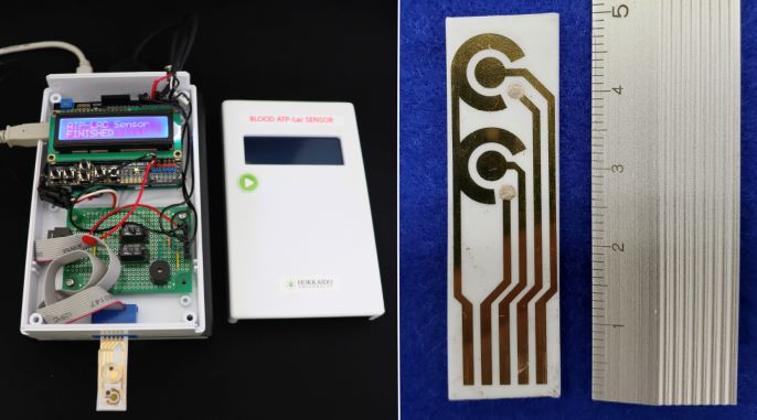
The prototype of the ATP and lactate sensor developed in the study (left); and the integrated sensor chip that detects ATP and lactate levels (right). Credit: Akihiko Ishida, Hokkaido University
Scientists at Hokkaido University have developed a prototype sensor that could help doctors rapidly measures levels of adenosine triphosphate (ATP) and lactate in blood samples from patients, aiding in the rapid assessment of the severity of conditions such as sepsis.
ATP is a molecule found in every living cell that stores and carries energy. In red blood cells, ATP is produced by a biochemical pathway called the Embden–Meyerhof pathway. Severe illnesses such as multiple organ failure, sepsis and influenza reduce the amounts of ATP produced by red blood cells.
As such, the severity of these illnesses could be gauged by monitoring the amounts of ATP and lactates in a patient’s blood. “In 2013, our co-authors at Tokushima University proposed the ATP-lactate energy risk score (A-LES) for measuring ATP and lactate blood levels to assess acute influenza severity in patients,” explained Akihiko Ishida, an applied chemist at Hokkaido University. “However, current methods to measure these levels and other approaches for measuring disease severity can be cumbersome, lengthy or not sensitive enough. We wanted to develop a rapid, sensitive test to help doctors better triage their patients.”
The researchers developed a biosensor that can detect levels of ATP and lactate in blood with great high sensitivity in as little as five minutes. The process is straightforward. Chemicals are added to a blood sample to extract ATP from red blood cells. Enzymes and substrates are then added to convert ATP and lactate to the same product that can be detected by specially modified electrodes on a sensor chip; the amount of by-product present in the sample increases the electrical current measured.
Schematic representation of the proposed sensor for sequentially detecting ATP and lactate levels in the blood. Through a series of chemical reactions, ATP and lactate are converted to hydrogen peroxide, the breakdown of which to water H2O causes the sensor chip to generate a signal that is detected by the sensor.
The team conducted parallel tests and found that other components present in blood, such as ascorbic acid, pyruvic acid, adenosine diphosphate (ADP), urate and potassium ions, don’t interfere with the ability of the electrodes to accurately detect ATP and lactate. They also compared their sensor with those currently available and found it allowed for the relatively simple and rapid measurement of the two molecules.
“We hope our sensor will enable disease severity monitoring and serve as a tool for diagnosing and treating patients admitted to intensive care units,” said Ishida.
The researchers plan to further simplify the measurement process by integrating an ATP extraction method into the chip itself, as well as reducing the size of the sensor system.