Tag: obesity

Late Night Snacks Impact Hunger, Metabolism and Adipose Tissue

Photo by Aaron Burden on Unsplash

While popular diets discourage midnight snacking, few studies have examined the simultaneous effects of late eating on the weight gain trifecta regulation of calorie intake, the number of calories burnt, and molecular changes in fat tissue. Now, a new study published in Cell Metabolism has found that timing of food intake significantly impacts energy expenditure, appetite, and molecular pathways in adipose tissue.

“We wanted to test the mechanisms that may explain why late eating increases obesity risk,” explained senior author Frank A. J. L. Scheer, PhD, Director of the Medical Chronobiology Program in the Brigham’s Division of Sleep and Circadian Disorders. “Previous research by us and others had shown that late eating is associated with increased obesity risk, increased body fat, and impaired weight loss success. We wanted to understand why.”

“In this study, we asked, ‘Does the time that we eat matter when everything else is kept consistent?'” said first author Nina Vujovic, PhD, a researcher in the Medical Chronobiology Program in the Brigham’s Division of Sleep and Circadian Disorders. “And we found that eating four hours later makes a significant difference for our hunger levels, the way we burn calories after we eat, and the way we store fat.”

Vujovic, Scheer and their team studied 16 patients with a body mass index (BMI) in the overweight or obese range. Each participant completed two laboratory protocols: one with a strictly scheduled early meal schedule, and the other with the exact same meals, each scheduled about four hours later in the day. In the last two to three weeks before starting each of the in-laboratory protocols, participants maintained fixed sleep and wake schedules, and in the final three days before entering the laboratory, they strictly followed identical diets and meal schedules at home. In the lab, participants regularly documented their hunger and appetite, provided frequent small blood samples throughout the day, and had their body temperature and energy expenditure measured. To measure how eating time affected molecular pathways involved in adipogenesis, or how the body stores fat, investigators collected biopsies of adipose tissue from a subset of participants during laboratory testing in both the early and late eating protocols, to enable comparison of gene expression patterns/levels between these two eating conditions.

Results revealed that eating later had profound effects on hunger and appetite-regulating hormones leptin and ghrelin, which influence our drive to eat. Specifically, levels of the hormone leptin, which signals satiety, were decreased across the 24 hours in the late eating condition compared to the early eating conditions. When participants ate later, they also burned calories at a slower rate and exhibited adipose tissue gene expression towards increased adipogenesis and decreased lipolysis, which promote fat growth. Notably, these findings convey converging physiological and molecular mechanisms underlying the correlation between late eating and increased obesity risk.

Vujovic explained that these findings are not only consistent with a large body of research suggesting that eating later increases risk of developing obesity, but they shed new light on how this might occur. By using a randomised crossover study, and tightly controlling for behavioural and environmental factors such as physical activity, posture, sleep, and light exposure, investigators were able to detect changes the different control systems involved in energy balance, a marker of how our bodies use the food we consume.

Future studies will include more female participants. Despite only five female participants, the study was set up to control for menstrual phase, reducing confounding but making recruiting women more difficult. Going forward, Scheer and Vujovic are also interested in better understanding the effects of the relationship between meal time and bedtime on energy balance.

“This study shows the impact of late versus early eating. Here, we isolated these effects by controlling for confounding variables like caloric intake, physical activity, sleep, and light exposure, but in real life, many of these factors may themselves be influenced by meal timing,” said Scheer. “In larger scale studies, where tight control of all these factors is not feasible, we must at least consider how other behavioural and environmental variables alter these biological pathways underlying obesity risk. “

Source: Brigham and Women’s Hospital
Link Found Between Dementia Indicators and Metabolism

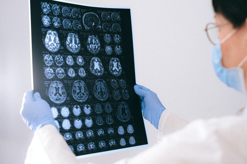
MRI images of the brain
Photo by Anna Shvets on Pexels

A world-first study published in the journal Diabetes, Obesity and Metabolism, has uncovered an association between metabolism and dementia-related brain measures, providing valuable insights about the disease.

Analysing UK Biobank data from 26 239 people, University of South Australia researchers found that those with obesity related to liver stress, or to inflammation and kidney stress, had the most adverse brain findings.

The study measured associations of six diverse metabolic profiles and 39 cardiometabolic markers, using MRI brain scan measures of brain volume, brain lesions, and iron accumulation, to identify early risk factors for dementia.

Participants with metabolic profiles associated with obesity were more likely to have adverse MRI profiles showing lower hippocampal and grey matter volumes, greater burden of brain lesions, and higher accumulation of iron.

UniSA researcher, Dr Amanda Lumsden, says the research adds a new layer of understanding to brain health.

“Dementia is a debilitating disease that affects more than 55 million people worldwide,” Dr Lumsden said.

“Understanding metabolic factors and profiles associated with dementia-related brain changes can help identify early risk factors for dementia.

“In this research, we found that adverse neuroimaging patterns were more prevalent among people who had metabolic types related to obesity.

“These people also had the highest Basal Metabolic Rate (BMR) -how much energy your body requires when resting in order to support its basic functions — but curiously, BMR seemed to contribute to adverse brain markers over and above the effects of obesity.”

Senior investigator Professor Elina Hyppönen said that the finding presents a new avenue for understanding brain health.

“This study indicates that metabolic profiles are associated with aspects of brain health. We also found associations with many individual biomarkers which may provide clues into the processes leading to dementia,” said Prof Hyppönen.

“The human body is complex, and more work is now needed to find out exactly why and how these associations arise.”

Source: University of South Australia

Obesity and Diabetes in Pregnancy may Raise Child’s ADHD Risk

Boy hanging from tree
Photo by Annie Spratt on Pexels

A recent study has found that children born to women with gestational diabetes and obesity may have twice the risk of developing attention-deficit/hyperactivity disorder (ADHD) compared to those born to mothers without obesity. The findings, published in the Journal of Clinical Endocrinology & Metabolism, also found found that in women with a healthy weight gain during pregnancy, this risk increase was not seen.

ADHD is a growing problem. According to data from 2016-2019, 6 million children aged 3–17 years have received an ADHD. Maternal obesity is a major risk factor for ADHD in children, and roughly 30% of women have obesity at their first doctor’s visit during pregnancy, rising to 47% in women with gestational diabetes. Excessive weight gain during pregnancy in this population is a risk factor for children developing ADHD.

“Our study found pregnant women with obesity and gestational diabetes had children with long-term mental health disorders such as ADHD,” said Verónica Perea, MD, PhD, of the Hospital Universitari Mutua Terrassa in Barcelona. “We did not find this association when these women gained a healthy amount of weight during pregnancy.”

Studying 1036 children born to women with gestational diabetes, the researchers found that 13% of these children were diagnosed with ADHD. When compared to mothers without obesity, the researchers found children of women with gestational diabetes and obesity were twice as likely to have ADHD compared to those born to mothers without obesity.

Notably, this association was only seen in women with gestational diabetes, obesity and excessive weight gain during pregnancy. There was no increased risk of ADHD in children of women with gestational diabetes and obesity if the amount of weight these women gained during pregnancy was within the normal range.

“It’s important for clinicians to counsel their patients on the importance of healthy weight gain during pregnancy,” Perea said.

Source: The Endocrine Society

C-section Delivery Linked to Later CVD Risk

Photo by Christian Bowen on Unsplash

A new Australian and New Zealand Journal of Public Health study has found that Australian children who were born via caesarean section (C-section) have a greater risk of cardiovascular disease (CVD) and obesity. These findings have prompted a call to limit the increasingly popular practice.

According to a Lancet review, C-sections are already known to have a number of negative outcomes, with evidence higher rates of maternal mortality and morbidity than after vaginal birth. C-sections are further associated with an increased risk of uterine rupture, abnormal placentation, ectopic pregnancy, stillbirth, and preterm birth. Short-term risks of C-section include altered immune development, an increased likelihood of allergy, atopy, and asthma, and reduced gut microbiome diversity. Associations of C-section with greater incidence of late childhood obesity and asthma are frequently reported.

Researchers used data from the Longitudinal Study of Australian Children to analyse the health outcomes of children delivered by C-section.

“C-section births have risen across the world with a disproportionately higher rate in developed countries. In Australia, the C-section birth rate has increased from 18.5% in 1990 to 36% in 2019 and nearly half of Australian babies are projected to be caesarean born by 2045,” said study author Dr Tahmina Begum.

A relationship was discovered between C-section births and certain cardiovascular disease (CVD) risk factors in children.

“Four out of six individual CVD risk components and the composite index of the five CVD risk components showed a positive association with C-section birth. Our study also provided a direct relationship between C-section and increased overweight and obesity among children at 10–12 years of age,” said Dr Fatima.

A biologically plausible link involved the gut microbiome, she said. “There’s an altered microbial load from C-section birth as compared to vaginal birth. This altered microbial ecosystem hampers the ‘gut-brain axis’ and releases some pathogenic toxins that cause metabolic damage.”

Other possible causes included foetal stress from physiological or pharmacological induction of labour during a C-section. She said the study provides important insights into health care policy and the strategic direction towards chronic disease risk reduction.

“Growing rates of C-sections conducted for non-clinical reasons is a major public health concern that calls for a reduction in the rate of unnecessary C-sections and their associated human and economic costs,” said Dr Begum.

Source: James Cook University

Why Only Some Obese Patients Develop Diabetes

A 3D map of the islets in the human pancreas. Source: Wikimedia

Oregon State University researchers have used a new analytical method to shed light on an enduring mystery in type 2 diabetes: why some obese patients develop diabetes and others don’t. The reason is down to a genetic pathway linking diet and gut microbiota to macrophages and white adipose tissue. Their findings appear in the Journal of Experimental Medicine.

Type 2 diabetes is frequently associated with obesity. Ins some patients, that means insulin resistance. Later stages of the disease sees the pancreas producing insufficient insulin to maintain normal glucose levels.

In either case, hyperglycaemia is the result, which, if left untreated, impairs many major organs, sometimes to disabling or life-threatening degrees. Overweight status is a key risk factor for type 2 diabetes, often a result of eating too much fat and sugar in combination with low physical activity.

Associate Professors Andrey Morgun and Natalia Shulzhenko of OSU and Giorgio Trinchieri of the National Cancer Institute developed a novel analytical technique, multi-organ network analysis, to explore the mechanisms behind early-stage systemic insulin resistance.

The scientists sought to learn which organs, biological pathways and genes are playing roles.

The findings showed that a particular type of gut microbe leads to white adipose tissue containing macrophage cells associated with insulin resistance.

“Our experiments and analysis predict that a high-fat/high-sugar diet primarily acts in white adipose tissue by driving microbiota-related damage to the energy synthesis process, leading to systemic insulin resistance,” said Morgun. “Treatments that modify a patient’s microbiota in ways that target insulin resistance in adipose tissue macrophage cells could be a new therapeutic strategy for type 2 diabetes.”

The human gut microbiome is incredibly complex, comprising more than 10 trillion microbial cells from about 1000 different bacterial species.

Associate Profs Morgun and Shulzhenko, in earlier research developed a computational method, transkingdom network analysis, that predicts specific types of bacteria controlling the expression of mammalian genes connected to specific medical conditions such as diabetes.

“Type 2 diabetes is a global pandemic, and the number of diagnoses is expected to keep increasing over the next 10 years,” Associate Prof Shulzhenko said. “The so-called ‘western diet’ – high in saturated fats and refined sugars – is one of the primary factors. But gut bacteria have an important role to play in mediating the effects of diet.”

In the new study, the scientists made use of transkingdom network analysis and multi-organ network analysis. Mouse experiments examined the intestine, liver, muscle and white adipose tissue, and the molecular signature (gene expression) of white adipose tissue macrophages in obese human patients.

“Diabetes induced by the western diet is characterised by microbiota-dependent mitochondrial damage,” Associate Prof Morgun said. “Adipose tissue has a predominant role in systemic insulin resistance, and we characterised the gene expression program and the key master regulator of adipose tissue macrophage that are associated with insulin resistance. We discovered that the Oscillibacter microbe, enriched by a western diet, causes an increase of the insulin-resistant adipose tissue macrophage.”

The researchers add, however, that Oscillibacter is likely not the only microbial regulator for expression for the genetic pathway they discovered, while clearly instrumental, is probably not the only important pathway, depending on which gut microbes are present.

“We previously showed that Romboutsia ilealis worsens glucose tolerance by inhibiting insulin levels, which may be relevant to more advanced stages of type 2 diabetes,” Shulzhenko said.

Source: Oregon State University

Bariatric Surgery Reduces Cancer Risks with Obesity

Obesity
Image source: Pixabay CC0

A study published in JAMA shows that weight loss through bariatric surgery for adults with obesity was associated with a 32% lower risk of developing cancer and a 48% lower risk of cancer-related death compared with those who did not have the surgery.

Rising obesity numbers are being seen all over the world. The International Agency for Research on Cancer describes 13 types of cancer as obesity-associated cancers such as endometrial cancer, postmenopausal breast cancer, and cancers of the colon, liver, pancreas, ovary and thyroid.

Lead author of the study, Ali Aminian, MD, at Cleveland Clinic, said that bariatric surgery is currently the most effective treatment for obesity. “Patients can lose 20 to 40% of their body weight after surgery, and weight loss can be sustained over decades. The striking findings of this study indicate that the greater the weight loss, the lower the risk of cancer,” said Dr Aminian.

From 2004 and 2017, the SPLENDID (Surgical Procedures and Long-term Effectiveness in Neoplastic Disease Incidence and Death) study matched a group of 5053 adult patients with obesity who had bariatric surgery 1:5 to a control group of 25 265 patients with obesity who did not undergo the surgery.

After 10 years, 2.9% of patients in the bariatric surgery group and 4.9% of patients in the non-surgical group developed an obesity-associated cancer.

After 10 years, 0.8% of patients in the surgery group and 1.4% of patients in the non-surgical group died from cancer, indicating that bariatric surgery is associated with a 48% lower cancer mortality risk.

Researchers noted that the benefits of bariatric surgery were seen in a wide range of study participants in terms of age, sex and race. In addition, benefits were similarly observed after both gastric bypass and gastric sleeve operations.

“According to the American Cancer Society, obesity is second only to tobacco as a preventable cause of cancer in the United States,” said the study’s senior author, Steven Nissen, MD, Chief Academic Officer of the Heart, Vascular and Thoracic Institute. “This study provides the best possible evidence on the value of intentional weight loss to reduce cancer risk and mortality.”

Numerous studies have shown the health benefits of bariatric or weight-loss surgery in patients with obesity. The Cleveland Clinic-led STAMPEDE study showed that following bariatric surgery, significant weight loss and control of type 2 diabetes last over time. The SPLENDOR study showed that in patients with fatty liver, bariatric surgery decreases the risk of the progression of liver disease and serious heart complications.

The SPLENDID study adds important findings to the literature focused on the link between obesity and cancer. Given the growing epidemic of obesity worldwide, these findings have considerable public health implications.

“Based on the magnitude of benefit shown in our study, weight loss surgery can be considered in addition to other interventions that can help prevent cancer and reduce mortality,” said Jame Abraham, M.D., chairman of the Hematology and Medical Oncology Department at Cleveland Clinic. “Further research needs to be done to understand the underlying mechanisms responsible for reduced cancer risk following bariatric surgery.”

Source: Cleveland Clinic

Obesity in Women Linked to Increased Fracture Risk

Obesity
Image source: Pixabay CC0

Women with obesity and overweight, particularly women with high waist circumference, are more susceptible to fractures than those with normal weight, according to new research presented at the European Congress on Obesity (ECO). In men, however, underweight, not overweight, is associated with a greater risk of broken bones.

Obesity has long been thought to help protect against fractures. This is because mechanical loading on bones, which increases with body weight, helps increase bone mineral density, an important determinant of bone strength.

However, recent studies have suggested that the relationship between obesity and fracture risk varies depending on sex, the skeletal site studied and definition of obesity used (body mass index [BMI] vs waist circumference).

To find out more, Dr Anne-Frederique Turcotte, Endocrinology and Nephrology Unit, CHU de Quebec Research Centre, Quebec City, Canada, and colleagues, analysed data from CARTaGENE, a prospective population-based cohort of almost 20 000 individuals aged 40-70 years from Quebec, Canada.

In women, greater waist circumference (WC) was linearly associated with an increased risk of fracture. For each 5cm (two inch) increase in WC, the risk of fracture at any site was 3% higher and the risk of a distal lower limb fracture was 7% higher.  The association between WC and ankle fractures was particularly strong.

In women, greater BMI was associated with a greater risk of distal lower limb fractures. Compared with women with a BMI of 25 kg/m², those with a BMI of 27.5-40 kg/m² showed a greater risk of distal lower limb fractures.  The increase in risk rose linearly from 5% in those with a BMI of 27.5 kg/m², to 40% in those with a BMI of 40 kg/m².

Women with a BMI of 22.5 kg/m² had a 5% lower risk of distal lower limb fractures than those with a BMI of 25 kg/m².

It isn’t known why obesity is associated with a higher risk of fractures in women.  However, most fractures are a result of a fall and falls are more common in people with obesity.  The ankle, unlike the hip and thighbone, is not protected by soft tissue, which could make it more prone to breaking during a fall.

Dr Turcotte added: “Waist circumference was more strongly associated with fractures in women than BMI.  This may be due to visceral fat – fat that is very metabolically active and stored deep within the abdomen, wrapped around the organs – secreting compounds that adversely affect bone strength.

“We also know that people with obesity take longer to stabilise their body, when they trip, for example.  This is particularly pronounced when weight is concentrated at the front of the body, suggesting that individuals with distribution of body fat in the abdominal area may be at higher risk of falling.” 

In men, increases in BMI and WC were not significantly associated with fractures.  However, men with underweight were at higher risk of distal upper limb fractures than those with normal weight.  Men with a BMI ≤17.5 kg/m² were twice as likely to have distal upper limb fracture as men with a BMI of 25 kg/m².

The researchers say a larger number of fractures in men is needed to determine whether this is a true result or whether the pattern for men follows that for women.

The analyses were adjusted for a number of potential confounders: age, menopausal status, ethnicity, marital status, education, income, area of residence, smoking status, alcohol consumption, physical activity level, supplemental calcium and vitamin D intake, history of fracture and comorbidities and medications known to influence fracture risk.

The study authors said: : “Our findings show that the relationship between obesity and fractures is complex and varies by sex. In women, there was a linear relationship between waist circumference and the incidence of fracture at any site and at the distal lower limb, particularly at the ankle.

“Similar results were observed for women with a BMI between 27 and 40 kg/m². In men however, there was no relationship between obesity and the risk of fracture, although a BMI in the underweight range was associated with a higher risk of some fractures.”

Source: EurekAlert!

Obesity in Mice Causes AD Treatments to Backfire

Mouse
Photo by Kanasi on Unsplash

In a new study published in Nature, researchers found that treatments that were effective for atopic dermatitis (AD) in lean mice actually worsened the condition in obese mice.

Tracking the development of AD in obese and lean mice, the researchers found that obese mice developed more inflammation and more severe AD. This increased inflammation was present even after obese mice lost weight. There were similar results in an experimental model of asthma, with obese mice developing more inflammation.

The researchers next looked in detail at immune cells called T cells in lean and obese mice with AD. Lean mice had more TH2 cells, a class of T cells known to play a role in the development of AD. Obese mice had more of a class of T cells called TH17. These cells trigger a different type of inflammation.

Similar trends were seen in blood samples taken from people. Markers of TH17 cell activity increased along with body mass index (BMI) in a database of serum collected from people with AD. Conversely, in samples from patients with severe asthma, TH2 cell activity decreased as BMI increased.

Drugs that block TH2 cell activity are used in the treatment of severe AD as well as asthma and other inflammatory conditions. The researchers tested antibodies to block TH2 cell activity in lean and obese mice with severe AD. While the antibodies reduced skin inflammation in lean mice as expected, they made the condition worse in obese mice. Analysis of immune cells suggested that blocking TH2 cell activity in the obese mice worsened other forms of inflammation.

Obese mice were also found to have less activity of peroxisome proliferator-activated receptor-γ (PPARɣ) in their TH2 cells. When lean mice were engineered to lack PPARɣ, their inflammatory response resembled that of obese mice.

Drugs that increase PPARɣ activity increase insulin sensitivity and are approved for the treatment of type 2 diabetes. The researchers found that giving one of these drugs to obese mice changed their inflammatory response to resemble that of lean mice. It also restored their sensitivity to the antibodies that block TH2 cell activity.

“Our findings demonstrate how differences in our individual metabolic states can have a major impact on inflammation, and how available drugs might be able to improve health outcomes,” said Dr Ronald Evans from the Salk Institute, who helped lead the work.

Source: National Institutes of Health

Weekly Prednisone Could Reduce Obesity and Help in Muscular Dystrophy

Source: Pixabay CCO

In a study on obese mice fed a high-fat diet, receiving prednisone once weekly improved their exercise endurance and strength, and their reduced weight. The study was published in the Journal of Experimental Medicine. A previous study had found that weekly prednisone was helpful in muscular dystrophy.

“These studies were done in mice. However, if these same pathways hold true in humans, then once-weekly prednisone could benefit obesity,” explained senior author Dr Elizabeth McNally.

“Daily prednisone is known to promote obesity and even metabolic syndrome – a disorder with elevated blood lipids and blood sugar and weight gain,” Dr McNally said. “So, these results, in which we intermittently ‘pulse’ the animals with once-weekly prednisone, are strikingly different. Obesity is a major problem, and the idea that once-weekly prednisone could promote nutrient uptake into muscle might be an approach to treating obesity.”

The once-weekly prednisone, a glucocorticoid steroid, promoted nutrient uptake into the muscles. The researchers also found these mice had increased adiponectin levels, an adipocyte-secreted hormone involved in protecting against diabetes and insulin resistance. Mice that were already obese from eating a high-fat diet were also found to benefit after once-weekly prednisone, experiencing increased strength, running capacity and lower blood glucose.

Most knowledge on steroids like prednisone is derived from studies of daily doses of prednisone

“We see a very different outcome when it is taken once a week,” Dr McNally said. “We need to fine tune dosing to figure out the right amount to make this work in humans, but knowing adiponectin might be one marker could provide a hint at determining what the right human dose is.”

Dr McNally described the weekly dose as “a bolus, or spike, of nutrients going into your muscle.”

“We think there is something special about promoting this spike of nutrients into muscle intermittently, and that it may be an efficient way to improve lean body mass,” she added.

“What is exciting to me about this work is the finding that a simple change in the dosing frequency can transform glucocorticoid drugs from inducers to preventers of obesity,” said corresponding author Mattia Quattrocelli. “Chronic once-daily intake of these drugs is known to promote obesity. Here we show that dosing the same type of drug intermittently – in this case, once weekly – reverses this effect, promotes muscle metabolism and energy expenditure, and curtails the metabolic stress induced by a fat-rich diet.”

Many patients take prednisone daily for different immune conditions, which has side effects including weight gain and even muscle atrophy with weakness. Investigators want to determine whether patients can get the same immune benefit with intermittent prednisone dosing, which could be much more beneficial to the muscle.

Dr McNally’s team previously found that intermittent prednisone administration was helpful for muscular dystrophy, showing once weekly prednisone improved strength, recently reporting that a pilot trial in humans with muscular dystrophy in which one weekly dose of prednisone improved lean mass.

McNally wants to identify biomarkers most critical to measure a beneficial response to prednisone.

“If we can determine how to choose the right dose of prednisone that minimises atrophy factors and maximises positive markers like adiponectin, then we can really personalise the dosing of prednisone,” she said.

The group also recently showed that weekly prednisone uses strikingly different molecular pathways to strengthening the muscle in male versus female mice, based on a new study just published in the Journal of Clinical Investigation by Isabella Salamone, a graduate student in Dr McNally’s lab.

The benefits of weekly prednisone are linked to circadian rhythms, according to another new study published in Science Advances. Human cortisol and steroid levels spike early in the morning before awakening.

“If you don’t give the drug at the right time of day, you don’t get the response,” Quattrocelli said. “In mice, we obtained good effects with intermittent prednisone in muscle mass and function when we dose them at the beginning of their daytime. Mice have a circadian rhythm inverted to us, as they generally sleep during the daytime and are active at night. This could mean that the optimal dosing time for humans during the day could be in the late afternoon/early evening, but this needs to be appropriately tested.”

Conducting these studies in mice is a major limitation, Dr McNally said.

“While we are encouraged by the pilot study in humans with muscular dystrophy, mouse muscles have more fast-twitch fibres than humans, and slow-twitch muscle could be different,” Dr McNally said. “More studies are needed to try to better understand whether these same mechanisms work in human muscles.”

Source: Northwestern University

High Lipid Levels Even More Damaging than Previously Believed

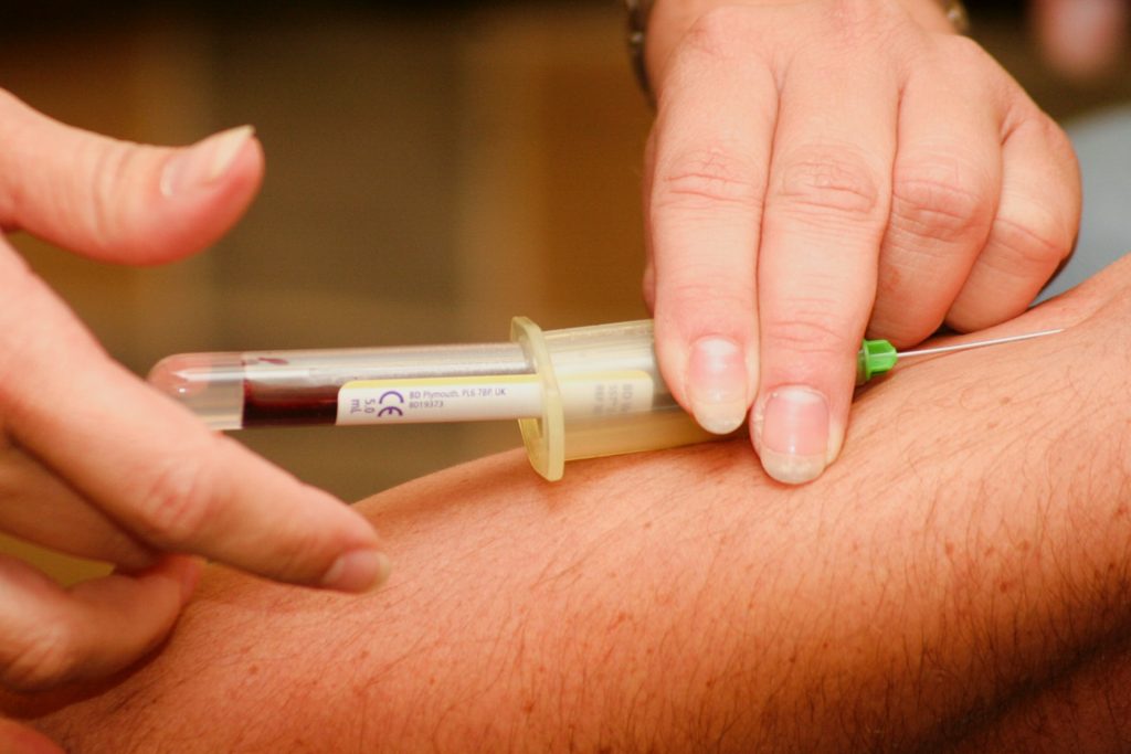
Blood sample being drawn
Photo by Hush Naidoo Jade Photography on Unsplash

High lipid levels in people with type 2 diabetes and obesity are more harmful than previously thought, according to findings from a new study which found that stressed cells can damage nearby cells.

In patients with metabolic diseases, elevated lipid levels in the blood create stress in muscle cells – a reaction to changes outside the cell which damage their structure and function.

The study, published in Nature Communications, shows that these stressed-out cells give off a signal which can be passed on to other cells.

The signals, known as ceramides, may confer a short-term protective benefit, because they are part of a mechanism designed to reduce stress in the cell. But in long term conditions such as metabolic diseases, the signals can actually kill the cells and worsen symptoms and the illness.

High lipid levels have long been known to damage tissues and organs, contributing to the development of cardiovascular and metabolic diseases including type 2 diabetes, a condition which can be caused by obesity.

Professor Lee Roberts, who supervised the research, said: “Although this research is at an early stage, our discovery may form the basis of new therapies or therapeutic approaches to prevent the development of cardiovascular and metabolic diseases such as diabetes in people with elevated blood fats in obesity.”

In the lab, the team replicated the blood lipid levels observed in humans with metabolic disease by exposing skeletal muscle cells to palmitate, a fatty acid. The cells began to transmit the ceramide signal.

When these cells were mixed with others which had not been previously exposed to lipids, the researchers found that they communicated with each other, transporting the signal in packages called extracellular vesicles.

The experiment was reproduced in human volunteers with metabolic diseases and gave comparable results. The findings provide a completely new angle on how cells respond to stress, with important consequences for our understanding of certain metabolic diseases including obesity.

Professor Roberts said: “This research gives us a novel perspective on how stress develops in the cells of individuals with obesity, and provides new pathways to consider when looking to develop new treatments for metabolic diseases.

“With obesity an ever-increasing epidemic, the burden of associated chronic disease such as type 2 diabetes necessitates new treatments. We hope the results of our research here open a new avenue for research to help address this growing concern.”

Source: University of Leeds