Targeting Immune Suppression to Overcome Melanoma Resistance

For patients with advanced melanoma without BRAF mutation who no longer respond to immune checkpoint inhibitors, treatment options remain frustratingly limited. A new study from Vanderbilt researchers led by Professor Emerita of Pharmacology Ann Richmond outlines a promising therapeutic strategy that may re-sensitise these resistant tumours to immunotherapy.
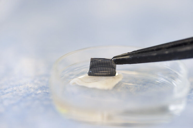
The research introduces a three-drug combination that enhances immune activity and suppresses tumour-promoting immune cells by leveraging a low dose of the MEK inhibitor trametinib and multi-kinase inhibitor rigosertib alongside a CD40 agonist to shift the tumour microenvironment toward immune activation. Notably, all three agents have been either approved by the U.S. Food and Drug Administration or are currently in clinical trials, which may speed their path to patient testing.
“While agonist CD40 therapy can be helpful for treatment of melanoma, this therapy also induces the CD11b+ B regulatory cells that suppress the T cell response to tumours,” Richmond said. “We showed that combining CD40 therapy with trametinib and rigosertib prevents the induction of these B regulatory cells.”
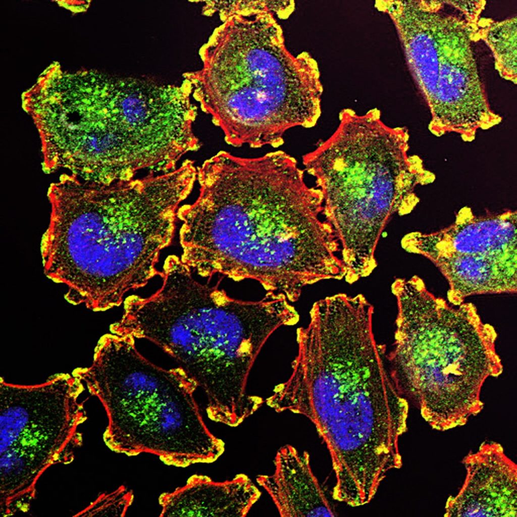
Immune checkpoint inhibitors have become a mainstay of melanoma treatment, working by releasing the molecular “brakes” that prevent T cells from attacking cancer. But resistance to ICI is common in metastatic melanoma, especially in tumours that evolve immune-suppressive microenvironments. While CD40 agonists can activate immune cells, this therapy also unexpectedly expands CD11b+ regulatory B cells.
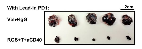
By combining CD40 activation with MEK and PI3K inhibition, the researchers blocked the expansion of suppressive B cells while retaining the benefits of CD40 stimulation. In preclinical mouse models of melanoma, the triple combination not only suppressed tumour growth but also restored responsiveness to checkpoint blockade.
Key findings
- B cells as a resistance mechanism: CD40 therapy alone induced regulatory B cells that dampen T cell–mediated tumor immunity.
- Triple combination prevents immune suppression: Co-treatment with trametinib and rigosertib blocked the agonist CD40 induction of regulatory B cells, allowing immune responses to proceed.
- ICIs regain effectiveness: The drug cocktail slowed tumor progression and re-sensitized resistant melanomas to anti-PD-1 therapy.
Translational promise
Because trametinib, rigosertib, and CD40 agonists are already in human trials or approved for other indications, this therapeutic strategy may advance more quickly than approaches requiring new drug development. Richmond’s team sees potential for testing the triple therapy in clinical trials for melanoma patients who have progressed on ICI.
“This approach provides a new route to enhance antitumor immunity in patients with tumors that no longer respond to immunotherapy,” Richmond said.
Source: Vanderbilt University