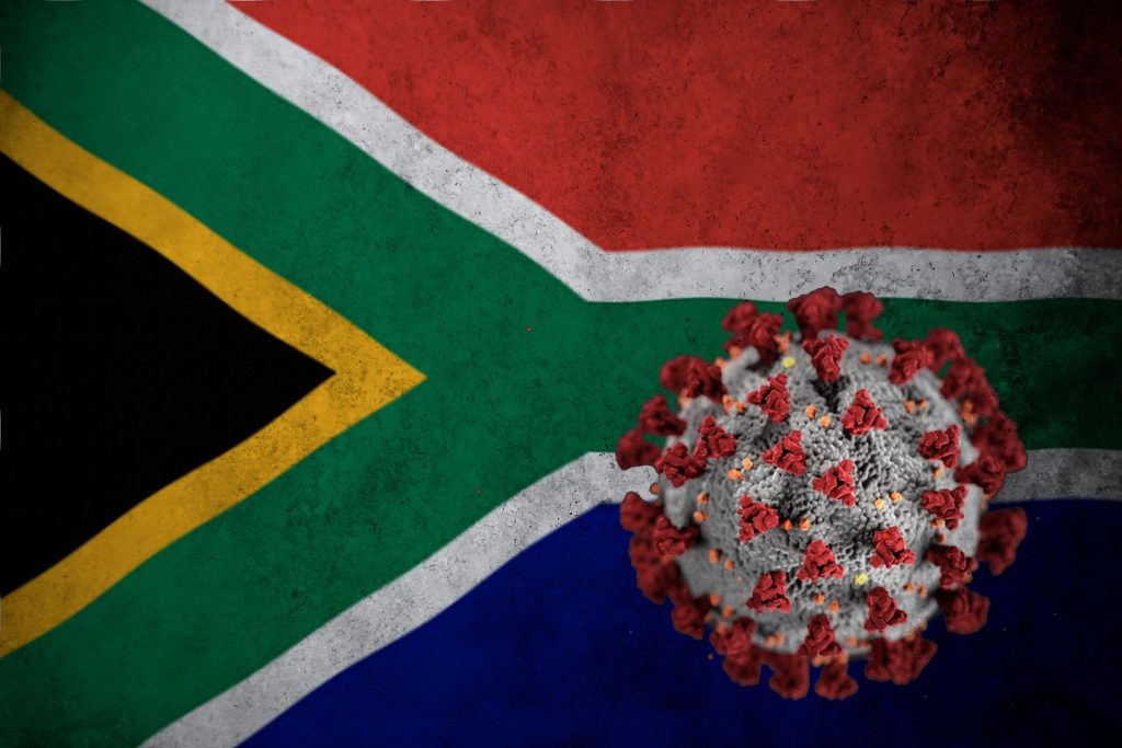
The SARS-CoV-2 virus can infect cardiac pacemaker cells, causing the cells to undergo self-destruct by ferroptosis according to a preclinical study reported in Circulation Research. This may explain the heart arrhythmias that are commonly observed in COVID patients.
In the study, the researchers used an animal model as well as human stem cell-derived pacemaker cells to show that SARS-CoV-2 can readily infect pacemaker cells and trigger a process called ferroptosis, where cells self-destruct, releasing damaging reactive oxygen molecules.
“This is a surprising and apparently unique vulnerability of these cells — we looked at a variety of other human cell types that can be infected by SARS-CoV-2, including even heart muscle cells, but found signs of ferroptosis only in the pacemaker cells,” said study co-senior author Professor Shuibing Chen.
Arrhythmias, including tachycardia and bradycardia, has been observed in some COVID patients, and multiple studies link these arrhythmias to worse COVID outcomes. But how the coronavirus caused these remained unclear.
In the new study, the researchers examined golden hamsters (one of the only lab animals that reliably develops COVID-like signs from SARS-CoV-2 infection) and found evidence that following nasal exposure, the virus can infect the sinoatrial node, which is the natural cardiac pacemaker.
The researchers then induced human embryonic stem cells to mature into cells closely resembling sinoatrial node cells. They showed that these induced human pacemaker cells can be infected by SARS-CoV-2 as they express ACE2 receptors. Large increases in inflammatory immune gene activity were also seen in the infected cells.
The team’s most surprising finding, however, was that the pacemaker cells, in response to the stress of infection, showed clear signs of a cellular self-destruct process called ferroptosis, which involves accumulation of iron and the runaway production of reactive oxygen molecules. The scientists were able to reverse these signs in the cells using compounds that are known to bind iron and inhibit ferroptosis.
“This finding suggests that some of the cardiac arrhythmias detected in COVID patients could be caused by ferroptosis damage to the sinoatrial node,” said co-senior author Dr Robert Schwartz
While COVID patients could in principle be treated with ferroptosis inhibitors specifically to protect sinoatrial node cells, antiviral drugs that block the effects of SARS-CoV-2 infection in all cell types would be preferable, the researchers said.
The researchers plan to continue to use their cell and animal models to investigate sinoatrial node damage in COVID and other settings.
“There are other human sinoatrial arrhythmia syndromes we could model with our platform,” said co-senior author Dr. Todd Evans. “And, although physicians currently can use an artificial electronic pacemaker to replace the function of a damaged sinoatrial node, there’s the potential here to use sinoatrial cells such as we’ve developed as an alternative, cell-based pacemaker therapy.”
Researchers have revealed biological reasons for how disease progression happens and why a certain population of asthma patients are less susceptible to severe COVID.
This research, published in PNAS, shows the importance of the well-known cytokine interleukin-13 (IL-13) in protecting cells against SARS-CoV-2, something which helps explain why people with allergic asthma fare better than the general population despite having a chronic lung condition. However, the same cannot be said for individuals with other diseases, such as chronic obstructive pulmonary disease (COPD) or emphysema, who are at very high risk of severe COVID.
“We knew there had to be a bio-mechanistic reason why people with allergic asthma seemed more protected from severe disease,” said Assistant Professor Camille Ehre, PhD, senior author of the paper. “Our research team discovered a number of significant cellular changes, particularly due to IL-13, leading us to conclude that IL-13 plays a unique role in defence against SARS-CoV-2 infection in certain patient populations.”
Although cytokines like IL-13 cannot be used as therapies because they trigger inflammation, it is important to understand natural molecular pathways that cells use to protect themselves from pathogen invasion, as these studies have the potential to reveal new therapeutic targets.
Many health factors increase a person’s risk of severe COVID, but during the pandemic, epidemiologists found that people with allergic asthma were less susceptible to severe disease.
“These are patients with asthma caused by allergens, such as mould, pollen, and dander,” said A/Prof Ehre. “To find out why they are less susceptible, we investigated specific cellular mechanisms in primary human airway epithelial cell cultures.”
Genetic analysis human airway cell cultures infected with SARS-CoV-2 revealed that the expression of ACE2 governed which cell types were infected and their viral load.
Electron microscopy (EM) identified an intense exodus of virus from infected ciliated cells, which move mucus along the airway surface. EM also revealed severe cytopathogenesis – changes inside human cells due to viral infection. And these changes culminating in ciliated cells (packed with virions) shedding away from the airway surface.
“This shedding is what provides a large viral reservoir for spread and transmission of SARS-CoV-2,” A/Prof Ehre said. “It also seems to increase the potential for infected cells to relocate to deeper lung tissue.”
Further experiments on infected airway cells revealed that a major mucus protein called MUC5AC was depleted inside cells, likely because the proteins were secreted to try to trap invading viruses. But the virus load kept increasing because the cells tasked with producing MUC5AC were overwhelmed in the face of a rampant viral infection.
The researchers knew from epidemiological studies that allergic asthma patients—known to overproduce MUC5AC—were less susceptible to severe COVID. A/Prof Ehre and colleagues also knew the cytokine IL-13 increased MUC5AC secretion in the lungs when asthma patients faced an allergen.
The scientists decided to mimic asthmatic airways by treating human airway cells with IL-13. They then measured viral titres, viral mRNA, the rate of infected cell shedding, and the overall number of infected cells. Each one was significantly decreased. They found this remained true even when mucus was removed from the cultures, suggesting other factors were involved in the protective effects of IL-13 against SARS-CoV-2.
Bulk RNA-sequencing analyses revealed that IL-13 upregulated genes that control glycoprotein synthesis, ion transport, and antiviral processes – all of which are important in airway immune defence. They also showed that IL-13 reduced the expression of the viral receptor, ACE2, as well as reducing the amount of virus inside cells and cell-to-cell viral transmission.
Taken together, these findings indicate that IL-13 significantly affected viral entry into cells, replication inside cells, and spread of virus, thus limiting the virus’s ability to find its way deeper into the airways to trigger severe disease.
“We think this research further shows how important it is to treat SARS-CoV-2 infection as early as possible,” A/Prof Ehre said. “And it shows just how important specific mechanisms involving ACE2 and IL-13 are, as we try our best to protect patients from developing severe infections.”
New computational simulations of the behaviour of the SARS-CoV-1 and SARS-CoV-2 spike proteins before they fuse with human cell receptors show that SARS-CoV-2, is in fact more stable and slower changing than SARS-CoV-1 that caused the SARS epidemic in 2003.
Though severe acute respiratory syndrome coronaviruses 1 and 2 (SARS-CoV-1 and SARS-CoV-2) have striking similarities, why the latter is more transmissible remains unclear.
The spike proteins of each, which bind to host cell angiotensin converting enzyme 2 (ACE-2), otherwise known as the human cell receptor, have been proposed as the reason for their difference in transmissibility. A more detailed understanding of the spike proteins prior to binding could lead to the development of better vaccines and medications.
The new finding, which appears in the Journal of Biological Chemistry, does not necessarily mean that SARS-CoV-2 is more likely to bind to cell receptors, but it does mean that its spike protein has a better chance of effective binding.
“Once it finds the cell receptor and binds to it, the SARS-CoV-2 spike is more likely to stay bound until the rest of the necessary steps are completed for full attachment to the cell and initiation of cell entry,” explained Associate Professor Mahmoud Moradi, of the Fulbright College of Arts and Sciences.
To determine differences in conformational behaviour between the two versions of the virus, the researchers performed equilibrium and nonequilibrium simulations of the molecular dynamics of SARS-CoV-1 and SARS-CoV-2 spike proteins, leading up to binding with cell angiotensin converting enzyme 2.
Equilibrium simulations allow the models to evolve spontaneously on their own time, while nonequilibrium simulations change according to external input. The former is less biased, but the latter is faster and allows for many more simulations to run. Both methodological approaches provided a consistent picture, independently demonstrating the same conclusion that the SARS-CoV-2 spike proteins were more stable.
The models revealed other important findings, namely that the energy barrier associated with activation of SARS-CoV-2 was higher, meaning the binding process happened slowly. Slow activation allows the spike protein to evade human immune response more efficiently, because remaining in an inactive state longer means the virus cannot be attacked by antibodies that target the receptor binding domain.
Researchers understand the importance of the receptor-binding domain (RBD), which viruses use to gain entry to human cells. The team’s modelling confirms the importance of the RBD but also suggest that other domains, such as the N-terminal domain, could play a crucial role in the different binding behaviour of SARS-CoV-1 and -2 spike proteins.
N-terminal domain of a protein is a domain located at the N-terminus or simply the start of the polypeptide chain, as opposed to the C-terminus, which is the end of the chain. Though it is near the receptor-binding domain and is known to be targeted by some antibodies, the function of the N-terminal domain in SARS-CoV-1 and -2 spike proteins is not fully understood. Moradi’s team is the first to find evidence for potential interaction of the N-terminal domain and the receptor binding domain.
“Our study sheds light on the conformational dynamics of the SARS-CoV-1 and SARS-CoV-2 spike proteins,” Moradi said. “Differences in the dynamic behaviour of these spike proteins almost certainly contribute to differences in transmissibility and infectivity.”
New research published in the journalJCI Insight shows that immune responses to the Pfizer-BioNTech mRNA vaccine differ significantly in individuals depending on whether or not they had a prior COVID infection. Notably, those who had COVID before vaccination produced a surge of antibodies after the first dose, with little or no increase seen after the second dose. The opposite pattern was observed in infection-naïve individuals.
“Our study shows that the presence of immune memory induced by prior infection alters the way in which individuals respond to SARS-CoV-2 mRNA vaccination,” explained first author Professor Steven G. Kelsen. “The lack of response after the second vaccine dose in previously infected individuals is especially relevant, because it could mean that some people may require only one dose or could potentially skip the booster shot.”
Prof Kelsen and colleagues carried out the study in health care workers, some having previously tested positive for SARS-CoV-2 infection and others never having been infected. The researchers measured levels of neutralising antibodies in blood samples taken at three different time points, including before vaccination and after each vaccine dose. They also performed qualitative assessment for local reactions and systemic symptoms, such as fever, headache, and fatigue, associated with vaccination.
While levels of neutralising antibodies hit their maximum in some people with prior COVID after the first vaccine dose, individuals with no history of infection showed massive responses after the second dose. Those high levels also plummeted quickly, while the COVID group retained longer lasting immunity, despite the lack of response to a second dose. However, prior infection was also linked to more frequent and longer-lasting adverse reactions to the vaccine.
“Previous studies had similarly reported long-lasting immunity and strong immune reactions in COVID patients,” Prof Kelsen said. “We now provide new information on how prior infection interacts with vaccination in terms of measurable immune response and how individuals react to mRNA vaccines based on infection history.”
The next steps for Prof Kelsen and collaborators are to modify their neutralising antibody assay to detect Omicron and other SARS-CoV-2 variants. “We also are interested in understanding how long protection from a booster dose of the vaccine lasts,” he said.
Olfactory dysfunction in COVID patients is common. Researchers recently searched the medical literature for studies reporting changes in olfactory structures detected through imaging tests of patients with COVID, and found that swelling in nasal passages is responsible for some temporary olfactory dysfunction. Their results in were published in The Laryngoscope.
A recent meta-analysis based on 83 studies provided high-quality evidence of a combined prevalence of olfactory dysfunction in 47.9% of COVID patients. Olfactory dysfunction is diagnosed more commonly among female patients and outpatients.
The researchers observed abnormality in olfactory clefts, which provide a crucial channel for airborne molecules to reach sensory olfactory neurons. The rate of abnormalities was nearly 16-fold higher in patients with COVID and olfactory dysfunction (63%) compared with controls (4%).
“Before this study, most scientists thought that the loss of smell in COVID was mainly due to inflammation and damage to the olfactory nerves. Now, we have compiled evidence from medical imaging that COVID loss of smell is also due to swelling and blockage of the passages in the nose that conduct smells,” said senior author Neville Wei Yang Teo, MRCS, MMed, of Singapore General Hospital.
“We think this is good news for patients who want to recover their sense of smell, since these blockages are expected to resolve with time, while nerve damage in comparison would likely be more difficult to recover from,” added lead author Claire Jing-Wen Tan, of the National University of Singapore. “These findings may not fully account for those who suffer from prolonged olfactory dysfunction, however, and further studies that evaluate patients in this group may provide more information.”
In an interview with 702, CSIR senior researcher Dr Ridhwaan Suliman warned that 20% of South Africans still do not have any immunity to COVID, either from vaccination or prior infection. Since the start of the pandemic in South Africa, he has been tweeting his graphs of infections and explaining the science to the public.
Explaining the figure, he said: “It’s an extrapolation of recent research studies that show sero-prevalence levels across the country, up to about 80% currently. That means levels of immunity of people having previous infections, so natural immunity, plus acquired immunity through vaccination. So based on the 80% sero-prevalence levels, it means there’s still 20% that are susceptible… and 20% of South Africa’s population leaves 12 million people who don’t have levels of immunity or sero-prevalence currently.”
This number is however greatly increased from a previous survey conducted in January 2021, which reported a 19.1% rate.
He pointed out that this leave South Africa with a large number of people who are immune-naïve to COVID, who are therefore at risk of more severe consequences such as hospitalisation and death. However, the high levels of immunity means that, as seen in the Omicron wave, outcomes are reduced in severity.
Interviewer Bruce Whitfield asked Dr Suliman his opinion of prospective waves, as evidenced by increasing infections in countries like the US and China, and indeed, his own observations of queues starting to build up outside mobile COVID testing stations.
Dr Suliman replied that although the infections have been driven by the highly transmissible Omicron B.A.2 variant which is also dominant in South Africa, it “hasn’t resulted in a further uptick or resurgence following our fourth wave of the original Omicron strain, and so we’re currently in an ‘inter-wave’ period with low levels of transmission.”
He says that the situation is encouraging, with hospitalisations at their lowest levels seen since before the first wave in May 2020, but that this is still an inter-wave period.
Regarding when the fifth wave would be, Dr Suliman pointed out that each of the previous four waves “have been very cyclical or regular, with three months in between each wave.” Based on this, he said that he expects another wave is likely around the end of April or beginning of May.
However, he said that this should not be looked on with fear “because even with a surge in high levels of infection we do have high levels of population immunity which we hope will continue, and we will have less severe outcomes of hospitalisation and death even with those high levels.”
COVID vaccination during pregnancy is not associated with a higher risk of pregnancy complications, according to a large scale Swedish and Norwegian study published in the journal JAMA.
The study, which comprised almost 160 000 pregnancies, found there to be no increase in the risk of preterm birth, growth retardation, low Apgar scores at birth or the need for neonatal care after vaccination against COVID during pregnancy.
“The results are reassuring and can hopefully make pregnant individuals more willing to get vaccinated,” said co-first author Anne Örtqvist Rosin, researcher at the Department of Medicine, Karolinska Institutet.
Studies have shown that, compared to non-pregnant peers, pregnant women are at risk of serious COVID requiring intensive care with a higher risk of death. Preterm births are also more likely in pregnant women with severe COVID. COVID vaccines have been available in Sweden and Norway since January 2021, and in May 2021 Sweden recommended all pregnant individuals to have a COVID jab, followed in August by Norway.
“We’re still seeing that vaccination rates are lower than in the rest of the population, so it’s likely that there’s some concern about how the vaccines affect the pregnant individual and the foetus,” explained Dr Örtqvist Rosin. “When the vaccines were produced, pregnant women were not included in the large clinical studies, and until now there have been no population-based data about any risk there might be to them.”
The researchers linked Sweden’s Pregnancy Register and Norway’s Medical Birth Register to each country’s vaccination register to obtain data on if and when pregnant individuals were vaccinated and with which vaccine. The study included a total of 157 521 individuals who gave birth between January 2021 and January 2022, of whom 18% had been vaccinated. It was found that vaccinated individuals were at no higher risk than unvaccinated of developing one of the studied complications.
The majority of the pregnant individuals included in the study were vaccinated after week 12 in accordance with current recommendations, and 95% received an mRNA vaccine. This should be factored in when interpreting the results, which were similar for the different mRNA vaccines regardless of whether one or two doses were given. Vaccination during the third trimester and vaccination with the Moderna vaccine was associated with a slightly lower risk of needing neonatal care.
A possible benefit of vaccination during pregnancy is that the antibodies generated pass through the placenta, conferring a certain degree of protection against COVID to the newborn baby.
“We’re now planning to study how long this protection lasts, and if SARS-CoV-2 infection or vaccination during pregnancy has any other lasting effects on the child’s health,” said joint last author Professor Olof Stephansson at the Karolinska Institutet .
At-home monitoring of blood oxygen levels is a safe way for people with COVID to see if their condition is deteriorating, putting them in need emergency and hospital care, according to new research.
Pulse oximeters are readily available, relatively inexpensive devices. Studies have shown that a drop in blood oxygen levels is a critical indicator that a COVID patient’s health is deteriorating.
The study, published in Lancet Digital Health, carried out the first extensive evidence review of pulse oximetry and its potential in home monitoring for people with COVID.
Thirteen studies involving almost 3000 participants across five countries, were analysed, most of which were carried out during the first pandemic wave.
The investigators found that home oxygen monitoring, with medical guidance, was a safe and effective ‘safety net’ for who were ill with COVID at home, picking up early signs of deterioration and escalating care where necessary, thereby reducing the burden on strained clinical resources.
There was however a note of caution due to the lack of research on darker-skinned patients, in whom oximetry may be less accurate than in lighter-skinned people.
The researchers proposed some key recommendations to help standardise the practice of at-home oximetry for COVID monitoring.
The study makes the recommendation the use of a defined cutoff point in blood oxygen levels (92%), which will enable healthcare professionals to determine when a patient needs to go to hospital for treatment, or whether they can rule out the need for further care at the time.
Dr Ahmed Alboksmaty, Research Associate from the Institute of Global Health Innovation, said: “Throughout the pandemic, concern among the public has shifted from ‘Have I got COVID?’ to ‘If I got COVID, do I need to go to hospital?’. Our study shows that people with COVID can safely keep an eye on their blood oxygen levels at home using pulse oximetry. If their oxygen levels drop below a certain point, then this indicates that they need to seek professional medical care.
“Pulse oximetry is easy to self-use, affordable in cost, widely available, and as we have shown, a useful way to identify health deterioration in COVID patients.”
Some smartphones and mobile apps can also measure blood oxygen levels, which the researchers identify as a potentially widely accessible monitoring tool. However, though some studies have shown similar accuracies to conventional oximeters, the researchers say there is not enough evidence to support their use.
Current knowledge gaps also included insufficient data on whether pulse oximetry can improve the health outlook for patients.
Dr Ana Luisa Neves, Advanced Research Fellow from the Institute of Global Health Innovation, said: “Our research has demonstrated how the use of pulse oximetry in remote patient monitoring could help ease the strains on health systems during the COVID pandemic. However, it’s vital to ensure that the current lack of research in racially and ethnically diverse populations is addressed. It’s therefore critical to provide support to ensure this technology reduces, rather than entrenches, existing health inequalities.”
The American College of Cardiology has issued an expert consensus decision pathway for the evaluation and management of adults with key cardiovascular consequences of COVID. The document discusses myocarditis and other types of myocardial involvement, patient-centred approaches for long COVID and guidance on resumption of exercise following COVID. The clinical guidance was published today in the Journal of the American College of Cardiology.
“The best means to diagnose and treat myocarditis and long COVID following SARS-CoV-2 infection continues to evolve,” said Ty Gluckman, MD, MHA, co-chair of the expert consensus decision pathway. “This document attempts to provide key recommendations for how to evaluate and manage adults with these conditions, including guidance for safe return to play for both competitive and non-competitive athletes.”
Myocarditis
Myocarditis is a condition defined by the presence of cardiac symptoms such as chest pain, an elevated cardiac troponin, and abnormal ECG, cardiac imaging and/or cardiac biopsy findings.
Although rare, myocarditis with COVID is more commonly seen in men, and since it is associated with a higher risk of cardiac complications, a proactive management plan should be in place. For mild or moderate myocarditis, hospitalisation is recommended to closely monitor for worsening symptoms, while undergoing follow-up testing and treatment. Patients with severe myocarditis should ideally be hospitalised at appropriately equipped centres.
Myocarditis following COVID-19 mRNA vaccination is also rare and the benefits outweigh the risks. It is most commonly seen in younger males (40.6 cases per million for ages 12–29). Although most cases of myocarditis following COVID mRNA vaccination are mild, it should be diagnosed and treated similarly to myocarditis following COVID infection.
Long COVID
Post-acute sequelae of SARS-CoV-2 infection (PASC), or long COVID, is reported by up to 10-30% of infected individuals. It is defined by a constellation of new, returning or persistent health problems experienced by individuals four or more weeks after COVID infection. While individuals with this condition may experience wide-ranging symptoms, tachycardia, exercise intolerance, chest pain and shortness of breath represent some of the symptoms that draw increased attention to the cardiovascular system.
The writing committee has proposed two terms to better understand potential aetiologies for those with cardiovascular symptoms:
PASC-CVD, or PASC-Cardiovascular Disease, refers to a broad group of cardiovascular conditions (including myocarditis) that manifest at least four weeks after COVID infection.
PASC-CVS, or PASC-Cardiovascular Syndrome, includes a wide range of cardiovascular symptoms without objective evidence of cardiovascular disease following standard diagnostic testing.
Generally, patients with long COVID and cardiovascular symptoms should undergo evaluation with laboratory tests, ECG, echocardiogram, ambulatory rhythm monitor and/or additional pulmonary testing based on the clinical presentation. Cardiology consultation is recommended for abnormal test results, with additional evaluation based on the suspected clinical condition (eg, myocarditis).
Because multiple factors likely underlie PASC-CVS, evaluation and management may be best driven by the predominant cardiovascular symptom(s). For those with tachycardia and exercise intolerance, increased bedrest and/or a decline in physical activity may trigger cardiovascular deconditioning with progressive worsening of symptoms.
“There appears to be a ‘downward spiral’ for long COVID patients. Fatigue and decreased exercise capacity lead to diminished activity and bedrest, in turn leading to worsening symptoms and decreased quality of life,” said Nicole Bhave, MD, co-chair of the expert consensus decision pathway. “The writing committee recommends a basic cardiopulmonary evaluation performed upfront to determine if further specialty care and formalized medical therapy is needed for these patients.”
For PASC-CVS patients with tachycardia and exercise intolerance, upright exercise (walking or jogging) should be replaced with recumbent or semi-recumbent exercise (rowing, swimming or cycling) to avoid worsening fatigue. Exercise intensity and duration should be low initially, with gradual increases in exercise duration over time. Transition back to upright exercise can be done as symptoms improve. Additional interventions (increased salt and fluid intake, elevation of the head during sleep, support stockings) and pharmacological treatments (beta-blockers) should be considered on a case-by-case basis.
Return to Play
Concerns arose about return to play for athletes after COVID due to observations of cardiac injury among some hospitalised COVID patients, along with uncertainty around cardiovascular sequelae after mild illness. However, data do not show a low prevalence of clinical myocarditis and no increase of cardiac events.
For athletes recovering from COVID with ongoing cardiopulmonary symptoms or those requiring hospitalisation with increased suspicion for cardiac involvement, further evaluation with triad testing (ECG, cardiac troponin and echocardiogram) should be performed. For those with abnormal test results, further evaluation with cardiac MRI should be considered. Individuals diagnosed with clinical myocarditis should abstain from exercise for three to six months.
Cardiac testing is not recommended for asymptomatic individuals following COVID infection. Individuals should abstain from training for three days to ensure that symptoms do not develop. For those with mild or moderate non-cardiopulmonary symptoms (fever, lethargy, muscle aches), training may resume after symptom resolution. For those with remote infection (≥ three months) without ongoing cardiopulmonary symptoms, a gradual increase in exercise is recommended without the need for cardiac testing.
Based on the low prevalence of myocarditis observed in competitive athletes with COVID-19, the authors note that these recommendations can be reasonably applied to high-school athletes (aged ≥ 14 years) along with adult recreational exercise enthusiasts. Future study is needed, however, to better understand how long cardiac abnormalities persist following COVID infection and the role of exercise training in long COVID.
With the spread of omicron infections in young children, doctors have observed the rise of a previously unrecognised COVID complication: croup. Published in Pediatrics, physicians at Boston Children’s Hospital reported on 75 children admitted to the emergency department (ED) with croup and COVID.
The children appeared at the ED from from March 1, 2020 through January 15, 2022. Some cases were surprisingly severe, requiring hospitalisation and more medication doses compared to croup caused by other viruses. Just over 80% occurred during the omicron period. The report was published March 8 in a pre-publication in.
“There was a very clear delineation from when omicron became the dominant variant to when we started seeing a rise in the number of croup patients,” said Ryan Brewster, MD, first author of the report.
Laryngotracheitis, commonly known as croup, is a common respiratory illness in babies and young children. It is marked by a distinctive barking cough and sometimes stridor. It happens when viral infections cause swelling around the upper respiratory tract. In severe cases, including some seen at Boston Children’s, it can dangerously constrict breathing.
COVID studies in animals have found that the omicron strain ‘prefers’ the upper airway more than earlier variants, which mainly targeted the lower respiratory tract. This may account for the sudden appearance of croup during the omicron surge, said Dr Brewster.
In keeping with the general pattern of croup, most of the children with COVID and croup were under two years old, and 72% were boys. Except for one child with a common cold virus, none had a viral infection other than SARS-CoV-2.
Although all the children survived, nine of the 75 children with COVID-associated croup (12%) required hospitalisation and four of them (44%, or 5%of the total) required intensive care. (By comparison, before COVID, fewer than 5% of children with croup were hospitalised, and of those, only 1 to 3% required intubation.)
Overall, 97% of the children were treated with dexamethasone, a steroid. All of those who were hospitalised received racemic epinephrine via nebuliser, which is reserved for moderate or severe cases, as did 29% of children treated in the ED. Those who were hospitalised needed a median of six doses of dexamethasone and 8 nebulised epinephrine treatments to control their symptoms.
“Most cases of croup can be managed in the outpatient setting with dexamethasone and supportive care,” said Dr Brewster. “The relatively high hospitalisation rate and the large number of medication doses our COVID croup patients required suggests that COVID might cause more severe croup compared to other viruses. Further research is needed to determine the best treatment options for these children.”