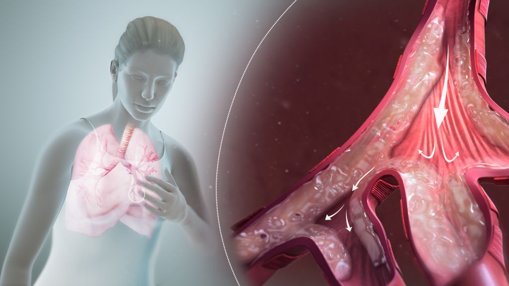
People who have had COVID are at increased risk of developing certain inflammatory diseases of the airways, such as asthma, hay fever and chronic sinusitis. However, vaccination against the SARS-CoV-2 virus appears to reduce the risk, according to a comprehensive epidemiological study led by researchers at Karolinska Institutet.
The international research team used an electronic health database in the United States, TriNetX, to investigate the link between COVID and so-called type-2 inflammatory diseases, a group of chronic conditions in which the immune system overreacts to allergens or infections.
The researchers compared 973 794 people who had had COVID with 691 270 people who had been vaccinated against the SARS-CoV-2 virus and 4 388 409 healthy controls with no documented infection or vaccination.
Inflammation in the airways
The results are presented in The Journal of Allergy and Clinical Immunology. People who had had COVID had a 66% higher risk of developing asthma, a 74% higher risk of chronic sinusitis and a 27% higher risk of hay fever compared with healthy controls. However, no increased risk was seen for the skin disease atopic eczema or for eosinophilic oesophagitis, an inflammation of the oesophagus.
“Our results suggest that COVID-19 can trigger type-2 inflammation in the airways, but not in other organs,” says Philip Curman, a physician and researcher at the Department of Medical Epidemiology and Biostatistics at Karolinska Institutet, Sweden, who led the research.
Vaccination against the virus had the opposite effect. The risk of asthma was 32% lower among vaccinated individuals compared with healthy unvaccinated individuals. The risk of sinusitis and hay fever was also slightly lower.
More than twice the risk
When people who had had COVID were compared with vaccinated individuals, an even clearer effect was seen. Infected individuals had more than twice the risk of developing asthma or chronic sinusitis and a 40% higher risk of developing hay fever compared with those who had been vaccinated.
“It is interesting to see that vaccination not only protects against the infection itself, but also appears to provide good protection against certain respiratory complications,” says Philip Curman.
The study is retrospective, i.e. based on data that has already been collected. This means that the researchers cannot draw any firm conclusions about causal links. Another limitation is that some infections may have gone undiagnosed, especially if they were detected through self-testing.
The research was conducted in close collaboration with the University of Lübeck and the Lübeck Institute of Experimental Dermatology in Germany, the Technical University of Madrid in Spain and Bar-Ilan University in Israel. It was mainly funded by the German Research Foundation (Deutsche Forschungsgemeinschaft), Region Stockholm and Karolinska Institutet. Two researchers received travel grants from TriNetX, which provides the database used in the study, and one of the authors is employed by the company.
Researchers at the Icahn School of Medicine at Mount Sinai have found that prenatal exposure to paracetamol may increase the risk of neurodevelopmental disorders, including autism spectrum disorder and attention-deficit/hyperactivity disorder (ADHD), in children. The study, published in BMC Environmental Health, is the first to apply the rigorous Navigation Guide methodology to systematically evaluate the rigour and quality of the scientific literature.
Paracetamol (known as acetaminophen in the US and Canada) is the most commonly used over-the-counter pain and fever medication during pregnancy and is used by more than half of pregnant women worldwide. Until now, acetaminophen has been considered the safest option for managing headache, fever, and other pain. Analysis by the Mount Sinai-led team of 46 studies incorporating data from more than 100 000 participants across multiple countries challenges this perception and underscores the need for both caution and further study.
The Navigation Guide Systematic Review methodology is a gold-standard framework for synthesising and evaluating environmental health data. This approach allows researchers to assess and rate each study’s risk of bias, such as selective reporting of the outcomes or incomplete data, as well as the strength of the evidence and the quality of the studies individually and collectively.
“Our findings show that higher-quality studies are more likely to show a link between prenatal acetaminophen exposure and increased risks of autism and ADHD,” said Diddier Prada, MD, PhD, Assistant Professor of Population Health Science and Policy, and Environmental Medicine and Climate Science, at the Icahn School of Medicine at Mount Sinai. “Given the widespread use of this medication, even a small increase in risk could have major public health implications.”
The paper also explores biological mechanisms that could explain the association between acetaminophen use and these disorders. Paracetamol is known to cross the placental barrier and may trigger oxidative stress, disrupt hormones, and cause epigenetic changes that interfere with foetal brain development.
While the study does not show that paracetamol directly causes neurodevelopmental disorders, the research team’s findings strengthen the evidence for a connection and raise concerns about current clinical practices.
The researchers call for cautious, time-limited use of paracetamol during pregnancy under medical supervision; updated clinical guidelines to better balance the benefits and risks; and further research to confirm these findings and identify safer alternatives for managing pain and fever in expectant mothers.
“Pregnant women should not stop taking medication without consulting their doctors,” Dr Prada emphasised. “Untreated pain or fever can also harm the baby. Our study highlights the importance of discussing the safest approach with health care providers and considering non-drug options whenever possible.”
With diagnoses of autism and ADHD increasing worldwide, these findings have significant implications for public health policy, clinical guidelines, and patient education. The study also highlights the urgent need for pharmaceutical innovation to provide safer alternatives for pregnant women.
Korean children with early life exposure to antibiotics were not diagnosed with autoimmune diseases at higher rates
Photo by Chayene Rafaela on Unsplash
The global incidence of autoimmune diseases among children has increased over the past few decades. A study published August 21st in the open-access journal PLOS Medicine by Ju-Young Shin at Sungkyunkwan University, Republic of Korea, and colleagues suggests that early life antibiotic exposure is not associated with an increased risk of autoimmune diseases in children.
Previous research has suggested that exposure to antibiotics as a foetus or infant may contribute to the development of autoimmune diseases among children. However, confounding variables limit the validity of prior studies and the association of antibiotics to autoimmune disease remains poorly understood.
In order to investigate whether antibiotics may increase risk of autoimmune diseases, researchers conducted a retrospective cohort study comprised of over 4 million children born in the Republic of Korea between April 1, 2009, and December 31, 2020. They accessed a mother-child linked insurance claims database from the South Korea National Health Insurance Service-National Health Insurance Database (NHIS-NHID) to identify children whose mothers had received antibiotic prescriptions during pregnancy or while breastfeeding their infant. The researchers then retrospectively analysed the health outcomes of each cohort for a period of over 7 years, tracking all diagnoses of Type 1 diabetes, Juvenile idiopathic arthritis, Inflammatory bowel disease (ulcerative colitis, Crohn’s disease), Systemic lupus erythematosus, and Hashimoto’s thyroiditis.
The researchers found no relationship between antibiotic exposure during pregnancy or early infancy and the overall incidence of autoimmune diseases in children. Future research is needed, however, to replicate the outcomes in other populations and to further investigate potential effects on subgroups.
According to the authors, “Our findings suggest no association between antibiotic exposure during the prenatal period or early infancy and the development of autoimmune diseases in children. This observation contrasts with several previous studies reporting increased risks and underscores the importance of carefully considering the underlying indications for antibiotic use and genetic susceptibility when interpreting such associations. While the potential benefits of antibiotic treatment in managing infections during pregnancy or early infancy likely outweigh the minimal risk of autoimmune outcomes, our findings also highlight the need for cautious and clinically appropriate use of antibiotics during these critical developmental periods in specific subgroups.”
The authors note, “Exposure to antibiotics during pregnancy or early infancy was not associated with an increased risk of autoimmune diseases in children. Nevertheless, the importance of follow-up studies to confirm and extend these findings cannot be overstated.”
3D structure of a melanoma cell derived by ion abrasion scanning electron microscopy. Credit: Sriram Subramaniam/ National Cancer Institute
Increased activity in a specific biological pathway may explain why many patients with a deadly form of skin cancer do not respond to the latest cancer treatments, a new study shows.
Publishing in the journal Cancer Research, the study featured data generated from experiments with human tissues and cells from patients with advanced melanoma that were implanted into mice. Results uncovered therapeutic targets that could limit melanoma growth in patients whose cancer failed to respond to initial treatment with immune checkpoint inhibitors.
Led by researchers at NYU Langone Health and its Perlmutter Cancer Center, the study focused on a subgroup of melanoma patients with mutations in the neurofibromin 1 (NF1) gene. NF1 mutations are just one type among several mutations, including those in the BRAF, NRAS, and PARP genes, that are linked to many cases of cancer, particularly melanoma. As many as 27% of melanoma patients are estimated to have NF1 mutations.
While immunotherapy, which stimulates the immune system to attack cancer cells as it would an invading virus, has proved to be a successful treatment, it does not work well for more than half of NF1-mutant melanoma patients.
“There is a pressing need for new drug therapies for melanoma patients with neurofibromin 1 mutations that do not respond to the latest immunotherapy, and for which there are no subsequent effective treatment options,” said study lead investigator Milad Ibrahim, PhD. Ibrahim is a postdoctoral fellow in the Dr Iman Osman Laboratory in the Ronald O. Perelman Department of Dermatology at the NYU Grossman School of Medicine.
To investigate why these patients were treatment resistant, investigators examined tumour cells from 30 melanoma patients who did not respond to immunotherapy. NF1 mutations were found in 40% of these melanoma samples. The samples came from NYU Langone’s extensive repository from more than 6000 melanoma patients.
Molecular testing showed that the signalling pathway built around a protein called epidermal growth factor receptor (EGFR) was more active in NF1 mutant melanoma cells than in cells with other melanoma-gene mutations. Increased EGFR activity has long been linked to abnormal cell growth in tumours and shorter survival with various cancers. The researchers also found that NF1 mutant melanoma cells depended on increased EGFR activity for survival, regardless of the presence of other mutations.
Because EGFR-inhibiting drugs are already used to treat some head and neck cancers, as well as colorectal and lung cancers, researchers then tested two drugs in the class, cetuximab and afatinib, in both NF1 mutant cell cultures and cancer cell lines without NF1 mutations. After transplanting both tumour cell types into mice and treating them with these drugs, results showed that both EGFR inhibitors were effective against cells and transplanted tumours with NF1 mutations, and they had no effect on melanomas without NF1 mutations.
“Our study results reveal a unique vulnerability in melanoma patients with neurofibromin 1 mutations, that an overexpression of the epidermal growth factor receptor pathway is essential for their survival and growth,” said the study’s senior investigator, Professor Iman Osman, MD.
“While further tests are needed, our results support a novel approach of deploying EGFR inhibitors either alone or in combination with other immunotherapies for treatment of melanoma patients whose tumours harbour NF1 mutation,” said the study’s co-senior investigator, Associate Professor Markus Schober, PhD.
However, Schober says this requires further testing in a clinical trial, which the research team plans to develop. He adds that if trial findings prove successful, the team’s research could provide a lifeline for many of these melanoma patients.
Alcohol withdrawal syndrome (AWS) is a potentially life-threatening condition that may complicate patients’ recovery after surgery.
Previous studies have estimated that up to 50% of hospitalised patients with AUD will develop some degree of AWS. Up to 7% of these patients may progress to severe withdrawal, including delirium tremens (DT) that can range in severity from irritability and confusion to tremors, nausea, vomiting and seizures.
Of those patients, 16 504 (0.5%) were diagnosed with AWS, including 6591 (0.2%) with DT.
“We found that alcohol withdrawal syndrome is linked with poorer surgical outcomes, extended hospitalisations and increased costs. These findings underscore the need for standardised perioperative screening and targeted management strategies to reduce these risks,” said study lead author Timothy Pawlik, MD, PhD, professor and chair of Ohio State’s Department of Surgery.
Patients with AWS were generally younger, male and more likely to have Medicaid, according to Pawlik, who holds the Urban Meyer III and Shelley Meyer Chair for Cancer Research at The Ohio State University College of Medicine.
AWS raises the risk of postoperative complications, especially respiratory failure and sepsis. The study found that patients with AWS had longer hospital stays (median 11 vs 6 days) and higher costs ($44 300 vs $28 800).
AWS was associated with a $10 030 higher adjusted hospitalisation cost per patient undergoing surgical care, contributing to an overall excess cost of $165.6 million, said study first author Azza Sarfraz, MBBS, a surgical oncology fellow at Ohio State.
“The lack of standard screening delays early detection and intervention,” Pawlik said. “Developing strategies for early identification, inpatient withdrawal management, and perioperative risk stratification may improve surgical outcomes, lower healthcare costs, and enhance patient care.”
A schematic of the eye’s anterior segment, demonstrating the anatomical placement of the microstent. The stent diverts aqueous humour from the anterior chamber to the suprachoroidal space through the flexible tube, creating a subconjunctival bleb supported by the expanding element. Credit: Yunlan Zhang, Zhong You, Jared Ching.
A team of researchers at the University of Oxford have unveiled a pioneering ‘microstent’ which could revolutionise treatment for glaucoma, a common but debilitating condition. The study has been published in The Innovation, Cell Press.
Glaucoma is a leading cause of vision loss, second only to cataracts. Globally, 7.7 million people were blind or visually impaired due to glaucoma in 2020. The condition can cause irreversible damage to the optic nerve, due to increased pressure within the eyeball. Current treatment options – principally surgery to create openings in the eye or insert tubes to drain fluid – are highly invasive, carry risk of complications, and have limited durability.
‘Our deployable microstent represents a significant advancement in glaucoma treatment,’ said lead author Dr Yunlan Zhang (University of Oxford at the time of the study/University of Texas). ‘Current surgical implants for this type of glaucoma have been shown to have limited long-term effectiveness, being susceptible to failure due to fibrosis (scarring) in the eye.’
The new microstent features a unique structural shape that allows it to expand once in the eye. At 200µm, less than a quarter of a millimetre, the stent’s tiny diameter enables it to fit within the needle of a standard hypodermic syringe, for minimally-invasive insertion. Once in place and expanded, the microstent spans the fluid-filled space between the white of the eye and the membrane that covers it.
By supporting this space, the stent reduces the excessive fluid buildup and resulting intraocular pressure in the eye which is responsible for the most common type of glaucoma, primary open-angle glaucoma. Initial trials carried out in rabbits found that the microstents lowered eye pressure in less than a month with minimal inflammation and scarring. Furthermore, the microstent achieved a greater reduction of eye pressure than a standard tubular implant.
This development has the potential to transform the landscape of glaucoma therapy. By offering an enhanced solution in the minimally invasive glaucoma surgery field that combines mechanical innovation with biocompatibility, we hope to improve patient outcomes and quality of life.
Senior co-author Dr Jared Ching (Department of Engineering Science, University of Oxford).
Senior co-author, Professor Zhong You (Department of Engineering Science, University of Oxford) said: ‘Our microstent is made from a durable and super-flexible nickel-titanium alloy called nitinol, renowned for its proven long-term safety for ocular use. Its unique material and structural properties help prevent subsequent movement, improve durability, and ensure long-term efficacy.’
The research team used advanced modelling techniques to guide the microstent’s design and ensure compatibility with the anatomy of the eye. The device’s superelastic properties enable it to accommodate how the eye changes and stretches over time without permanent deformation, enhancing its durability and functionality.
Colourised transmission electron micrograph of an HIV-1 virus particle (yellow/gold) budding from the plasma membrane of an infected H9 T cell (purple/green).
A new study in Nature shows that delivering a single injection of gene therapy at birth may offer years-long protection against HIV, taking advantage of a critical window in early life that could reshape the fight against paediatric infections in high-risk regions.
This study is among the first to show that the first weeks of life, when the immune system is naturally more tolerant, may be the optimal window for delivering gene therapies that would otherwise be rejected at older ages.
“Nearly 300 children are infected with HIV each day,” said first author Amir Ardeshir, associate professor of microbiology and immunology at the Tulane National Primate Research Center, who conducted the study alongside fellow researchers at the California National Primate Research Center. “This approach could help protect newborns in high-risk areas during the most vulnerable period of their lives.”
“This is a one-and-done treatment that fits the critical time when these mothers with HIV in resource-limited areas are most likely to see a doctor.”
Amir Ardeshir
In the study, nonhuman primates received a gene therapy that programs cells to continuously produce HIV-fighting antibodies. Timing proved critical to the one-time treatment offering long-term protection.
Those that received the treatment within their first month of life were protected from infection for at least three years with no need for a booster, potentially signifying coverage into adolescence in humans. In contrast, those treated at 8–12 weeks showed a more developed, less tolerant immune system that did not accept the treatment as effectively.
“This is a one-and-done treatment that fits the critical time when these mothers with HIV in resource-limited areas are most likely to see a doctor,” Ardeshir said. “As long as the treatment is delivered close to birth, the baby’s immune system will accept it and believe it’s part of itself.”
More than 100 000 children acquire HIV annually, primarily through mother-to-child transmission after birth from breastfeeding. Antiretroviral treatments have shown success in suppressing the virus and limiting transmission. However, adherence to treatment and access to doctors both decline after childbirth, particularly in areas with limited access to healthcare.
To deliver the treatment, researchers used an adeno-associated virus (AAV), a harmless virus that can act as a cargo truck to deliver genetic code to cells. The virus was sent to muscle cells, unique in their longevity, and delivered instructions to produce broadly neutralising antibodies, or bNAbs, which are capable of neutralising multiple strains of HIV.
This approach solved a longstanding problem with bNAbs. Previous studies found them effective at fighting HIV, but they required repeated infusions, which are costly and pose logistical challenges in low-resource settings.
“Instead, we turn these muscle cells – which are long-lived – into micro factories that just keep producing these antibodies,” Ardeshir said.
Newborns showed greater tolerance and expressed high levels of bNAbs, which successfully prevented infection, while older infants and juveniles were more likely to have produced anti-drug antibodies that shut down the treatment.
Researchers also found that exposing fetuses to the antibodies before birth helped older infants accept the gene therapy later, avoiding the immune rejection that often occurs with age.
Still, Ardeshir said a one-time injection at birth offered a more cost-effective and feasible real-world solution, while putting less burden on the mother for a follow-up visit.
Questions remain as to how the results translate to human infants and children, who may be less susceptible to AAV-delivered treatments. The study also used one strain of simian–human immunodeficiency virus, which doesn’t reflect the variety of HIV strains.
If successful, however, this treatment could dramatically reduce mother-to-child HIV transmission rates in high-risk regions such as sub-Saharan Africa, where 90% of paediatric HIV cases can be found. It may also be adapted to protect against other infectious diseases like malaria, which disproportionately affects young children in low-income countries.
“Nothing like this was possible to achieve even 10 years ago,” Ardeshir said. “This was a huge result, and now we have all the ingredients to take on HIV.”
Study finds sensory cells that detect tissue damage, irritants also rein in harmful immune responses to protect the lungs
Clusters of mouse vagus nerve sensory cells reveal the presence of TRPV1, a molecular sensor that detects irritants, heat, and inflammation. A new HMS study reveals nerve cells with this sensor play a central role in taming inflammation and tissue damage in the lung during flu infection. Image: Chiu Lab.
A group of nerve cells known for their role in detecting chemical irritation, tissue damage, heat, and pressure now emerge as critical defenders against the worst ravages of the flu caused by an overactive immune response, according to new research by scientists at Harvard Medical School and the Harvard T.H. Chan School of Public Health.
The cells, called TRPV1 vagal nociceptors, live in the vagus nerve, which sends signals from internal organs – including the heart, lungs, and gut – to the brain to help regulate heart rate, breathing, digestion, and other functions. In the lungs, these cells trigger the protective cough reflex that forces the airways to expel foreign particles, mucus, and other irritants.
But the new research, published in Science Immunologyand conducted in mice, shows that in the setting of flu, these cells do much more – they rein in the immune system and avert the smoldering inflammation that often occurs in the aftermath of a viral infection and can injure healthy tissue.
Each year, the flu sickens millions and kills between 290 000 and 650 000 people worldwide, according to the World Health Organization. While the immune system helps fight off the virus, an excessive inflammatory response can inflict tissue damage and worsen illness. The findings are especially relevant in the wake of the COVID-19 pandemic, which revealed how an aberrant immune response following viral infection can sometimes lead to serious organ damage and even organ failure.
“Our research shows that the infected lung is a battleground where nerves and immune cells engage in a delicate dance to safeguard our health,” said co-senior study author Isaac Chiu, professor of immunology in the Blavatnik Institute at HMS. “Understanding this powerful neuro-immune signaling axis will be increasingly important as we design better ways to prevent and treat immune-mediated damage in viral infections, which can sometimes be worse than the direct damage caused by the virus itself.”
The findings, he added, raise the possibility that vagus nerve function may be one variable that explains why some people with the flu go on to develop long-lasting and devastating immune-driven damage in their lungs while others recover once the initial infection is resolved.
“Flu infections are highly variable in severity and there is a need to understand why certain groups of people, such as the elderly, experience worse disease,” said study first author Nicole Almanzar, a doctoral student in the Chiu Lab. “Our study demonstrates that the nervous system is actively involved in regulating the response of the lungs to infection, offering a new perspective on viral infections that could help explain why particular factors increase risk of severe infections.”
The new study illuminates the complex interaction between body and brain and adds to a growing body of research showing that the nervous and immune systems engage in highly orchestrated crosstalk during infection to modulate body defenses.
One of Chiu’s earlier studies found that during bacterial infections of the lung, the same set of vagal nerve neurons suppressed the immune defences. In the new study, however, the immune-taming function of these cells worked to shield the lung from excessive damage during viral infection.
“Context clearly matters,” Chiu said.
Disabling the neurons worsened flu damage in the lungs
In a set of experiments, researchers exposed a group of mice with genetically disabled or chemically silenced TRPV1 neurons to influenza A virus. Mice without these nerve cells fared notably worse than mice with functioning TRPV1 cells. Even though the overall amount of virus in the lungs was the same in both groups, mice lacking TRPV1 neurons suffered more severe lung pathology, higher levels of harmful inflammation, and lower survival rates. Interestingly, Almanzar noted, even though the overall viral load remained the same, the spread of the virus within the lobes of the lungs was more pronounced in mice without these protective neurons.
The researchers also found that the absence of TRPV1 neurons altered the lung’s immune landscape. The lungs of mice lacking these neurons had an overabundance of neutrophils and macrophages – two types of immune cells that, in excess, can worsen tissue damage. At the same time, interferon signalling – one of the body’s most important viral-defence pathways – was seriously impaired in these immune cells.
In another experiment, the researchers used an antibody to deplete inflammation-driving cells in flu-infected mice lacking TRPV1 neurons. These animals had notably better survival than untreated mice lacking these protective neurons. The observation further underscores how nerve cells help prevent harmful immune reactions that can sometimes be more dangerous than the virus itself.
The researchers noted that they do not yet know precisely how TRPV1 neurons restrain the march of inflammatory cells at the molecular level – a question they plan to explore in subsequent work.
“The vagus nerve is powerfully controlling inflammation, but how it does so remains a mystery to be solved,” Chiu said. “But we’re excited that it plays such a strong role in viral infections.”
Harnessing the immune brake for therapy
Moving forward, this insight opens the door to exciting new avenues for therapy. Instead of only targeting the flu virus or dampening immune activity, the research team said, future treatments could mimic the function of nerve cells to ensure the delicate balance between protective and damaging immune responses is not thrown off.
The idea is not that far-fetched, the researchers said, noting that the FDA recently approved a therapy for rheumatoid arthritis by vagus nerve stimulation.
“Imagine if you could harness this brake to control inflammation in the lungs and beyond,” Chiu said. “By stimulating related circuits where the vagus nerve shuts down immune cells, one could envision treating immune-mediated dysfunction of many kinds, including that caused by viral infections.”
New research, led by Queen Mary University of London and published in Clinical Psychological Science, has revealed that highly sensitive people (HSP) are more likely to experience mental health problems compared to individuals who are less sensitive.
The meta-analysis of 33 studies, the first of its kind, looked at the relationship between sensitivity and common mental health problems such as depression and anxiety. Researchers found there was a significant, positive relationship between the two, concluding that highly sensitive people are more likely to experience depression and anxiety compared to those who are less sensitive.
In the study, sensitivity was defined as a personality trait that reflects people’s capacity to perceive and process environmental stimuli such as bright lights, subtle changes in the environment and other peoples’ moods. Often overlooked in mental health studies and clinical practice, which tend to focus on neuroticism and its association with mental health conditions, this research shows that understanding a person’s sensitivity level is important and can have therapeutic implications.
People with sensitive personality traits may benefit from different treatment plans
For example, people with more sensitive personality traits may be more likely to benefit from treatment plans which involve techniques such as applied relaxation and mindfulness, which can also prevent relapse.
Tom Falkenstein, a psychotherapist and a PhD student at Queen Mary University of London, said: “This is the most extensive systematic review on sensitivity and mental health in adolescents and adults to date, and is the first ever meta-analysis on the topic to estimate the impact of this relationship. We found positive and moderate correlations between sensitivity and various mental health problems such as depression, anxiety, post-traumatic stress disorder, agoraphobia and avoidant personality disorder. Our findings suggest that sensitivity should be considered more in clinical practice which could be used to improve diagnosis of conditions.”
“In addition, our findings could help improve treatment for these individuals. Around 31% of the general population are considered highly sensitive, and, as our findings show, are more likely to respond better to some psychological interventions than less sensitive individuals. Therefore, sensitivity should be considered when thinking about treatment plans for mental health conditions. Our work shows it is crucial that the awareness of sensitivity is improved among mental health care professionals, so clinicians and practitioners can recognise the trait in their patients, and tailor treatment to their sensitivity.”
Michael Pluess, Professor in Developmental Psychology at University of Surrey and Visiting Professor at Queen Mary University of London said: “This is the first meta-analysis providing robust evidence that highly sensitive people are more prone to common mental health problems. However, it is important to remember that highly sensitive people are also more responsive to positive experiences, including psychological treatment. Our results provide further evidence that sensitive people are more affected by both negative and positive experiences and that the quality of their environment is particularly important for their well-being.”
The systematic review and meta analysis of 33 studies was carried out by an academic team from several universities including Queen Mary University and the University of Surrey.
Results show no clear evidence of benefit for ketamine in chronic pain and identified an increased risk of adverse effects such as delusions, delirium, paranoia, nausea, and vomiting
Photo by Towfiqu Barbhuiya on Unsplash
The off-label use of ketamine to treat chronic pain is not supported by scientific evidence, a new Cochrane review has found.
Ketamine is an anaesthetic commonly used for procedural sedation and short-term pain relief. Ketamine is also frequently prescribed off-label to manage chronic pain conditions such as nerve pain, fibromyalgia and complex regional pain syndrome. It is one of several NMDA receptor antagonists – a group of drugs thought to reduce pain by blocking certain brain receptors involved in pain signalling.
The review, conducted by researchers from UNSW Sydney, Neuroscience Research Australia (NeuRA), and Brunel University of London, examined 67 trials involving over 2300 adult participants. It assessed five NMDA receptor antagonists: ketamine, memantine, dextromethorphan, amantadine, and magnesium.
Results show no clear evidence of benefit for ketamine in chronic pain and identified an increased risk of adverse effects such as delusions, delirium, paranoia, nausea, and vomiting. Evidence was rated low to very low certainty, due to small study sizes and poor methodological quality.
“We want to be clear – we’re not saying ketamine is ineffective, but there’s a lot of uncertainty,” said Michael Ferraro, Doctoral Candidate at UNSW and NeuRA, first author of the review. “The data could point to a benefit or no effect at all. Right now, we just don’t know.”
Researchers looked at the effects across various chronic pain conditions and dosing strategies but found no clear evidence of benefit in any specific condition or dose. Side effects were a major concern, particularly with intravenous use.
“The most common adverse events we saw were psychotomimetic effects such as delusions, delirium and paranoia, as well as nausea and vomiting.” said Ferraro. “These effects are distressing for many patients. Clinicians often try to balance the dose for pain relief without triggering those symptoms, but this isn’t always achieved.”
The review also found no studies that reported on two key outcomes: whether ketamine reduced depressive symptoms or opioid use. This is notable, as ketamine is often proposed for patients with depressive symptoms or opioid tolerance.
“This group of drugs, and ketamine in particular, are in relatively common use for chronic pain around the world. Yet we have no convincing evidence that they are delivering meaningful benefits for people with pain, even in the short term,” said Neil O’Connell, Professor at Brunel University of London, co-senior author of the review. “That seems a good reason to be cautious in the clinic and clearly indicates an urgent need to undertake high quality trials.”
The authors hope the review will help inform patients and clinicians weighing up potential benefits and harms, and guide future research. While more evidence is needed, this review highlights the importance of high-quality trials to understand whether ketamine has a role in chronic pain care.
“We’ve seen the harm that can come from taking medicines developed for acute pain and applying them to chronic pain, opioids are a prime example. Now we’re seeing a similar pattern with ketamine,” said co-senior author James McAuley, Professor at UNSW and senior researcher at NeuRA . “As opioid prescribing is slowly reduced, there’s a growing demand for alternatives, but we need to be careful not to rush into widespread use without strong evidence.”