After a Stroke, Muscles Lose Basic ‘Building Blocks’

Muscle sarcomeres (consecutive green lines), the smallest functional unit of muscle, from inside a living human. Credit: Northwestern University

In a new study of stroke patients, researchers have discovered that, in an attempt to adapt for an unusable arm, muscles actually lose sarcomeres — their smallest, most basic building blocks.

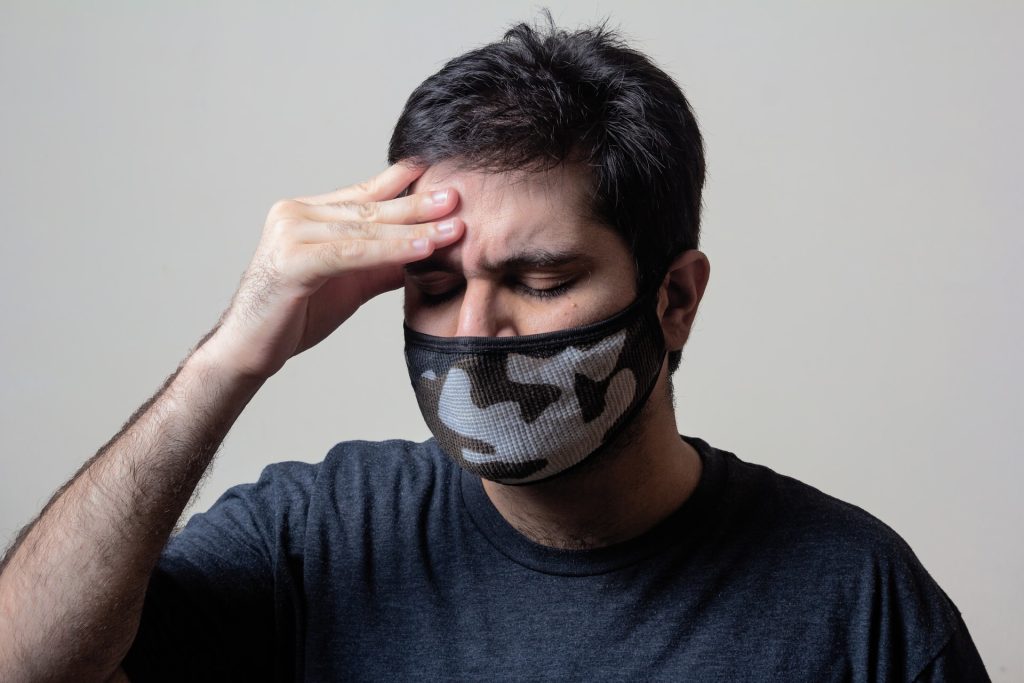
Patients that have suffered a stroke are often unable to use the arm on their affected side. Sometimes, they end up holding it close to their body, with the elbow flexed. Northwestern University and Shirley Ryan AbilityLab researchers found out why this happens.

Stacked end to end (in series) and side to side (in parallel), sarcomeres form the length and width of muscle fibres. By imaging biceps muscles with three noninvasive methods, the researchers found that stroke patients had fewer sarcomeres along the length of the muscle fibre, resulting in the muscle structure being shorter overall.

This finding is consistent with the common patient experience of abnormally tight, stiff muscles that resist stretching, and it suggests that changes in the muscle potentially amplify existing issues caused by stroke, which is a brain injury. The team hopes this discovery can help improve rehabilitation techniques to rebuild sarcomeres, ultimately helping to ease muscle tightening and shortening.

“This is the most direct evidence yet that chronic impairments, which place a muscle in a shortened position, are associated with the loss of serial sarcomeres in humans,” said senior author Wendy Murray. “Understanding how muscles adapt following impairments is critical to designing more effective clinical interventions to mitigate such adaptations and to improve function following motor impairments.”

Murray is a professor of biomedical engineering at Northwestern’s McCormick School of Engineering, a professor of physical medicine and rehabilitation at the Northwestern University Feinberg School of Medicine and research scientist at the Shirley Ryan AbilityLab. The research was completed in collaboration with Julius Dewald, professor of physical therapy and human movement sciences and of physical medicine and rehabilitation at Feinberg, professor of biomedical engineering at McCormick, and research scientist at Shirley Ryan AbilityLab.

Measuring just 1.5 to 4.0 micrometres in length, sarcomeres are made up of two main proteins: actin and myosin. When these proteins work together, they enable a muscle to contract and produce force. Even though previous animal studies have found that serial sarcomeres are lost from muscles after a limb is immobilised in a cast, the phenomenon had never before been demonstrated in humans. The animal studies found that the shorter muscles due to lost serial sarcomeres also became stiffer.

There is a classic relationship between force and length,” explained first author Amy Adkins, a PhD student in Murray’s laboratory. “Given that the whole muscle is composed of these building blocks, losing some of them affects how much force the muscle can generate.”

To conduct the study in humans, the researchers combined three non-invasive medical imaging techniques: MRI to measure muscle volume, ultrasound to measure bundles of muscle fibers and two-photon microendoscopy to measure the microscopic sarcomeres.

Imaging opens new possibilities
Combining these technologies, the researchers imaged biceps from seven stroke patients and four healthy participants. As stroke patients are more affected on one side of their body, the researchers compared imaging from the patients’ affected side to their unaffected side as well as to images from the healthy participants.

In the stroke patients’ affected biceps, researchers found less volume, shorter muscle fibres and comparable sarcomere lengths. After combining data across scales, they found that affected biceps had fewer sarcomeres in series compared to the unaffected biceps. Greater differences between stroke patients’ arms than healthy participants’ arms were seen, indicating that stroke was the cause.

By combining medical imaging to better view muscle structure, the study also establishes that it is possible to study muscle adaptations in sarcomere number in humans. Prior to two-photon microendoscopy, human studies were limited either to examining dissected tissues in anatomy labs, which give imperfect insight into how muscles adapt to injury and impairment, measuring sarcomere lengths during surgery or from a muscle biopsy, which restricts who can participate in the study.

“In almost every facet of our world, there is an important relationship between how something is put together (its structure) and how it works (its function),” the researchers said. “Part of the reason medical imaging is such a valuable resource and clinical tool is that this is also true for the human body, and imaging gives us an opportunity to measure structure.”

Source: Northwestern University

Journal information: Adkins, A.N., et al. (2021) Serial sarcomere number is substantially decreased within the paretic biceps brachii in individuals with chronic hemiparetic stroke. PNAS. doi.org/10.1073/pnas.2008597118.

Do Heart Hormones Drive Nighttime Hypertension?

Photo by Marcelo Leal on Unsplash

In a new series of studies, University of Alabama at Birmingham researchers have described the reasons behind low levels of natriuretic peptides (NPs) in obese individuals. 

First reported six decades ago, NPs are beneficial hormones produced by the heart, and are responsible for blood pressure regulation and the overall cardiovascular and metabolic health of humans. This study also addresses how the disturbance of an individual’s diurnal rhythm of these hormones contributes to poor cardiovascular health in obese individuals.

High blood pressure at nighttime is seen commonly in obese individuals, who already have higher risk of hypertension and poor cardiovascular outcomes. This can contribute to outcomes such as stroke, heart failure, heart attack and cardiac death. But why this impairment of this day-night blood pressure rhythm is not well understood — however, scientists believe that part of the reason lies with NPs.

“All the hormones in the human body have a day-night rhythm,” noted Vibhu Parcha, MD, a clinical research fellow in the Division of Cardiovascular Disease and the first author of both the studies. “It has been hypothesised the NP hormones should also have this rhythm, but this had not yet been demonstrated in humans. Our clinical trial assessed the 24-hour cycle of the NP hormones and compared it to the 24-hour cycle of blood pressure. We also studied how these cycles differ between lean and obese individuals and studied the reasoning behind why obese individuals experience lower levels of NPs.”

Following a rigorous clinical trial of healthy individuals, researchers found that NP hormones have a diurnal rhythm with higher levels in the afternoon and lower levels at nighttime — similar to the 24-hour cycle of blood pressure. In obese individuals however, researchers observed that the relationship between NPs and blood pressure does not function the same way. This leads to higher nighttime blood pressure and increased risk of cardiovascular disease. The low production of NPs combined with a relatively higher elimination of NPs from an obese individual’s system leads to low levels of these beneficial hormones in circulation, which may explain the NP deficiency.

“This is the first time we have seen that NPs, like other hormones, have a 24-hour rhythm,” said senior author Pankaj Arora, MD, a physician-scientist in UAB’s Division of Cardiovascular Disease. “These studies give us a better understanding of NPs and of the reasoning behind the NP deficiency in obese individuals. We now have an FDA-approved medication (LCZ696) that improves circulating NP levels. This medication is considered a first-line treatment for heart failure and may be used to increase NP levels.”

This medication could specifically target NPs and blood pressure if given at the right time of day and could control hypertension with precision, Dr Arora added. These findings point to using a physiologically-driven precision ‘chronopharmacotherapy’ approach to improve the diurnal blood pressure profile in obese individuals.

Source: University of Alabama at Birmingham

Journal information: Vibhu Parcha et al, Chronobiology of Natriuretic Peptides and Blood Pressure in Lean and Obese Individuals, Journal of the American College of Cardiology (2021). DOI: 10.1016/j.jacc.2021.03.291

Dopamine Involved in Both Autistic Behaviour and Motivation

Dopamine can help explain both autistic behaviours and men’s need for motivation or ‘passion’ in order to succeed compared to women’s ‘grit’, according to a new study.

Men – more often than women – need passion to succeed at things. At the same time, boys are diagnosed as being on the autism spectrum four times as often as girls. Both statistics may be related to dopamine, one of our body’s neurotransmitters.

“This is interesting. Research shows a more active dopamine system in most men” than in women, says Hermundur Sigmundsson, a professor at the Norwegian University of Science and Technology’s (NTNU) Department of Psychology.

He is behind a new study addressing gender differences in key motivating factors to excel in something. The study uses men’s and women’s differing activity in the dopamine system as an explanatory model. The study enrolled 917 participants aged 14 to 77, consisting of 502 women and 415 men.

“We looked at gender differences around passion, self-discipline and positive attitude,” said Prof Sigmundsson. The study refers to these qualities as passion, grit and mindset. The researchers also applied theories to possible links with dopamine levels. Dopamine, a neurotransmitter that is released in the brain, is linked to learning, attention and our ability to focus. It can contribute to a feeling of satisfaction.

Men generally secrete more dopamine, but it plays a far more complex role than simply being a ‘happy hormone’. Dopamine is linked to learning, attention and our ability to focus.Previous studies on Icelandic students have shown that men are more dependent on passion in order to succeed at something. This study confirms the earlier findings. In six out of eight test questions, men score higher on passion than women.

However, the association with dopamine levels has not been established previously.

“The fact that we’ve developed a test to measure passion for goal achievement means that we can now relate dopamine levels to passion and goal achievement,” explained Prof Sigmundsson.

Women, on the other hand, may have greater self-discipline – or grit – and be more conscientious, according to other studies. Their level of passion may not be as pronounced in general, but they are also able to use this to excel.

The results for the women, however, are somewhat more ambiguous than men’s need to have a passion for something, and this study found no such gender difference. Nor did the researchers find any difference between the sexes in terms of growth mindset.

Previous studies have associated the dopamine system with many different conditions, such as ADHD, psychoses, manias and Parkinson’s disease. However, it may also be related to a certain form of autistic behaviour.

Some individuals with autism may develop a deep interest in certain topics, something which others may find strange or even off putting. People on the autism spectrum can focus intensely on these topics or pursuits, at least for a while, and dopamine may play a role in this.

“Other research in neuroscience has shown hyperactivity in the dopamine system in individuals with autism, and boys make up four out of five children on the autism spectrum. This, and dopamine’s relationship to passion, might be a mechanism that helps to explain this behaviour,” concluded Prof Sigmundsson.

Source: Norwegian University of Science and Technology


Journal reference: 
Sigmundsson, H., et al. (2021) Passion, grit and mindset: Exploring gender differences. New Ideas in Psychology. doi.org/10.1016/j.newideapsych.2021.100878.

In the Immune Battle, MRSA Uses Toxins to Fight Dirty

Scanning electron micrograph of methicillin-resistant Staphylococcus aureus and a dead human neutrophil. Credit: NIAID

Researchers have uncovered a novel trick employed by the bacterium Staphylococcus aureus — MRSA uses toxins to ‘fight dirty’ and stifle the immune response. This finding is a step towards one day producing a vaccine against MRSA.

Every year, there are some 700 000 deaths due to the emerging global threat of antimicrobial resistance (AMR). Turning the tables against AMR requires immediate action, and the development of novel vaccines to prevent such infections in the first place, are an attractive and potentially very effective option.

Staphylococcus aureus is the causative agent of the infamous MRSA ‘superbug’, one of the chief concerns of AMR. Immunologists from Trinity College Dublin, working with scientists at GSK, discovered the deadly bacteria’s new trick to foil the immune system. They found that the bacterium interferes with the host immune response by causing toxic effects on white blood cells, preventing them from carrying out their infection-fighting jobs.

The study also showed that the toxicity could be lessened following vaccination with a mutated version of a protein specifically engineered to throw a spanner in the MRSA works. This could one day lead to a vaccine for humans.

Rachel McLoughlin, Professor in Immunology in Trinity’s School of Biochemistry and Immunology and the Trinity Biomedical Sciences Institute (TBSI), said: “As a society we are witnessing first-hand the powerful impact that vaccination can have on curbing the spread of infection. However, in the backdrop of the COVID epidemic we must not lose sight of the fact that we are also waging war on a more subtle epidemic of antimicrobial resistant infection, which is potentially equally deadly.

“In this study we have identified a mechanism by which a protein made by the bacterium – known as Staphylococcal Protein A (SpA) – attacks and rapidly kills white blood cells. This protein has been widely studied for its immune evasion capacity and has a well-documented role in rendering antibodies raised against the bacterium non-functional.

“Here we uncover a previously undocumented strategy by which SpA forms immune complexes through its interaction with host antibodies, that in turn exert toxic effects on multiple white blood cell types. This discovery highlights how important it will be for effective vaccines to be capable of disarming the effects of protein A.”

Dr Fabio Bagnoli, Director, Research & Development Project Leader, GSK, said: “Our collaboration with Trinity College Dublin and in particular with Professor Rachel McLoughlin, a worldwide recognised expert on staphylococcal immunology, is critical for increasing our knowledge on protective mechanisms against S. aureus.”

The study documents the latest discovery made by this group at Trinity under an ongoing research agreement with GSK Vaccines (Siena, Italy). Overall, this collaboration aims to increase understanding of the immunology of Staphylococcus aureus infection to advance development of next-generation vaccines to prevent MRSA infections.

Source: Trinity College Dublin

Journal information: Fox, P. G., et al. (2021) Staphylococcal Protein A Induces Leukocyte Necrosis by Complexing with Human Immunoglobulins. Scientific Reports. doi.org/10.1128/mBio.00899-21.

Study Uncovers a Key Brain Building Block

Astrocytes (red) from a rat brain. Credit: Jeffrey C. Smith Lab, National Institute of Neurological Disorders and Stroke, NIH

A new study from Duke and UNC scientists has discovered a crucial protein involved in the communication and coordination between astrocytes as they build synapses — essentially a brain building block. 

Astrocytes, specialised, star-shaped glial cells that outnumber the neurons they support over fivefold and which make up about half the mass of a human brain, are increasingly being viewed as having a critical role in shaping the development of the brain.  Astrocytes tile the entire central nervous system (CNS) and exert many essential complex functions in the healthy CNS, including guiding development of the brain. 

The researchers found that a molecule, called hepaCAM, is a key component of this process. Without it, astrocytes aren’t as sticky as they should be, and tend to stick to themselves rather than forming connections with their neighbouring astrocytes.

This finding, in studies on mice with the gene for hepaCAM deleted from their astrocytes, helps in the understanding of several brain disorders, including cognitive decline, epilepsy and autism spectrum disorders.

One rare brain disorder, called megalencephalic leukoencephalopathy (MLC) is also known to be caused by a mutation in the hepaCAM gene, and this work might provide answers about what exactly has gone wrong. MLC is a developmental disorder that grows progressively worse, causing macrocephaly (a large head), swelling of the brain’s white matter, intellectual disability and epilepsy.

By deleting hepaCAM from astrocytes to see what it does, “we sort of made the cells into introverts,” explained senior author Cagla Eroglu, an associate professor of cell biology at the Duke University School of Medicine. “They’re normally wanting to reach out, but without hepaCAM, they started to hug themselves instead.”

“If the astrocyte makes junctions to its neighbours, then you start to have a network,” Prof Eroglu said. “To make a functional brain, you need a functional astrocytic network.”

The researchers zeroed in on hepaCAM by searching for highly active genes in astrocytes, and which have been implicated in brain dysfunction. They partnered with another group working on hepaCAM at the University of Barcelona, but that group has been  looking at the molecule for its role in regulating chloride signaling channels in astrocytes.

The Duke group found that deleting hepaCAM from astrocytes led to a synaptic network that was too easily excited and not as well dampened. “The effect on the inhibitory synapses was the strongest,” said first author Katie Baldwin, who recently became an assistant professor of cell biology and physiology at the University of North Carolina at Chapel Hill. “You’re putting the inhibition down and the excitation up, so that really could point to a mechanism for epilepsy.”

Prof Baldwin plans to test whether hepaCAM-deficient mice have behavioural differences or changes in learning and memory, or whether they exhibit the stress and social anxiety that are markers of autism spectrum disorders. She said they might also reintroduce the disease-mutation versions of the protein to mice that were born without it to see what effects it has.

“We know hepaCAM is interacting with itself between two astrocytes, but we don’t know what it’s interacting with at the synapse,” Prof Baldwin said. “We don’t know if it could be interacting with hepaCAM which is also found in the neurons, or if it could be some other protein that we don’t know about yet.

Source: Duke University School of Nursing

Journal information: Katherine T. Baldwin et al, HepaCAM controls astrocyte self-organization and coupling, Neuron (2021). DOI: 10.1016/j.neuron.2021.05.025

Artificial Sweeteners Can Turn Gut Bacteria Bad

Source: Breakingpic on Pexels

Scientists have found that common artificial sweeteners can turn previously healthy gut bacteria pathogenic, invading the gut wall and potentially leading to serious health issues.

This study is the first to show the pathogenic effects of some of the most widely used artificial sweeteners (saccharin, sucralose, and aspartame) on two types of gut bacteria, Escherichia coli and Enterococcus faecalisE. faecalis is capable of crossing the intestinal wall to enter the bloodstream and congregate in the lymph nodes, liver, and spleen, causing a number of infections including septicaemia. To top it off, this commensal bacteria has emerged as a multi-drug resistant pathogen.

Previous studies have shown that artificial sweeteners can affect the composition of gut bacteria, but this new molecular research, led by academics from Anglia Ruskin University (ARU), has shown that sweeteners can also induce pathogenic features in certain bacteria. It found that these pathogenic bacteria can latch onto, invade and kill epithelial Caco-2 cells lining the intestinal wall.

This new study discovered that at a concentration equivalent to two cans of diet soft drink, all three artificial sweeteners significantly increased the adhesion of both E. coli and E. faecalis to intestinal Caco-2 cells, and differentially increased biofilm formation. Bacteria growing in biofilms are less sensitive to antimicrobial resistance treatment and are more likely to secrete toxins and express disease-causing virulence factors.

Additionally, all three sweeteners caused the pathogenic gut bacteria to invade Caco-2 cells found in the wall of the intestine, save for saccharin, which had no significant effect on E. coli invasion.

Senior author Dr Havovi Chichger, Senior Lecturer in Biomedical Science at ARU, said: “There is a lot of concern about the consumption of artificial sweeteners, with some studies showing that sweeteners can affect the layer of bacteria which support the gut, known as the gut microbiota.

“Our study is the first to show that some of the sweeteners most commonly found in food and drink—saccharin, sucralose and aspartame—can make normal and ‘healthy’ gut bacteria become pathogenic. These pathogenic changes include greater formation of biofilms and increased adhesion and invasion of bacteria into human gut cells.

“These changes could lead to our own gut bacteria invading and causing damage to our intestine, which can be linked to infection, sepsis and multiple-organ failure.

“We know that overconsumption of sugar is a major factor in the development of conditions such as obesity and diabetes. Therefore, it is important that we increase our knowledge of sweeteners versus sugars in the diet to better understand the impact on our health.”
Source: EurekAlert!

Journal reference: Shil, A & Chichger, H (2021) Artificial Sweeteners Negatively Regulate Pathogenic Characteristics of Two Model Gut Bacteria, E. coli and E. faecalis. International Journal of Molecular Sciences. doi.org/10.3390/ijms22105228.

The Origin Mystery of SARS-CoV-2 Deepens

SARS-CoV-2 viruses emerging from a human cell. Credit: NIAID

Australian researchers studying SARS-CoV-2 have discovered that the virus is most ideally adapted to infect human cells — instead of bat or pangolin cells, prompting renewed questions about its origin.

The scientists, from Flinders University and La Trobe University, described how they used high-performance computer modelling of SARS-CoV-2’s structure at the beginning of the pandemic to predict its ability to infect humans and a range of 12 domestic and exotic animals.

They were hoping to identify an intermediate animal vector that may have played a role in transmitting a bat virus to humans, and to understand any risk posed by the susceptibilities of pets and livestock.

Using genomic data from 12 animal species, the researchers painstakingly built computer models of the key ACE2 protein receptors for each species. These models were then used to calculate how strongly the SARS-CoV-2 spike protein bound to each species’ ACE2 receptor.

Surprisingly, the results showed that SARS-CoV-2 bound to ACE2 on human cells more tightly than any of the tested animal species, including bats and pangolins. If one of the animal species tested was the origin, it would normally be expected to show the highest binding to the virus.

“Humans showed the strongest spike binding, consistent with the high susceptibility to the virus, but very surprising if an animal was the initial source of the infection in humans,” said Professor David Winkler at La Trobe University.

The findings, originally released on the ArXiv preprint server, have now been peer reviewed and published in Scientific Reports.

“The computer modelling found the virus’s ability to bind to the bat ACE2 protein was poor relative to its ability to bind human cells. This argues against the virus being transmitted directly from bats to humans. Hence, if the virus has a natural source, it could only have come to humans via an intermediary species which has yet to be found,” says Flinders affiliated Professor Nikolai Petrovsky.

The team’s computer modelling also showed fairly strong binding of SARS-CoV-2 to ACE2 from pangolins, which are occasionally used as food or in traditional medicines. Professor Winkler noted that pangolins displayed the highest spike binding energy of all the animals in the study – significantly higher than bats, monkeys and snakes.

“While it was incorrectly suggested early in the pandemic by some scientists that they had found SARS-CoV-2 in pangolins, this was due to a misunderstanding and this claim was rapidly retracted as the pangolin coronavirus they described had less than 90% genetic similarity to SARS-CoV-2 and hence could not be its ancestor,” Prof Petrovsky said.

Similarity in spike proteins

As shown in this and other studies, the specific part of the pangolin coronavirus spike protein that binds to ACE2 was almost identical to its SARS-CoV-2 counterpart.

“This sharing of the almost identical spike protein almost certainly explains why SARS-CoV-2 binds so well to pangolin ACE2. Pangolin and SARS-CoV-2 spike proteins may have evolved similarities through a process of convergent evolution, genetic recombination between viruses, or through genetic engineering, with no current way to distinguish between these possibilities,” Prof Petrovsky said.

“Overall, putting aside the intriguing pangolin ACE2 results, our study showed that the COVID-19 virus was very well adapted to infect humans.”

“We also deduced that some domesticated animals like cats, dogs and cows are likely to be susceptible to SARS-CoV-2 infection too,” Prof Winkler added.

The question of how the virus came to infect humans currently has two main explanations. The virus may have jumped to humans from bats through an intermediary animal which remains to be identified. The other explanation making headlines in the media is an accidental release from a virology lab, where it perhaps was created in ‘gain of function‘ tests, which are carried out around the world to better understand pathogens. A number of organisations and governments, including the World Health Organization and the United States have urged further investigation to find out which of these is correct — though a definitive answer may take years.
How and where the SARS-CoV-2 virus adapted to become such an effective human pathogen remains a mystery, the researchers concluded, adding that finding the origins of the disease will help efforts to protect humanity against future coronavirus pandemics.

Source: EurekAlert!

Journal information: Sakshi Piplani et al, In silico comparison of SARS-CoV-2 spike protein-ACE2 binding affinities across species and implications for virus origin, Scientific Reports (2021). DOI: 10.1038/s41598-021-92388-5

New Machine Learning Tools Could Save Teeth

Photo by Kevin Bation on Unsplash

Machine learning tools could help identify those at greatest risk for tooth loss and refer them for further dental assessment for early interventions to avert or delay the conditions.

The study by researchers at the Harvard School of Dental Medicine compared five different algorithms using various combinations of variables to screen for risk. The results showed those that factored medical characteristics and socioeconomic variables, including race, education, arthritis, and diabetes, outperformed algorithms that relied on dental clinical indicators alone.

“Our analysis showed that while all machine-learning models can be useful predictors of risk, those that incorporate socioeconomic variables can be especially powerful screening tools to identify those at heightened risk for tooth loss,” said study lead investigator Hawazin Elani, assistant professor of oral health policy and epidemiology at HSDM.

The approach could be used to screen people globally and in a variety of health care settings even by non-dental professionals, she added. This approach could be applied around the world, even allowing non-dental professionals to screen patients.

Tooth loss can affect quality of life, well-being, nutrition, and social interactions. It is also associated with dementia. If the earliest signs of dental disease are identified, then the process can be delayed or averted with prompt treatment. However, many people with dental disease may not see a dentist until the process is too far gone. This is where screening tools could help identify those at highest risk and refer them for further assessment, the team said.

For the study, the researchers used data on nearly 12 000 adults from the National Health and Nutrition Examination Survey to design and test five machine-learning algorithms and assess their predictions for both complete and incremental tooth loss among adults based on socioeconomic, health, and medical characteristics.

A key point is that algorithms were designed to assess risk without a dental exam, though anyone at risk for tooth loss would still need one. The study’s findings point to the importance of socioeconomic factors.

“Our findings suggest that the machine-learning algorithm models incorporating socioeconomic characteristics were better at predicting tooth loss than those relying on routine clinical dental indicators alone,” Elani said. “This work highlights the importance of social determinants of health. Knowing the patient’s education level, employment status, and income is just as relevant for predicting tooth loss as assessing their clinical dental status.”

Low socioeconomic status populations have long been known to have greater rates of tooth loss, likely due to lack of regular access to dental care, among other reasons, the team said.

“As oral health professionals, we know how critical early identification and prompt care are in preventing tooth loss, and these new findings point to an important new tool in achieving that,” said Jane Barrow, associate dean for global and community health and executive director of the Initiative to Integrate Oral Health and Medicine at HSDM. “Dr. Elani and her research team shed new light on how we can most effectively target our prevention efforts and improve quality of life for our patients.”

Source: Harvard Medical School

Journal information: Hawazin W. Elani et al, Predictors of tooth loss: A machine learning approach, PLOS ONE (2021). DOI: 10.1371/journal.pone.0252873

Double Threat of Flu and S. Pneumoniae Unravelled

Streptococcus pneumoniae bacteria. Image by CDC on Unsplash

Researchers have found a further reason for why flu and Streptococcus pneumonia are such a deadly combination, by a surface protein causing it to stick to dead or dying lung cells. The finding by University of Alabama at Birmingham (UAB) follows thirty years after the discovery of the surface protein, called pneumococcal surface protein A, or PspA.

This new mechanism had been overlooked because it facilitates bacterial adherence only to dead or dying lung epithelial cells, not to living cells. Previously, researchers typically used healthy lung cell monolayers to search for bacterial adhesins that aid infection. In flu, the virus killing off lung cells was found to set the stage for S. pneumonia attachment to the airway, thereby worsening disease and pneumonia.

Study leaders Carlos Orihuela, PhD, and David Briles, PhD, professor at UAB, said their findings provide further explanation for how an infection by influenza A flu virus — followed by S. pneumoniae superinfection — causes severe pneumonia and a high death rate. Understanding of this mechanism could also lead to improvements for disease treatment and vaccination.

A historical example of the deadly synergy of flu infection followed by S. pneumoniae superinfection is found in banked lung samples from the 1918 Spanish influenza pandemic that killed 40 million to 50 million people — the vast majority of these samples showed co-infection or secondary infection with S. pneumonia.

The UAB research on PspA began with puzzling results from experimental lung infections of mice with influenza A, followed by either wild-type S. pneumonia that has the intact PspA gene, or a mutant S. pneumoniae that lacks PspA. Lung homogenates from mice infected with the wild-type had much higher numbers of S. pneumonia bacteria than lungs infected with the mutant. However, when researchers washed the interiors of the lungs and collected that bronchoalveolar lavage fluid, they counted similar numbers of the wild-type S. pneumonia and the mutant.

“This unexpected result was interpreted to mean that wild-type S. pneumoniae were more resistant to dislodgement than S. pneumonia with a pspA gene deletion, and it served as rationale for further experimentation,” Dr Orihuela said.

From this, the researchers were then able to show that PspA functions as an adhesin to dying host cells, as well as its previously established virulence mechanisms. The researchers also detailed the molecular mechanism of this bacterial adherence.

Both influenza A infection and release of the S. pneumoniae toxin pneumolysin cause death of lung epithelial cells. As they are dying, cells’ phosphatidylserine residues wind up on the outer cell membrane, where they bind the host enzyme glyceraldehyde-3-phosphate dehydrogenase, or GAPDH. In turn, the S. pneumoniae PspA on the bacteria surface binds to the GAPDH. PspA-GAPDH-mediated binding to lung cells increased S. pneumoniae localisation in the lower airway, and this was enhanced by pneumolysin exposure or co-infection with influenza A virus.

One of the fragments of protein responsible for the binding was introduced into the lungs of influenza-infected mice and reduced the disease severity of S. pneumoniae superinfection, presumably through binding competition.

“Our findings support the targeting of regions of PspA for therapeutic and vaccine development against influenza A/Streptococcus pneumoniae superinfections,” Dr Orihuela said. “Importantly, and despite more than 30 years since its discovery, PspA was not previously shown to function as an adhesin. Thus, our finding of PspA’s role in adherence substantially advances our knowledge on the interactions of S. pneumoniae with its host.”

Source: University of Alabama at Birmingham

Journal information: Sang-Sang Park et al, Streptococcus pneumoniae binds to host GAPDH on dying lung epithelial cells worsening secondary infection following influenza, Cell Reports (2021). DOI: 10.1016/j.celrep.2021.109267

Encouraging News for Migraine Sufferers

Photo by Usman Yousaf on Unsplash

According to a new study from the University of Toronto, 63% of Canadians who suffer from migraine headaches are able to flourish, despite the painful condition.

“This research provides a very hopeful message for individuals struggling with migraines, their families and health professionals. The findings of our study have contributed to a major paradigm shift for me. There are important lessons to be learned from those who are flourishing,” said lead author Esme Fuller-Thomson, Professor at both the Factor-Inwentash Faculty of Social Work and the Department of Family & Community Medicine, University of Toronto.

Over the past decade, Prof Fuller-Thomson has published on negative mental health outcomes associated with migraines, including suicide attempts, anxiety disorders and depression.

Migraine is a complex,   genetically influenced disorder characterised by episodes of moderate-to-severe headache, usually unilateral and associated with nausea and increased sensitivity to light and sound. A migraine headache is the seventh most disabling disorder in the world. However, there is little research investigating factors associated with mental health and well-being among sufferers.

The University of Toronto study investigated optimal mental health in a nationally representative sample of 2186 Canadian adults diagnosed with migraines. The data were drawn from Statistics Canada’s Canadian Community Health Survey-Mental Health. To be defined in excellent mental health, respondents had to achieve three things: 1) almost daily happiness or life satisfaction in the past month, 2) high levels of social and psychological well-being in the past month, and 3) freedom from generalised anxiety disorder and depressive disorders, suicidal thoughts and substance dependence for at least the preceding full year.

“We were so encouraged to learn that more than three in every five migraineurs were in excellent mental health and had very high levels of well-being,” said Prof Fuller-Thomson.

Those experiencing migraines who had at least one person they could confide in were four times more likely to be in excellent mental health than those without. Additionally, those using religious or spiritual beliefs to cope with everyday difficulties had 86% higher odds of excellent mental health than those who did not use spiritual coping. Poor physical health, functional limitations, and a history of depression were also found to inhibit excellent mental health among those with migraines.

“Health professionals who are treating individuals with migraines need to consider their patients’ physical health needs and possible social isolation in their treatment plans” said co-author Marta Sadkowski, a recent nursing graduate from the University of Toronto.

Source: EurekaAlert!

Journal reference: Fuller-Thomson, E & Sadkowski, M., (2021) Flourishing Despite Migraines: A Nationally Representative Portrait of Resilience and Mental Health among Canadians. Annals of Headache Medicine. doi.org/10.30756/ahmj.2021.05.02.