Are We Wrong About Amyloid Plaques in Alzheimer’s?

A recent study sheds new light on the disease and the highly debated aducanumab, a new drug recently approved by the FDA that treats the amyloid plaques.

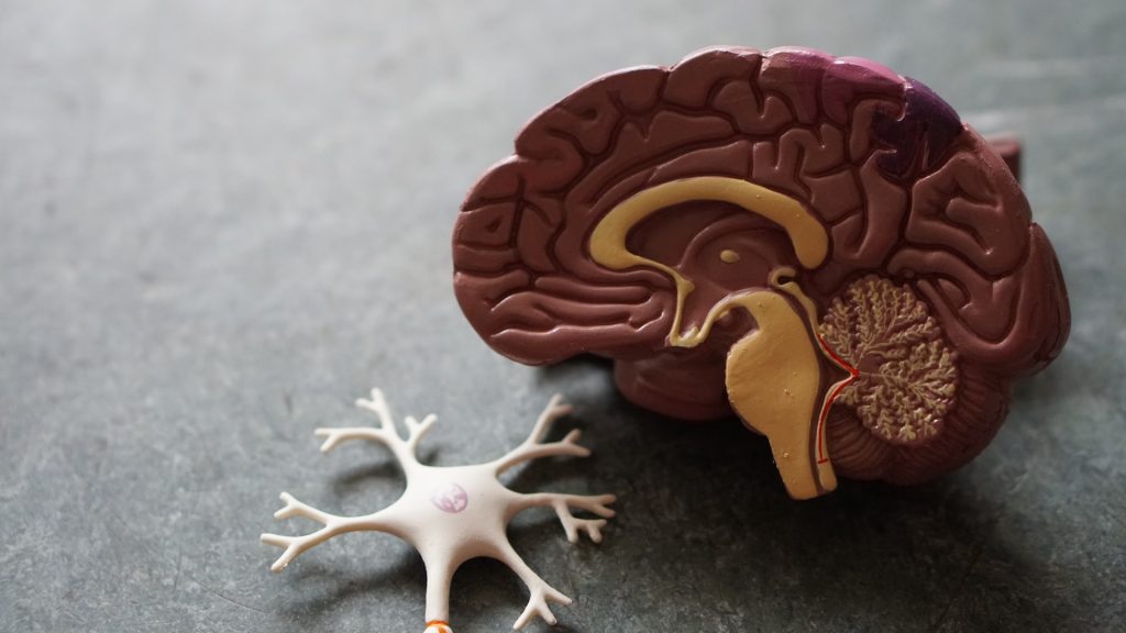
Led by the University of Cincinnati and conducted in collaboration with the Karolinska Institute in Sweden, the study claims that the treatment of Alzheimer’s disease might lie in normalising levels of a brain protein called amyloid-beta peptide. This protein is needed in its original, soluble form to keep the brain healthy, but it sometimes hardens into ‘brain stones’ or clumps, called amyloid plaques.

“It’s not the plaques that are causing impaired cognition,” said senior author Alberto Espay, professor of neurology at UC. “Amyloid plaques are a consequence, not a cause,” of Alzheimer’s disease, stated Prof Espay, who is also a member of the UC Gardner Neuroscience Institute.

Since its discovery, scientists have focused on treatments to eliminate the plaques. But the UC team, he said, viewed it differently: Cognitive impairment could be due to a decline in soluble amyloid-beta peptide instead of the corresponding accumulation of amyloid plaques. 
To test their hypothesis, they analyzed the brain scans and spinal fluid from 600 individuals enrolled in the Alzheimer’s Disease Neuroimaging Initiative study, who all had amyloid plaques. From there, they compared the amount of plaques and levels of the peptide in the individuals with normal cognition to those with cognitive impairment. They found that individuals with high levels of the peptide were cognitively normal, despite the numbers of plaques in their brains.

They also found that higher levels of soluble amyloid-beta peptide were associated with a larger hippocampus, the area of the brain most important for memory.

According to the authors, as we age most people develop amyloid plaques, but few people develop dementia. In fact, by the age of 85, 60% of people will have these plaques, but only 10% develop dementia.

“The key discovery from our analysis is that Alzheimer’s disease symptoms seem dependent on the depletion of the normal protein, which is in a soluble state, instead of when it aggregates into plaques,” said co-author Kariem Ezzat from the Karolinska Institute.

The most relevant future therapeutic approach for the Alzheimer’s program would then be to restore these brain soluble proteins to their normal levels, said Prof Espay.

The research team is now working to test their findings in animal models. If successful, future treatments may be very different from those tried over the last two decades. Treatment, says Espay, may consist of increasing the soluble version of the protein in a manner that keeps the brain healthy while preventing the protein from hardening into plaques.  

Source: University of Cincinnati 

Journal information: Andrea Sturchio et al, High cerebrospinal amyloid-β 42 is associated with normal cognition in individuals with brain amyloidosis, EClinicalMedicine (2021). DOI: 10.1016/j.eclinm.2021.100988

Connective Tissue Protein Has Immune Role

Photo by National Cancer Institute on Unsplash

A new study finds that a connective tissue protein also encourages immune responses that fight bacterial infections, while restraining responses that can be deadly in sepsis.

The study focuses on the extracellular matrix (ECM) of connective tissues, once viewed merely as structural material. It is now increasingly recognised as a signaling partner with nearby cells in normal function, as well as being involved in disease. Fibroblasts are important players in the ECM; these cells make tough structural matrix proteins like collagen. The study was published online June 28 in the Proceedings of the National Academy of Sciences.

The new analysis found that lumican, a protein-sugar combination (proteoglycan) secreted by fibroblasts, and known to partner with collagen in connective tissues, also promotes immune system responses in immune cells called macrophages that fight bacterial infections. The study also found that lumican protects tissues by holding back a different type of immune response that reacts to DNA, whether from an invading virus, or released from cell death.

Such inflammatory responses are a transition into healing, but in sepsis they grow out of control, causing damage to the body’s own tissues. Sepsis affects 48.9 million people worldwide, the authors said, but the ECM’s role the condition is largely unknown.

“Lumican may have a dual protective role in ECM tissues, promoting defense against bacteria on the one hand, and on the other, limiting immune overreactions to DNA that cause self-attack, or autoimmunity,” said corresponding study author Shukti Chakravarti, PhD, professor in the Department of Ophthalmology and the Department of Pathology at NYU Langone Health.

The findings suggest that connective tissue, and extracellular matrix proteins like lumican, usually operate outside of cells, but as disease or damage break down ECM, get sucked into and regulate immune cells homing in on the damage.

Lumican  interacts with two proteins on surfaces of immune cells that control the activity of toll-like receptors, which recognise structural patterns common to molecules made by invading microbes, said the researchers. As they are less specific than other parts of the immune system, toll-like receptors can also cause attacks by immune cells on the body’s own tissues if over-activated.

In this study, the researchers found that lumican promotes the ability of toll-like receptor (TLR)-4 on the surfaces of immune cells to recognise bacterial cell-wall toxins called lipopolysaccharides (LPS). Lumican, by attaching to two proteins, CD14 and Caveolin1, probably using collagen-covered regions, stabilises their interactions with TLR4 to increase its ability to react to LPS. This results in production of the signalling protein TNF alpha, which amplifies immune responses.

Along with describing the effect of lumican on the surfaces of immune cells, the new study finds that lumican is taken up from outside cells into membrane-bound pouches, called endosomes, and pulled into cells. Such compartments deliver ingested bacteria to other endosomes that destroy them, heighten inflammation, or produce protective interferon responses. Once pulled inside, the researchers found, lumican bolstered TLR4 activity by slowing down its passage into lysosomes, pockets where such proteins are broken down and recycled.

However, while it encouraged TLR4 activity on cell surfaces, lumican, once inside immune cells, had the opposite effect on toll-like receptor 9 (TLR9), which reacts to DNA instead of bacterial LPS.

Mice with the lumican gene deleted had trouble both fighting off bacterial infections (less cytokine response, slower clearance, greater weight loss), and trouble restraining the immune overreaction to bacteria (sepsis). Elevated lumican levels were also found in human sepsis patients’ blood plasma, and that human immune cells (blood monocytes) treated with lumican had elevated TLR4 activity but suppressed TLR9 responses.

“As an influencer of both processes, lumican-based peptides could be used as a lever, to tweak inflammation related to TNF-alpha, or endosomal interferon responses, to better resolve inflammation and infections,” suggested George Maiti, PhD, a postdoctoral fellow in Dr Chakravarti’s lab.

“Our results argue for a new role for ECM proteins at sites of injury. Taken up by incoming immune cells it shapes immune responses beyond the cell surface by regulating the movement and interaction of endosomal receptors and signaling partners,” said Dr Chakravarti.

Source: NYU Langone Health

Journal information: George Maiti et al., “Matrix lumican endocytosed by immune cells controls receptor ligand trafficking to promote TLR4 and restrict TLR9 in sepsis,” PNAS (2021). www.pnas.org/cgi/doi/10.1073/pnas.2100999118

Trauma Patients with COVID at Great Risk

Photo by Nate Isaac on Unsplash

The COVID pandemic has placed a great strain on healthcare resources, with a number of indirect impacts ranging from increased incidence of heart attacks to decreased cancer screenings, but also increased the risk of complications and death among trauma patients with COVID. 

The study revealed that the risk of death for COVID-positive patients in trauma centres across the US state of Pennsylvania was six times higher than non-COVID-negative patients with similar injuries. Complication risk in COVID-positive patients was doubled for venous thromboembolism, renal failure, need for intubation, and unplanned ICU admission, and was five times greater for pulmonary complications. In patients over age 65, the risks were even higher. The findings were recently published in The Journal of Trauma and Acute Surgery.  

“COVID had the largest impact on patients whose injuries were relatively minor, and who we would have otherwise expected to do well,” said lead author Elinore Kaufman, MD, MSHP, an assistant professor in the Division of Trauma, Surgical Critical Care and Emergency Surgery at Penn Medicine. “Our findings underscore how important it is for hospitals to consistently test admitted patients, so that providers can be aware of this additional risk and treat patients with extra care and vigilance.”

Researchers conducted a retrospective study of 15 550 patients admitted to Pennsylvania trauma centers from March 21, 2020, (when non-essential businesses statewide were ordered close) to July 31, 2020. Of the 15 550 patients, 8170 were tested for the virus, and 219 tested positive. During this period, the researchers evaluated length of stay, complications, and overall outcomes for patients who tested positive for COVID, compared to patients who did not have the virus. They found that rates of testing increased over time, from 34% in April 2020 to 56% in July. Centres had a great variability in testing, a median of 56.2% of the time with a range of 0 to 96.4%.

“First, we need to investigate how to best care for these high-risk patients, and establish standard protocols to minimise risks,” said senior author Niels D Martin, MD, chief of Surgical Critical Care and an associate professor in the division of Trauma, Surgical Critical Care and Emergency Surgery. “Second, we need more data on the risks associated with patients who present symptoms of COVID, versus those who are asymptomatic, so we can administer proven treatments appropriately and increase the likelihood of survival with minimal complications.”

Source: University of Pennsylvania

‘Far Too Many Oocytes’ Being Extracted in IVF

Photo by MART PRODUCTION from Pexels

A study has suggested that IVF clinics in the UK may be retrieving “far too many oocytes” and that most of them “may never be used and are probably discarded”, a finding that may well represent global practice.

Studies indicate that the optimal and safe number of oocytes needed for achieving an ongoing pregnancy is between six and 15. However, the use of egg freezing (such as to preserve fertility to a later age, known as social egg freezing), frozen embryo replacement (FER) cycles and aggressive stimulation regimes has raised this number in order to boost success rates in older women and in poor responders who produce fewer eggs. What is not known is the impact of numbers of eggs retrieved and of over-stimulation practices on the health of patients, and on their emotional state and finances.

Details of the analysis were presented online at the virtual Annual Meeting of ESHRE by Dr Gulam Bahadur from North Middlesex University Hospital, London.

More than 1.625 million eggs in the UK were retrieved from 147,274 women between 2015 and 2018. Although an average of 11 eggs was collected per patient, 16% of cycles were associated with 16-49 oocytes retrieved (per cycle) and 58 women each had over 50 eggs collected in a single egg retrieval procedure.

“Our observations suggests that the high oocyte number per retrieval procedure needs re-evaluation,” said Dr Bahadur. “In particular, this needs to focus on the side effects, including ovarian hyperstimulation syndrome and procedure-related complications, and on the fate of unused frozen oocytes and the costs associated with freezing them.

“Patients should be advised that it’s better to collect fewer eggs leading to good quality embryos which may go to term and result in a healthy baby.”

This report is based on all UK IVF clinics and relates to non-donor fertility treatment carried out between 2015 and 2018 during which 172 341 fresh oocyte retrieval cycles took place. All outcomes and patterns remained uniform over the four years.

A substantial number (n = 10 148) of cycles did not yield any oocytes. Over half (53%) of all IVF cycles were in the desired egg yield range of 6-15. In addition, a quarter of cycles yielded 1-5 eggs; 14% produced 16-25; and a minority (2%) resulted in 26-49 oocytes. The authors point out that multiple birth rates increase significantly from 6-15 oocytes onwards, which increases the risk of birth complications and low birth weight.

A total of 931 265 embryos resulted from all eggs retrieved – a fertilisation rate of 57%. Of the embryos created, more than one in five (22% or 209,080) were transferred into the uterus, while a slightly higher proportion (24% or 219, 563) were frozen.

The fate of the unfertilised oocytes (43%) is unknown, though they are usually discarded. Most of the embryos not transferred (54%) will likely be discarded after patients have paid for several years of storage.

“This comes with a financial and emotional cost,” said Bahadur. “Patients build an attachment with this frozen material and there’s insufficient counselling to support them. They should be given more information about the implications of freezing eggs and embryos.”

Source: European Society of Human Reproduction and Embryology

Medical Device Warning for Certain Apple Devices

Apple has released a list of its products that it advises should be kept a “safe distance” away from sensitive medical devices such as pacemakers and implanted defibrillators. These products are iPhone 12 models, Apple Watch and MacBook Pro. 

A number of consumer-electronic devices contain components, such as magnets, which are known to interfere with medical devices. A number of other manufacturers, for example Samsung and Huawei, have issued similar guidance for some of their products.

Heart health is a promoted feature of some Apple products; certain Apple Watches can make electrocardiogram tests and display the results to the user, as well as recording the data for later medical examination. A number of studies have shown that Apple watches can detect cardiovascular problems such as atrial fibrillation with a fairly high degree of sensitivity. However, the current notice warns of the risks posed by components in some products.

“Under certain conditions, magnets and electromagnetic fields might interfere with medical devices,” Apple wrote, noting “implanted pacemakers and defibrillators might contain sensors that respond to magnets and radios when in close contact”.

Implanted defibrillators send electrical pulses to regulate abnormal heart rhythms. Apple said the listed products should be kept more than 15cm away from medical devices, double that if they are wirelessly charging.

The support page that listed the devices, had said earlier this month that iPhone 12 models were “not expected to pose a greater risk of magnetic interference to medical devices” than other iPhones.

However, the website MacRumours, which first noted the list, pointed to research suggesting that the iPhone 12 could interfere with implanted devices.

A study published June 2 in the Journal of the American Heart Association found that “Apple’s iPhone 12 Pro Max MagSafe technology can cause magnet interference”, and so had the potential “to inhibit life-saving therapy”.

The researchers acknowledged the study’s small scale as a limitation, though in a press release lead investigator Dr Michael Wu wrote that they were surprised by the strength of the magnets in the iPhone 12.

“In general, a magnet can change a pacemaker’s timing or deactivate a defibrillator’s life-saving functions, and this research indicates the urgency for everyone to be aware that electronic devices with magnets can interfere with cardiac implantable electronic devices.”

However Marie Moe, a computer security consultant for Mnemonic, told the BBC she was not worried.

“These Apple gadgets are generally not emitting large magnetic fields, unlike heavy machinery, big concert speakers or welding equipment that anyone with a pacemaker should be more concerned about getting in close proximity to,” she said. She is a pacemaker user herself and studies their use.

Ms Moe added that magnets as strong as those in the iPhone 12 could only put the pacemaker into “a kind of safety mode where the pacing is constant”, which would revert back once the device was removed.

Jo Whitmore, senior cardiac nurse at the British Heart Foundation, agreed that devices kept at a safe distance were not cause for concern. “It’s perfectly OK to use a smartphone when you have a pacemaker, and they’re designed to return to normal settings once the magnet is moved away,” she said.

She added that patients should check the device instructions or talk to the manufacturer if they are concerned, and they could also contact their doctor or pacing clinic.

Source: BBC News

Chemical Fingerprints Improve Stem Cell Production

Photo by Louis Reed on Unsplash

Researchers in Japan have developed a new, noninvasive way to monitor the tricky art of stem cell production.

The current era of ethical stem cell research was ushered in by the 2012 Nobel prize-winning discovery that ordinary cells could be coaxed to revert to their earliest pluripotent stage ushered in. Suddenly, scientists could have an ethical, near-inexhaustible supply of pluripotent stem cells — the most versatile of stem cells — that can become any type of cell much like how embryonic stem cells function.

These reprogrammed cells called induced pluripotent stem cells (or iPS cells) hold great promise for regenerative medicine, where they can be used to develop tissue or organ replacement-based treatments for life-threatening diseases.

One key challenge is that it is a lengthy and delicate process to artificially induce ordinary cells to reset back to pluripotency. Obtaining iPS cells therefore is a matter of chance. However, knowing all they can about the complex chemical changes happening inside during reprogramming can help scientists increase the chances of successfully obtaining viable iPS cells for clinical applications. Current methods that track reprogramming status, however, use destructive and costly techniques.

A study led by Dr Tomonobu Watanabe, professor at Hiroshima University’s Research Institute for Radiation Biology and Medicine, showed that Raman spectroscopy could be a low-cost, simpler, and non-intrusive technique to monitor the cell’s internal environment as it transitions.

Dr Watanabe explained: “The quality evaluation and sorting of existing cells have been carried out by investigating the presence or absence of expression of surface marker genes. However, since this method requires a fluorescent antibody, it is expensive and causes a problem of bringing the antibody into the cells.”

He added that the “solution of these problems can accelerate the spread of safe and low-cost regenerative medicine using artificial tissues. Through our method, we provide a technique for evaluating and sorting the quality of iPS cells inexpensively and safely, based on scattering spectroscopy.”

Raman spectroscopy is an alternative to invasive approaches that require dyes or labels to extract biochemical information. It instead makes use of vibration signatures produced when light beams interact with chemical bonds in the cell. Since each chemical has its own distinct vibration frequency, scientists can use it to identify the cell’s molecular makeup.

The team used this spectroscopic technique to get the “chemical fingerprints” of mouse embryonic stem cells, the neuronal cells they specialised into, and the iPS cells formed from those neuronal cells. These data were then used to train an AI model to can track the reprogramming is progressing, and verify iPS cell quality by checking for a “fingerprint” match with the embryonic stem cell.

To measure the progress, they assigned the “chemical fingerprint” of neuronal cells as the transformation starting point and the embryonic stem cell’s patterns as the desired end goal. Along the axis, they used “fingerprint” samples collected on days 5, 10, and 20 of the neuronal cells’ reprogramming as reference points on how the process is advancing.

“The Raman scattering spectrum contains comprehensive information on molecular vibrations, and the amount of information may be sufficient to define cells. If so, unlike gene profiling, it allows for a more expressive definition of cell function,” Dr Watanabe said.

“We aim to study stem cells from a different perspective than traditional life sciences.”

Source: Hiroshima University

Journal information: Germond, A., et al. (2020) Following Embryonic Stem Cells, Their Differentiated Progeny, and Cell-State Changes During iPS Reprogramming by Raman Spectroscopy. Analytical Chemistry doi.org/10.1021/acs.analchem.0c01800.

Breakthrough AI Development for Premature Baby Care

Photo by Hush Naidoo on Unsplash

Researchers believe they have made a breakthrough in the science of keeping premature babies alive.

As part of her PhD work, James Cook University engineering lecturer Stephanie Baker led a pilot study that used a hybrid neural network to accurately predict how much risk individual premature babies face. This study was published in the journal Computers in Biology and Medicine.

Complications resulting from premature birth are the leading cause of death in children under five and over 50% of neonatal deaths occur in preterm infants, she said. In 2005, 12.9 million births, or 9.6% of all births worldwide, were preterm.

“Preterm birth rates are increasing almost everywhere. In neonatal intensive care units, assessment of mortality risk assists in making difficult decisions regarding which treatments should be used and if and when treatments are working effectively,” said Ms Baker.

To better guide their care, preterm babies are often given a score that indicates the risk they face.

“But there are several limitations of this system. Generating the score requires complex manual measurements, extensive laboratory results, and the listing of maternal characteristics and existing conditions,” noted Ms Baker.

She said the alternative was to measure variables that do not change (eg, birthweight) that prevents recalculation of the infant’s risk on an ongoing basis and does not show their response to treatment.

“An ideal scheme would be one that uses fundamental demographics and routinely measured vital signs to provide continuous assessment. This would allow for assessment of changing risk without placing unreasonable additional burden on healthcare staff,” said Ms Baker.

She said the JCU team’s research had culminated in the Neonatal Artificial Intelligence Mortality Score (NAIMS), a hybrid neural network that relies on simple demographics and trends in heart and respiratory rate to determine mortality risk.

“Using data generated over a 12 hour period, NAIMS showed strong performance in predicting an infant’s risk of mortality within 3, 7, or 14 days.

“This is the first work we’re aware of that uses only easy-to-record demographics and respiratory rate and heart rate data to produce an accurate prediction of immediate mortality risk,” said Ms Baker.

According to Ms Baker, the technique was fast with no invasive procedures or knowledge of medical histories needed.

“Due to the simplicity and high performance of our proposed scheme, NAIMS could easily be continuously and automatically recalculated, enabling analysis of a baby’s responsiveness to treatment and other health trends,” said Ms Baker.

She said NAIMS had proved accurate when tested against hospital mortality records of preterm babies and had the added advantage over existing schemes of being able to perform a risk assessment based on any 12 hour period of data gathered during the patient’s stay.

Ms Baker said the next step in the process was partnering with local hospitals to gather more data and undertake further testing.

“Additionally, we aim to conduct research into the prediction of other outcomes in neo-natal intensive care, such as the onset of sepsis and patient length of stay,” said Ms Baker.

Source: James Cook University

Journal information: Baker, S., et al. (2021) Hybridized neural networks for non-invasive and continuous mortality risk assessment in neonates. Computers in Biology and Medicine. doi.org/10.1016/j.compbiomed.2021.104521.

Lockdown Level 4; Third Wave Driven by Delta Variant


In response to the third wave driven by the delta variant, President Cyril Ramaphosa instituted a two-week Level 4 lockdown during a ‘family meeting’ address to the nation.

He warned that the healthcare system was facing a dire situation. “Our health facilities are stretched to the limit… ICU beds are in short supply,” he said

In a press briefing on Friday, the head of the World Health Organization said the COVID Delta variant, first seen in India, is “the most transmissible of the variants identified so far,” and warned it is now spreading in at least 85 countries.

“We are in the exponential phase of the pandemic with the numbers just growing very, very, extremely fast and (they) will keep growing in the next weeks,” said Tulio de Oliveira, a leading virologist in the country.

The Delta variant first seen in India now appears to be “dominating infections in South Africa,” de Oliveira of the Network for Genomic Surveillance in South Africa told a virtual briefing.

The Delta variant has emerged as dominant in South Africa. Source: Department of Science & Technology

Koleka Mlisana, the head of a government ministerial advisory committee on COVID, told the same briefing that there is “evidence that the Delta variant may actually be taking over”.

Acting Minister of Health, Mmamoloko Kubayi-Ngubane said that due to the prevalence of the Delta variant, infection numbers “are likely to surpass the second wave peak” in January.

Only about 2.4 million people have been immunised since February. Thousands of EFF activists rallied in Pretoria on Friday to demand a faster coronavirus vaccination rollout, including expedited approvals for the Sinovac vaccine from China and Russia’s Sputnik V.

Source: Medical Xpress

Preliminary Study Explains Why Delta Variant is So Infectious

Colorized scanning electron micrograph of an apoptotic cell (purple) heavily infected with SARS-COV-2 virus particles (yellow), isolated from a patient sample. Image captured at the NIAID Integrated Research Facility (IRF) in Fort Detrick, Maryland. Credit: NIAID

A preliminary study has possibly determined why the SARS-CoV-2 Delta variant is more infectious and pathogenic than its ancestor.

Through a series of in vitro experiments, researchers have discovered that variant’s enhanced ability to induce cell-to-cell fusion (syncytia) and reduced susceptibility to vaccine and infection-induced antibodies together help make the Delta variant more infectious than previously circulating variants. The study, which is yet to be peer reviewed, is currently available on the bioRxiv preprint server.

The SARS-CoV-2 virus has undergone more than 12 000 mutations since it was first detected in December 2019, most of which are neutral and do not contribute to viral evolution. However, the acquisition of specific mutations in structural and non-structural proteins has caused the emergence of novel, more virulent SARS-CoV-2 variants.

Spike protein mutations are particularly concerning as they can significantly influence viral infectivity, virulence, and immune evasion ability.

The B.1.617 lineage drove a massive surge in new COVID cases in India. This lineage is further divided into three sub-lineages, namely B.1.617.1, B.1.617.2, and B.1.617.3. Although these emerged first in India, the B.1.617.2 or Delta variant or soon became dominant in many countries, including South Africa where it has driven a new surge of infections, particularly in Gauteng Province. The World Health Organization (WHO) has designated the Delta variant as a ‘Variant of Concern’ (VOC) due to its significantly increased infectivity and pathogenicity.

In the current study, the scientists have evaluated the susceptibility of the Delta variant to neutralisation by vaccine or natural infection-induced antibodies.

Delta variant mutations 

The Delta variant’s spike protein contains nine mutations in the S1 subunit and one mutation in the S2 subunit. In the S1 subunit, five mutations are present in the N-terminal domain containing binding sites (epitopes) for neutralising antibodies. In addition, two mutations are present in the receptor-binding domain of the S1 subunit, which is known to influence antibody-mediated neutralisation and infectivity. Among the three remaining mutations, two are known to increase angiotensin-converting enzyme 2 (ACE2) binding, viral replication, and spike protein cleavage at the S1/S2 site.    

Delta variant host cell entry

Using African green monkey and human cells, the researchers found that Delta can enter kidney cells of both species with similar efficacy as the wild-type SARS-CoV-2. However, for human colon and lung cells, Delta showed 1.5-fold and 2-fold higher invading ability, respectively, compared to the wild-type virus. Since the Delta variant spike protein did not exhibit increased ACE2 binding, the scientists suggest that increased entry of B.1.617.2 into colon and lung cells is not mediated by enhanced ACE2 binding.

Besides inducing fusion between the viral envelope and host cell membrane, the spike protein triggers the fusion of infected cells with nearby cells to form large multinucleated cells, known as syncytia. Given the fact that spike-induced syncytia formation contributes to COVID pathogenesis, the scientists investigated whether Delta variant infection is associated with increased syncytia formation.

By conducting in vitro experiments on human lung cells expressing high levels of ACE2, they found that Delta spike expression leads to 2.5-fold higher and larger syncytia formation than the wild-type spike expression.

Delta variant’s immune evasion ability less than Beta?

The scientists tested the ability of four therapeutic monoclonal antibodies to neutralise the Delta variant, of which only Bamlanivimab failed. The other three antibodies exhibited similar efficacy in neutralising both wild-type virus and Delta variant.

Antibodies derived from COVID recovered patients, and BNT162b2-vaccinated individuals showed only slightly reduced efficacy in neutralising the Delta variant as compared to the wild-type virus. In contrast, the B.1.315 or Beta variant, first detected in South Africa, showed a significantly higher ability to evade infection- and vaccination-induced immunity.

In summary

The study showed that Delta’s increased ability to invade lung cells may enhance infectivity and pathogenicity. Though it has lower susceptibility to antibody-mediated neutralisation, it is possible that Delta may be effectively controlled by immunity developed in response to natural infection or vaccination.

Source: News-Medical.Net

Journal information: Arora P. 2021. Increased lung cell entry of B.1.617.2 and evasion of antibodies induced by infection and BNT162b2 vaccination. bioRxiv. https://www.biorxiv.org/content/10.1101/2021.06.23.449568v1

Performance Enhancers Linked to Criminality

Photo by Jonathan Borba on Unsplash

A new study shows that both anabolic steroid use as well as legal performance-enhancing substances is longitudinally associated with criminal offending.

Although anabolic steroid use was known to be associated with criminal offending, the possibility of a similar link between use of legal performance-enhancing substances, such as creatine, and criminal offending remained unknown. 

To address this, researchers analysed a sample of over 9000 US participants from the National Longitudinal Study of Adolescent Health (Add Health). The results show a need for more research on performance-enhancing substances to understand the complex social problems associated with their use.

“This is the first study to identify relationships between legal performance-enhancing substance use and criminal offending,” said lead author Kyle T. Ganson, PhD, MSW, assistant professor at the University of Toronto’s Factor-Inwentash Faculty of Social Work. “This finding is acutely salient because these substances are easily accessible and commonly used, particularly among young people.”

The study highlights the importance of clinical professionals screening for performance-enhancing substance use and assessing patterns of criminal offending among young people.

“We need more research to identify effective prevention and intervention techniques to ensure that we reduce the use of these substances, as well as curtail any connection with criminal offending,” said co-author Jason M. Nagata, MD, MSc, assistant professor at the University of California, San Francisco’s Department of Pediatrics.

“The associations found in this study are likely explained by an intersection of behavioral, psychological, and sociocultural influences,” says Ganson. “We therefore need to target this problem from a multitude of angles, including clinically and via public health and policy interventions.”

Source: EurekAlert!

Journal information: Ganson, K.T., et al. (2021) Performance-Enhancing Substance Use and Criminal Offending: A 15-Year Prospective Cohort Study. Drug and Alcohol Dependence. doi.org/10.1016/j.drugalcdep.2021.108832.