Results support the potential for dapagliflozin to benefit these patients
Human liver. Credit: NIH
The sodium glucose cotransporter 2 (SGLT-2) inhibitor drug dapagliflozin, widely used to treat type 2 diabetes, also shows improvements for patients with progressive liver disease, according to results of a clinical trial in China published by The BMJ.
The results show that treatment with dapagliflozin improved metabolic dysfunction-associated steatohepatitis (MASH) – a condition where excess fat accumulates in the liver, leading to inflammation – and liver fibrosis compared with placebo.
MASH affects more than 5% of adults, more than 30% of individuals with diabetes or obesity, and can progress to cirrhosis in up to 25% of individuals.
Several studies have reported that SGLT-2 inhibitors can improve liver fat content, liver enzymes, and liver stiffness, but no trial has been carried out among patients with MASH.
To address this, researchers enrolled 154 adults (average age 35; 85% men) diagnosed with MASH after a liver biopsy at six medical centres in China from November 2018 to March 2023.
Almost half (45%) had type 2 diabetes, and almost all had liver fibrosis (33% stage 1, 45% stage 2, 19% stage 3).
After an initial screening biopsy, participants were randomly assigned to receive 10 mg of dapagliflozin or matching placebo once daily for 48 weeks and attended health education sessions twice a year.
Various factors including body weight, blood pressure, blood glucose, liver enzymes, physical activity, diet, insulin, and lipids were also assessed at enrollment and throughout the trial.
MASH improvement was defined as a decrease of at least 2 points in non-alcoholic fatty liver disease activity score (NAS) or a NAS of 3 points or less.
After an end of study biopsy at week 48, 53% (41 of 78) participants in the dapagliflozin group showed improvement in MASH without worsening of fibrosis (defined as no increase in fibrosis stage) compared with 30% (23 of 76) in the placebo group.
Resolution of MASH without worsening of fibrosis occurred in 23% (18 of 78) participants in the dapagliflozin group compared with 8% (6 of 76) in the placebo group.
Fibrosis improvement without worsening of MASH was also reported in 45% (35 of 78) participants in the dapagliflozin group compared with 20% (15 of 76) in the placebo group.
The percentage of participants who discontinued treatment because of adverse events was 1% (1 of 78) in the dapagliflozin group and 3% (2 of 76) in the placebo group.
The researchers acknowledge that the trial was conducted in a Chinese population, which limits its broader generalisability, and that female and older patients were under-represented. But they point out that results were consistent after further analyses, suggesting they are robust.
As such, they conclude: “Our findings indicate that dapagliflozin may affect key aspects of MASH by improving both steatohepatitis and fibrosis.” Large scale and long term trials are needed to further confirm these effects, they add.
The coming years are expected to be particularly exciting in the field of pharmacological treatment for MASH, say researchers from Argentina in a linked editorial.
As more drugs become available, therapeutic decisions will likely become increasingly tailored to individual patient profiles, they write. “Ideally, such treatments should provide cardiovascular benefit, have an established safety profile, and be accessible to broad and diverse patient populations,” they conclude.
In the US, drugs like semaglutide are used by over 15 million adults in the U.S., or 4.5% of the population for diabetes and also weight loss. Despite their effectiveness, they have drawbacks. Their effect may not last after discontinuing use, and side effects including osteoporosis and muscle loss have raised concerns about long-term harms. They also induce nausea, which can make it difficult to stay the course of treatment.
Now Tufts researchers led by Professor Krishna Kumar, have designed a new, next-generation compound with hopes that it could be more effective with fewer side effects, which they report in a paper in the Journal of the American Chemical Society.
While weight loss drugs currently on the market and in development target one, two, or even three hormone receptors related to glucose metabolism and the desire to eat, the Tufts team has identified a fourth target that could potentially further enhance the control strategy.
“Obesity is linked to over 180 different disease conditions, including cancer, cardiovascular disease, osteoarthritis, liver disease, and type 2 diabetes, and affects over 650 million people worldwide,” said Kumar. “What drives us is the idea that we can design a single drug to treat obesity and simultaneously mitigate the risk of developing a long list of health problems plaguing society.”
How the Drugs Work
After a meal, the hormone glucagon-like peptide 1 (GLP-1) is released to help stimulate the production of insulin and the uptake of glucose in muscle and other tissues. With the cells now loaded with fuel, the level of glucose in the blood returns to normal. Semaglutide uses GLP-1 with slight modifications to increase its availability in the bloodstream. Its success in controlling blood glucose has prompted the American Diabetes Association to recommend it and other GLP-1-based drugs as the new first line injectable treatments for diabetes, ahead of insulin.
But GLP-1 also acts directly on the brain, prompting satiety after a meal, and it slows down the rate at which stomach contents are emptied into the intestines, evening out the release of nutrients and glucose into the bloodstream. That’s why it has also become extremely popular as a weight loss treatment.
It’s still not a perfect drug strategy for weight loss, though. “The biggest problem with GLP-1 drugs is that they have to be injected once a week, and they can induce a very strong feeling of nausea,” said Kumar. “As much as 40% of people using these drugs give up after the first month.”
A second hormone released after eating is glucose-dependent insulinotropic peptide (GIP). It also makes us feel full after a meal. GIP looks a lot like GLP-1, so rather than administer two drugs, researchers created one peptide that incorporates structural elements of both – what’s called in drug development a chimera. That drug, tirzepatide, has the added benefit of significantly reducing nausea. As a more tolerable treatment, it may overtake semaglutide in the weight loss market.
“And then there is a third hormone, glucagon,” said Kumar. “Paradoxically, it actually increases blood glucose, but at the same time increases the expenditure of energy in cells of the body, raises body temperature, and suppresses appetite.” By adding glucagon to the mix, GLP-1 and GIP end up neutralizing its glucose-enhancing effect, leaving the remaining functionalities of all three hormones working together to enhance weight loss.
Glucagon is also similar in structure to GLP-1 and GIP, so drug developers created a single chimera peptide that incorporates elements of all three hormones, which can be recognised by their three separate receptors. That drug, called retatrudide, is currently in clinical trials that indicate even greater achievable weight loss (up to 24%) compared to the original GLP-1 drugs (6-15%).
Going for the Weight Loss Gold Standard with a Fourth Target
“The goal that people are trying to shoot for is bariatric surgery,” said Kumar. That’s a surgical procedure significantly reducing the size of the stomach, which can achieve long-lasting weight loss up to 30%. “For individuals with persistent obesity and potential deadly associated conditions, it becomes a necessary but invasive treatment.”
Current injectable weight loss drugs still fall short of that gold standard, so the Tufts chemists are focused on a drug redesign that could match the 30% weight loss outcome.
“There is one more hormone we wanted to bring in to complete a weight control quartet,” said Tristan Dinsmore, a graduate student in the Kumar lab and the lead author of the study. “It’s called peptide YY (PYY). This molecule is also secreted by the gut after we eat a meal, and its job is to reduce appetite and slow the process of emptying food from the stomach, but via different mechanisms than either GLP-1 or GIP. It may also be involved in directly ‘burning off’ fat.”
PYY is from a separate and structurally unrelated class of hormones than the first three, so blending its structure into a chimeric peptide that also mimics GLP-1, GIP, and glucagon was not easy. Instead, the Tufts team was able to join two peptide segments end-to-end, creating a new ‘tetra-functional’ clinical candidate.
“One of the limitations of the current drugs is that individual variation, possibly including how people express target receptors or respond to their corresponding hormones, can lead to lesser than desired weight loss outcomes in many patients,” said Martin Beinborn, visiting scholar in the Department of Chemistry. “By hitting four different hormone receptors at the same time, we hope to improve the chances of averaging out such variation toward the goal of achieving greater and more consistent overall effectiveness.”
“A second issue is that patients tend to regain weight after discontinuing currently available GLP-1 related drugs,” said Beinborn, who notes that lifestyle changes should ideally be a complement to medication treatment. This two-pronged approach will not only support reaching and keeping one’s target weight, but may also help preserve bone and muscle mass.
“Recent studies indicate that weight rebound after drug discontinuation is delayed with the newer, more effective GLP-1 mimetics,” he said. “Extending from this observation, one may speculate that multi-chimeras along the lines of the one we discovered could get us closer to the bariatric surgery standard of lasting weight loss.”
New research published in Allergyindicates that certain environmental exposures may affect a child’s risk of developing atopic eczema, a condition characterised by dry, itchy, and inflamed skin. In other words, although some people may be genetically predisposed to eczema, certain environmental factors may increase or decrease that risk.
For the study, investigators analysed data from 16 European studies to test for interactions between the 24 most significant eczema-associated genetic variants and 18 early-life environmental factors. They applied their findings to an additional 10 studies and used lab modelling tests to assess their results.
The first analysis (including 25 339 individuals) showed suggestive evidence for interaction between 7 environmental factors (antibiotic use, cat ownership, dog ownership, breastfeeding, elder sibling, smoking, and washing practices) and at least one established genetic variant for eczema, with 14 interactions in total.
In the additional analysis (254 532 individuals), dog exposure interacted with a particular genetic risk variant on chromosome 5, near the gene that codes for the interleukin-7 receptor, a protein involved in immune cell function. Lab modelling tests showed that this variant affects expression of interleukin-7 receptor in human skin cells and that dog exposure modifies the genetic effect of this variant on the development of eczema, essentially providing a protective effect by suppressing skin inflammation.
Additional studies are needed to explore these lab findings and the other potential interactions identified in the first analysis.
“Our research aims to answer some of the most difficult questions that I am asked in clinic: ‘Why does my child have eczema?’ and ‘What can I do to help protect my baby?’ We know that genetic make-up affects a child’s risk of developing eczema and previous studies have shown that owning a pet dog may be protective, but this is the first study to show how this may occur at a molecular level,” said corresponding author Sara J. Brown, MD, PhD, FRCPE, of the University of Edinburgh. “More work is needed, but our findings mean we have a chance to intervene in the rise of allergic disease, to protect future generations.”
Looking to nature for answers to complex questions can reveal new and unprecedented results that can even affect cells on molecular levels. For instance, human cells oxidise glucose to produce ATP (adenosine triphosphate), an energy source necessary for life.
Cancer cells produce ATP through glycolysis, which does not utilise oxygen even under conditions where oxygen is present, and convert glucose into pyruvic acid and lactic acid. This method of producing ATP, known as the Warburg effect, is considered inefficient, thus raising questions as to why cancer cells choose this energy pathway to fuel their proliferation and survival.
In search for this energy catalyst, Associate Professor Akiko Kojima-Yuasa’s team at Osaka Metropolitan University’s Graduate School of Human Life and Ecology analysed the cinnamic acid ester ethyl p-methoxycinnamate, a main component of kencur ginger, and its mechanism of action. In previous research, the team discovered that ethyl p-methoxycinnamate has inhibitory effects on cancer cells. Furthering their study, the acid ester was administered to Ehrlich ascites tumour cells to assess which component of the cancer cells’ energy pathway was being affected.
Results revealed that the acid ester inhibits ATP production by disrupting de novo fatty acid synthesis and lipid metabolism, rather than through glycolysis as commonly theorised. Further, the researchers discovered acid ester-induced inhibition triggered increased glycolysis, which acted as a possible survival mechanism in the cells. This adaptability was theorised to be attributed to ethyl p-methoxycinnamate’s inability to induce cell death.
“These findings not only provide new insights that supplement and expand the theory of the Warburg effect, which can be considered the starting point of cancer metabolism research, but are also expected to lead to the discovery of new therapeutic targets and the development of new treatment methods,” stated Professor Kojima-Yuasa.
Authors of a recent Lancet report argue that obesity should not just be seen as a risk factor for other diseases – but in some cases, should be seen as a disease itself. The position could change how we treat obesity globally. In the first of this two-part Spotlight series, we break down the debate around the issue, and its implications for health policy.
In 1990, just 2% of all young people around the world aged 5 to 24 were living with obesity. By 2021, this figure had more than tripled to over 6%. This is according to a recent study, which relied on Body Mass Index (BMI) data from 180 countries and territories around the world. It estimates that the rise in obesity among children and young people will only continue in the coming decades.
South Africa certainly isn’t immune to the crisis. A survey conducted in 2021/2022 found that 16% of all children aged 6 to 18 were “severely overweight”. Meanwhile, World Health Organization (WHO) data suggests that about 30% of all adults in South Africa are living with obesity, meaning a BMI of over 30, which is almost double the global level.
BMI, which simply looks at a person’s weight in relation to their height, is a crude measure of obesity. For instance, a person may have a high BMI simply because they have a lot of muscle rather than fat. But while it is agreed that BMI is a flawed indicator at the individual level, many experts recommend using it as a rough proxy for “health risk at a population level”.
For instance, a study which collected data on nearly three million people found that those who had very high BMI levels were, on average, more likely to die at an early age. The study also found that this was true of people with very low BMI levels (those who were underweight). In this context, the above figures paint a concerning picture.
Given the rising rates, experts argue that we need health systems to be able to track and respond to obesity urgently. But, according to a Lancet Commission published in January, health systems around the world may struggle to do this, because of a failure to accurately conceptualise and measure what obesity actually is.
The Lancet commission was developed by 58 experts from different medical specialties and though it has been the subject of debate, it has since been widely endorsed as a new way to understand obesity. Spotlight takes a look at what it concluded.
Delaying treatment for no reason
Obesity is often regarded as a risk factor for other diseases, for instance, type 2 diabetes. But according to the commission, there are certain cases in which obesity is not just a risk factor, but a disease itself – one that should be immediately treated.
One of the reasons for this is that obesity not only contributes to the emergence of other conditions but sometimes leads to clinical symptoms directly. For example, the cartilage that protects the joints in a person’s knees can sometimes become eroded when adults carry too much weight. In this case, a person could suffer from joint pain, stiffness and reduced mobility where obesity is clearly the cause.
Take another example. If fat deposits build up in the abdomen, this may limit how much the lungs can expand, causing breathlessness. Similarly, a build-up of fat around the neck can narrow a person’s upper airways, which can cause sleep apnoea.
Thus, obesity is not simply something which increases the risk of developing a separate disease in the future – but something which can directly (and presently) affect the functioning of organs.
More broadly, the commission argues that by hindering a person’s “mobility, balance and range of motion” obesity can in certain cases “restrict routine activities of daily living”. In these instances, obesity is a disease by definition, according to the commission. This is given that it defines disease as a “harmful deviation from the normal structural or functional state of an organism, associated with specific signs and symptoms and limitations of daily activities”.
But why does this conceptual debate matter?
Because at present, people often have to wait for other diseases to crop up before insurers or public health systems cover them for weight loss drugs or bariatric surgery – a procedure to help with weight loss and improve obesity-related health conditions. And when they do cover these services, it is often only after severe delay. Because obesity is only considered to be a risk factor, it isn’t typically treated with the same urgency as life-threatening diseases, according to the authors of the commission.
Professor Frances Rubino, the lead author of the commission, details how this problem manifests in the healthcare system.
“I’ve been doing bariatric surgery for 25 years in four different countries; in America, Italy, France and the UK,” he tells Spotlight, “In all of those countries, to meet the criteria for surgery people very often have to undergo six to 12 months of weight monitoring before their surgery is covered. So systematically you delay treatment”.
He continues: “Someone who has clinical obesity and has heart failure as a result of it is waiting for a year for what reason? That condition will only worsen and if the patient is still alive, the treatment [is] going to cost the same amount to the payer but it’s going to be less effective.”
Can’t people just diet?
One of the reasons that some academics have historically been reluctant to classify obesity as a disease is because of a fear that this may reduce people’s agency – instead of taking proactive steps to diet and exercise, people with obesity may simply view themselves as afflicted by a disease.
The belief that people with obesity can simply diet their way out of their situation is in fact partially why Rubino’s patients were forced to wait long periods of time before receiving bariatric surgery.
Rubino explains: “In America, many private payers [i.e. medical insurance schemes] have required weight monitoring programmes, where patients do nothing else other than see a dietician for 12 months, and if they skip one appointment, they have to start all over again. I think that in some cases, this has been misguided by the idea that you want to see if obesity can be reversed by somebody going on a diet.”
This, according to him, is a “misconception”, arguing that if someone faces such severe levels of obesity that they require surgery, diet is unlikely to offer a solution.
Indeed, research has shown that it’s very rare for people with obesity to lose large amounts of weight quickly without surgery or medication. For instance, a study on over 176 000 patients in the UK found that among men with “simply obesity” or a BMI of 30-34.9, only 1 in 210 were able to achieve a “normal” weight level within a year. Among men with morbid obesity or BMI of 35 or more, the chance was less than 1 than in 1000. Chances for women were roughly twice as good as men’s – so still exceedingly small.
Thus, if someone is severely obese and their excess weight is causing life-threatening symptoms, putting them on a diet for a year is unlikely to result in the urgent changes that may be required for them to get better. In fact, Rubino argues that they may simply die of their condition in the interim.
Taking a medical approach more quickly is easier now than ever before due to the regulatory approval of GLP-1 agonists like semaglutide and tirzepatide – Spotlight previously reported on the availability of these new diabetes and weight loss medicines in South Africa. An article by WHO officials from December states that because of the approval of these medicines “[h]ealth systems across the globe now may be able to offer a treatment response integrated with lifestyle changes that opens the possibility of an end to the obesity pandemic”.
Not all people with obesity are ill
There is a more scientific argument against categorising obesity as a disease. This is that while obesity can sometimes result in the negative health symptoms discussed above (like respiratory issues or reduced mobility) it doesn’t always do this.
In fact, the commission acknowledges that some people with obesity “appear to be able to live a relatively healthy life for many years, or even a lifetime”. One of the reasons for this is that excess fat may be stored in areas that don’t surround vital organs. For instance, if fat is stored in the limbs, hips, or buttocks, then this may cause less harm than if it is stored in the stomach.
Since obesity doesn’t always cause health problems, it isn’t always a disease. In order to deal with this conceptual hurdle, the commission classifies obesity into two categories – clinical and preclinical obesity.
If a person has pre-clinical obesity, this means they have a lot of excess fat, but no obvious health problems that have emerged as a result. In this case, obesity is not classified as a disease, though it may still increase the chance of future health problems (depending on a range of factors, like family history).
For a person to have clinical obesity, they must have a lot of excess fat as well as health problems that have already been directly caused by this. It is this that the commission defines as a disease.
This classification system, according to Rubino, ensures not only that we urgently treat people living with clinical obesity, but also that we don’t overtreat people – since if a person falls into the pre-clinically obese group, then they may not need treatment.
But if we’re going to treat clinical obesity as a disease, we’ll need clear methods of diagnosing people. Since BMI is deeply flawed and provides little information about whether a person is ill at the individual level, health systems will need something else. In part 2 of this Spotlight special series, we’ll discuss the options offered by the commission, and how this all relates to the situation in South Africa.
Scanning electron micrograph of a T cell lymphocyte. Credit: NIH / NIAID
A study initiated by a University of Arizona Comprehensive Cancer Center physician-scientist has for the first time defined how loss of the Y chromosome in male immune cells negatively affects immune system function. The findings, published in Nature, may explain why loss of Y is associated with lower cancer survival rates.
In males, each cell in the body usually contains one X and one Y chromosome. “Loss of Y” is a common, nonhereditary genetic change in men in which an immune cell in the blood loses its Y chromosome. It is often associated with aging. Loss of Y has been linked to increased mortality from carcinomas for many years, though no one knew why.
This study is the first to identify and define the relationship between loss of Y in white blood cells, immune cells and tumours, providing insights as to why men with loss of Y have increased cancer risks and poorer outcomes.
“These findings represent a big step forward in our understanding of why men with loss of Y in their blood cells have a higher mortality from cancer. It turns out it’s because these cells make the immune system infiltrating the cancer less effective,” said Dan Theodorescu, MD, PhD, director of the Cancer Center and a professor in the College of Medicine – Tucson.
“We hope this provides a solid lead and framework for the nascent Y chromosome field to pursue so we can collectively better understand all the possible biological implications of this finding and how to use them to develop more effective approaches in prevention, treatment resulting in higher survival rates for patients.”
The research team discovered that loss of the Y chromosome – previously identified in malignant epithelial cells by the Theodorescu lab – also occurred in nearby noncancerous tissues, including connective tissue and immune cells.
Most notably, the team found that this chromosomal loss in helper and cytotoxic T cells, which are responsible for attacking cancer cells, was associated with a reduced ability to kill those cancerous cells. The findings suggest a mechanism by which tumours may evade immune detection and suppression.
Finally, the research team found that loss of Y in epithelial cells, combined with loss of Y in T cells, resulted in more aggressive cancers and lower survival rates in patients.
“The study has potential implications for current immunotherapies, including CAR T therapy,” Theodorescu said. “Further research is clearly needed but perhaps immunotherapies using cells from a patient’s immune system could be screened for loss of Y before being used in treatment.”
A breakthrough study from the Hebrew University of Jerusalem, published this week in the prestigious journal PNAS (Proceedings of the National Academy of Sciences USA), reveals a previously unknown peripheral mechanism by which paracetamol relieves pain.
The study was led by Prof Alexander Binshtok from the Hebrew University’s Faculty of Medicine and Center for Brain Sciences (ELSC) and Prof Avi Priel from its School of Pharmacy. Together, they uncovered a surprising new way that paracetamol, one of the world’s most common painkillers, actually works.
For decades, scientists believed that paracetamol relieved pain by working only in the brain and spinal cord. But this new research shows that the drug also works outside the brain, in the nerves that first detect pain.
Their discovery centres on a substance called AM404, which the body makes after taking paracetamol. The team found that AM404 is produced right in the pain-sensing nerve endings – and that it works by shutting off specific channels (called sodium channels) that help transmit pain signals. By blocking these channels, AM404 stops the pain message before it even starts.
“This is the first time we’ve shown that AM404 works directly on the nerves outside the brain,” said Prof Binshtok. “It changes our entire understanding of how paracetamol fights pain.”
This breakthrough could also lead to new types of painkillers. Because AM404 targets only the nerves that carry pain, it may avoid the numbness, muscle weakness, and side effects that come with traditional local anaesthetics.
“If we can develop new drugs based on AM404, we might finally have pain treatments that are highly effective but also safer and more precise,” added Prof Priel.
Cells taken from the lungs of people with chronic obstructive pulmonary disease (COPD) have a larger accumulation of soot-like carbon deposits compared to cells taken from smokers without COPD, according to a study published in ERJ Open Research. Carbon can enter the lungs via cigarette smoke, diesel exhaust and polluted air.
The cells, called alveolar macrophages, normally protect the body by engulfing any particles or bacteria that reach the lungs. But, in their new study, researchers found that when these cells are exposed to carbon they grow larger and encourage inflammation.
The research was led by Dr James Baker and Dr Simon Lea from the University of Manchester, UK. Dr Baker said: “COPD is a complex disease that has a number of environmental and genetic risk factors. One factor is exposure to carbon from smoking or breathing polluted air.
“We wanted to study what happens in the lungs of COPD patients when this carbon builds up in alveolar macrophage cells, as this may influence the cells’ ability to protect the lungs.”
The researchers used samples of lung tissue from surgery for suspected lung cancer. They studied samples (that did not contain any cancer cells) from 28 people who had COPD and 15 people who were smokers but did not have COPD.
Looking specifically at alveolar macrophage cells under a microscope, the researchers measured the sizes of the cells and the amount of carbon accumulated in the cells.
They found that the average amount of carbon was more than three times greater in alveolar macrophage cells from COPD patients compared to smokers. Cells containing carbon were consistently larger than cells with no visible carbon.
Patients with larger deposits of carbon in their alveolar macrophages had worse lung function, according to a measure called FEV1%, which quantifies how much and how forcefully patients can breathe out.
When the researchers exposed macrophages to carbon particles in the lab, they saw the cells become much larger and found that they were producing higher levels of proteins that lead to inflammation.
Dr Lea said: “As we compared cells from COPD patients with cells from smokers, we can see that this build-up of carbon is not a direct result of cigarette smoking. Instead, we show alveolar macrophages in COPD patients contain more carbon and are inherently different in terms of their form and function compared to those in smokers.
“Our research raises an interesting question as to the cause of the increased levels of carbon in COPD patients’ macrophages. It could be that people with COPD are less able to clear the carbon they breathe in. It could also be that people exposed to more particulate matter are accumulating this carbon and developing COPD as a result.
“In future, it would be interesting to study how this carbon builds up and how lung cells respond over a longer period of time.”
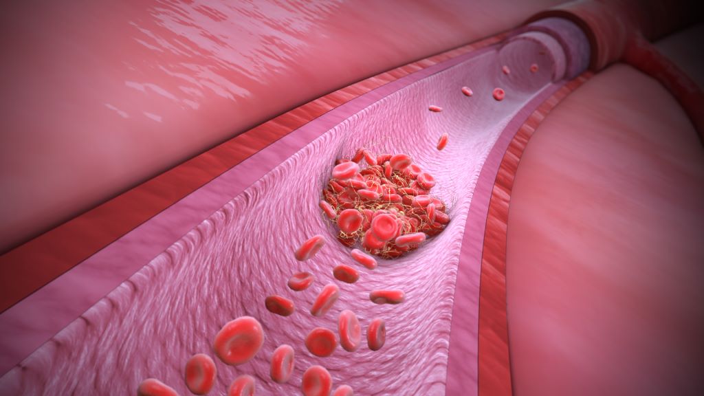
Though blood clots can form in both arteries and veins, the reasons behind them differ, as do the consequences and the chances of preventing blood clots. In Sweden, almost half of all cases of venous thrombosis have a genetic explanation. A team of researchers from Lund University in Sweden has now discovered three gene variants that increase the risk of blood clots in the leg by up to 180%.
There is a difference between arterial and venous blood clots. Blood clots in the arteries form when plaque in calcified vessels bursts and the body perceives it as an injury. This activates the platelets, which clump together and form a clot. In the worst case, it can lead to a stroke or heart attack. A venous thrombus, on the other hand, usually forms in the leg when the blood stagnates for too long. This can activate the body’s coagulation system, allowing the clotting system to be activated and the blood to clot, blocking blood flow. If the clot breaks loose and travels with the blood to the lungs, it can lead to pulmonary embolism, a life-threatening condition.
“Venous thrombosis is in fact one of the most common causes of death in the world. It is a common disease that has always been somewhat overshadowed by arterial blood clots,” says Bengt Zöller, a specialist in general medicine at Skåne University Hospital and professor of general medicine at Lund University.
In Sweden, more than 10 000 people suffer from venous thromboembolism each year and that number appears to be increasing. Several factors are contributing to this increase. One of the strongest risk factors is age, and as the number of older people in Sweden grows, the number of clots is also increasing. Ten per cent of 80-year-olds experience a blood clot at some point. The risk also increases if you are overweight or tall.
“The muscles control the blood flow in the veins and the legs become like columns of fluid where the force of gravity is strong. Too much sedentary and inactive behaviour, then, is harmful. Only the valves of the veins prevent backflow and if these are damaged, the risk of blood clots can increase. Therefore, tall people are more prone to blood clots, as their larger veins provide less blood flow, combined with the fact that blood must travel a greater distance back to the heart.”
Because the heart pumps blood out into the arteries, there is much higher blood pressure in the arteries than in the veins, which can contribute to atherosclerosis. High blood pressure, high levels of blood lipids and smoking are all risk factors for atherosclerosis of the arteries. But because the veins are a low-pressure system, the vessels do not become atherosclerotic. Therefore, neither high blood pressure nor blood lipids are associated with venous clots and smoking is considered only a weak to moderate risk factor. Being overweight, on the other hand, is a very significant culprit. Obesity has a negative impact on venous circulation, especially when combined with the fact that overweight people are often less active. Some clotting factors are also affected by obesity.
“In terms of diet, there are fewer studies, but ultra-processed foods have been associated with a slightly increased risk of blood clots, and plant-based, healthy foods with a reduced risk. In our studies, we have also seen that commercial fishermen have a lower risk, which may be due to a higher omega-3 content in their diet.”
There are also specific situations in which the risk of venous blood clots is particularly high. The risk of blood clots increases when blood flow is reduced, such as when travelling by air for long periods of time or when lying in bed for several days. Surgery or inflammation that damages the vessel wall can also lead to an increased tendency to clot. Particularly during pregnancy, blood clotting factors increase and levels of some protective proteins may decrease.
“In these risk situations, prophylaxis in the form of blood thinners may be particularly important if other risk factors are also present.”
Other risk factors are the genetic variants that affect different parts of the blood’s clotting ability. In Sweden, we have a high prevalence of APC (activated protein C) resistance due to an inherited mutation in the gene for coagulation factor V, called Factor V Leiden. About 10 per cent of Swedes have this mutation, which is considered the most common coagulation mutation among Indo-Europeans.
“Evolutionarily, bleeding less has been an advantage, but in our modern, sedentary society, APC resistance is becoming a risk factor.”
Bengt Zöller and his fellow researchers have now identified the strongest genetic risk factor since Factor V Leiden was discovered. They used data from the population-based Malmö Kost Cancer study, involving 30,000 Malmö residents. By selecting 27 genes previously associated with clotting disorders, they found three variants that, when taken together, were as significant a risk factor for venous blood clots as Factor V Leiden: ABO, F8, and VWF each increased the risk of venous blood clots by 10 to 30 percent.
“And the more of these variants a person has – the higher the risk. An individual with five of these gene variants has a 180 per cent higher risk of venous thrombosis. Unlike Factor V Leiden, which is only found in Indo-Europeans, these three different mutations are found in between five and fifty per cent of various populations around the globe.”
As these genetic variants are present in all populations, the next step is to investigate how the number of risk genes affects the duration of treatment with anticoagulants after a blood clot.
“I think tailoring treatment based on risk assessment will become increasingly important,” concludes Bengt Zöller.
A new study led by Keck Medicine of USC researchers may have uncovered an effective combination therapy for glioblastoma, a brain tumour diagnosis with few available effective treatments. According to the National Brain Tumor Society, the average survival for patients diagnosed with glioblastoma is eight months.
The study, which was published in the journal Med, finds that using Tumour Treating Fields therapy (TTFields), which delivers targeted waves of electric fields directly into tumours to stop their growth and signal the body’s immune system to attack cancerous tumour cells, may extend survival among patients with glioblastoma, when combined with immunotherapy (pembrolizumab) and chemotherapy (temozolomide).
TTFields disrupt tumour growth using low-intensity, alternating electric fields that push and pull key structures inside tumour cells in continually shifting directions, making it difficult for the cells to multiply. Preventing tumour growth gives patients a better chance of successfully fighting the cancer. When used to treat glioblastoma, TTFields are delivered through a set of mesh electrodes that are strategically positioned on the scalp, generating fields at a precise frequency and intensity focused on the tumour. Patients wear the electrodes for approximately 18 hours a day.
Researchers observed that TTFields attract more tumour-fighting T cells, which are white blood cells that identify and attack cancer cells, into and around the glioblastoma. When followed by immunotherapy, these T cells stay active longer and are replaced by even stronger, more effective tumour-fighting T cells.
“By using TTFields with immunotherapy, we prime the body to mount an attack on the cancer, which enables the immunotherapy to have a meaningful effect in ways that it could not before,” said David Tran, MD, PhD, chief of neuro-oncology with Keck Medicine, co-director of the USC Brain Tumor Center and corresponding author of the study. “Our findings suggest that TTFields may be the key to unlocking the value of immunotherapy in treating glioblastoma.”
TTFields are often combined with chemotherapy in cancer treatment. However, even with aggressive treatment, the prognosis for glioblastoma remains poor. Immunotherapy, while successful in many other cancer types, has also not proved effective for glioblastoma when used on its own.
However, in this study, adding immunotherapy to TTFields and chemotherapy was associated with a 70% increase in overall survival. Notably, patients with larger, unresected (not surgically removed) tumours showed an even stronger immune response to TTFields and lived even longer. This suggests that, when it comes to kick-starting the body’s immune response against the cancer, having a larger tumour may provide more targets for the therapy to work against.
Using alternating electric fields to unlock immunotherapy
Pembrolizumab, the immunotherapy used in this study, is an immune checkpoint inhibitor (ICI), which enhances the body’s natural ability to fight cancers by improving T cells’ ability to identify and attack cancer cells.
However, there are typically few T cells in and around glioblastomas because these tumours originate in the brain and are shielded from the body’s natural immune response by the blood-brain barrier. This barrier safeguards the brain by tightly regulating which cells and substances enter from the bloodstream. Sometimes, this barrier even blocks T cells and other therapies that could help kill brain tumours.
This immunosuppressive environment inside and around the glioblastoma is what makes common cancer therapies like pembrolizumab and chemotherapy significantly less effective in treating it. Tran theorised the best way to get around this issue was to start an immune reaction directly inside the tumour itself, an approach known as in situ immunisation, using TTFields.
This study demonstrates that combining TTFields with immunotherapy triggers a potent immune response within the tumour – one that ICIs can then amplify to bolster the body’s own defence against cancer.
“Think of it like a team sport – immunotherapy sends players in to attack the tumour (the offence), while TTFields weaken the tumour’s ability to fight back (the defence). And just like in team sports, the best defence is a good offence,” said Tran, who is also a member of the USC Norris Comprehensive Cancer Center.
Study methodology and results
The study analysed data from 2-THE-TOP, a Phase 2 clinical trial, which enrolled 31 newly diagnosed glioblastoma patients who had completed chemoradiation therapy. Of those, 26 received TTFields combined with both chemotherapy and immunotherapy. Seven of these 26 patients had inoperable tumours due to their locations – an especially high-risk subgroup with the worst prognosis and few treatment options.
Patients in the trial were given six to 12 monthly treatments of chemotherapy alongside TTFields for up to 24 months. The number and duration of treatments were determined by patients’ response to treatment. The immunotherapy was given every three weeks, starting with the second dose of chemotherapy, for up to 24 months.
Patients who used the device alongside chemotherapy and immunotherapy lived approximately 10 months longer than patients who had used the device with chemotherapy alone in the past. Moreover, those with large, inoperable tumours lived approximately 13 months longer and showed much stronger immune activation compared to patients who underwent surgical removal of their tumours.
“Further studies are needed to determine the optimal role of surgery in this setting, but these findings may offer hope, particularly for glioblastoma patients who do not have surgery as an option,” said Tran.
The researchers are now moving ahead to a Phase 3 trial.