For people with an injured anterior cruciate ligament (ACL) in the knee, surgical ACL reconstruction (ACLR) is an effective treatment for restoring joint stability, however, many treated patients still develop additional long-term knee problems, such as knee osteoarthritis. New research published in the Journal of Orthopaedic Research reveals that individuals exhibit an altered gait after ACLR, which can contribute to these problems.
For the study, investigators compared gait biomechanics between the ACLR and uninjured limbs of 58 patients who underwent ACLR and 58 uninjured control individuals.
Although gait biomechanics became more symmetrical in patients with ACLR over the first 12 months post‐ACLR, the ACLR and uninvolved limbs demonstrated persistent aberrant gait biomechanics compared with the uninjured control individuals.
“A persistent aberrant gait pattern following ACLR, like that observed in our study, can induce joint loads that may contribute to further long-term knee joint problems,” said corresponding author Christin Büttner, MS, of the University of North Carolina at Chapel Hill. Implementing early rehabilitative measures to normalise gait following ACLR could help to maintain long-term knee joint health in both the injured and uninjured limb.”
Obesity and type 2 diabetes are risk factors for various malignancies, including pancreatic cancer, which has a high death rate. A new analysis in Diabetes/Metabolism Research and Reviews suggests that metabolic-bariatric surgery may lower the risk of developing pancreatic cancer in people with obesity, especially in those who also have type 2 diabetes.
In the systematic review and meta-analysis, investigators identified 12 relevant studies that explored the effects of metabolic-bariatric surgery on pancreatic cancer incidence, with a total of 3 711 243 adults with obesity. Surgery was associated with a 44% reduction in pancreatic cancer risk among individuals with obesity but without type 2 diabetes and a 79% risk reduction in those with both obesity and type 2 diabetes.
“Metabolic-bariatric surgery not only has beneficial effects on obesity and type 2 diabetes but also may play a crucial role in reducing the risk of pancreatic cancer in these individuals,” said corresponding author Angeliki M. Angelidi, PhD, of the Broad Institute of MIT and Harvard. “These findings underscore the need for further research to elucidate the underlying mechanisms and understand the full spectrum of health benefits of metabolic-bariatric surgery beyond weight loss.”
Fluorescein angiography capable of assessing neural blood flow in chronic nerve compression neuropathy
Fluorescein-enhanced contrast imaging shows a rabbit’s normal sciatic nerve, left, and a damaged one. Credit: Osaka Metropolitan University
In the modern office, it’s a daily struggle against the onset of carpal tunnel syndrome. The worst case could mean needing surgery to alleviate compression of the nerves or to repair damaged nerves. Helping surgeons visually check the areas where neural blood flow has decreased due to chronic nerve compression can lead to improvements in diagnostic accuracy, severity assessments, and outcome predictions.
With this in mind, an Osaka Metropolitan University-led research team involving Graduate School of Medicine student Kosuke Saito and Associate Professor Mitsuhiro Okada investigated the use of fluorescein angiography, a method employed in neurosurgery and ophthalmology to highlight blood vessels, to visualise neural blood flow in chronic nerve compression neuropathies like carpal tunnel syndrome. The findings were published in Neurology International.
The team found that fluorescein angiography could detect a decrease in neural blood flow in rats and rabbits with chronic nerve compression neuropathy. The results also correlated with electrodiagnostic findings.
Then fluorescein angiography was used for human patients undergoing open carpal tunnel release surgery, and the data also correlated strongly with electrodiagnostic testing. The findings indicate that fluorescein angiography might possess high diagnostic capabilities to assess neural blood flow during surgery.
“In surgery for severe chronic nerve compression neuropathy, the surgeon’s experience plays a big role in judging whether the surgical range is appropriate or whether additional treatment is necessary,” graduate student Saito noted. “This research has shown that fluorescein angiography can visualise impaired areas and assess the impairment severity, so we believe that it has the potential to contribute to improving accuracy for related surgeries.”
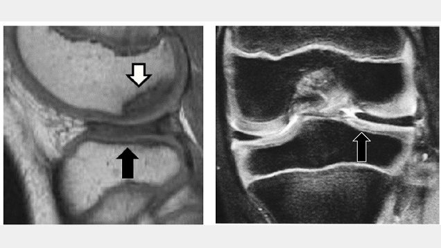
Discoid lateral meniscus and osteochondritis dissecans in adolescent patients. The black arrow represents DLM and the white arrow represents osteochondritis dissecans. Credit: Osaka Metropolitan University
Growing pains are common in maturing children, but sometimes this growth can be irregular and cause injury. Discoid lateral meniscus (DLM), a misshapen knee cartilage, is one such occurrence that can degenerate into osteochondritis dissecans, a joint disorder where the bone and joint begin to separate from the rest of the bones. It has been reported that osteochondritis dissecans of the femoral condyle occurs in approximately 14.5% of cases of DLM, but there has been little analysis of its treatment to date.
Dr Ken Iida and Specially Appointed Professor Yusuke Hashimoto’s team at Osaka Metropolitan University’s Graduate School of Medicine analysed the incidence of post-surgery osteochondritis dissecans. This analysis consisted of two groups, a pre-osteochondritis group with DLM and osteochondritis dissecans of the outer femoral epicondyle, and a non-osteochondritis dissecans DLM group. They studied 95 cases of DLM patients under the age of 15 who underwent surgery between 2003 and 2017 and had five years of post-surgery records. There were 15 cases in the pre-osteochondritis dissecans group and 80 non-osteochondritis dissecans cases.
Their analysis found that the surgical results for osteochondritis dissecans were good in pre-osteochondritis cases, but 28.5% had a recurrence of the joint disorder. In the non-osteochondritis dissecans group, 8.8% were diagnosed with the disorder after surgery. Additionally, age was found to be a risk factor for relapse or post-surgical osteochondritis dissecans, and surgery on patients ages 9 and under was also involved in the occurrence of osteochondritis dissecans.
“Patients with DLM accompanied by osteochondritis dissecans of the femoral condyle often have difficulty in deciding on a treatment method,” Dr Iida explained. “Based on the results of this study, we believe for patients ages 9 years or younger, it is necessary to consider conservative treatment methods rather than immediate surgery.”
A UCLA research team has created the Comorbid Operative Risk Evaluation (CORE) score to better account for the role chronic illness plays in patient’s risk of mortality after operation, allowing surgeons to adjust to patients’ pre-existing conditions and more easily determine mortality risk.
For almost 40 years, researchers have used two tools, the Charlson Comorbidity Index (CCI) and Elixhauser Comorbidity Index (ECI), to measure the impact of existing health conditions on patient outcomes. These tools use ICD codes that are input by medical professionals and billers to account for patient illness. These tools, however, were not designed for patients undergoing surgery and often address chronic illnesses that are not relevant to surgical populations. They often capture data from medical billing records and lack nuanced information regarding pre-existing health conditions.
A total of 699 155 patients were used to develop the model, of which 139 831 (20%) comprised the testing cohort. The researchers queried adults undergoing 62 operations across 14 specialties from the 2019 National Inpatient Sample (NIS) using International Classification of Diseases, 10th Revision (ICD-10) codes. They sorted ICD-10 codes for chronic diseases into Clinical Classifications Software Refined (CCSR) groups. They used logistic regression on CCSR with non-zero feature importance across four machine learning algorithms predicting in-hospital mortality, and used the resultant coefficients to calculate the Comorbid Operative Risk Evaluation (CORE) score based on previously validated methodology. The final score ranges from zero, representing lowest risk, to 100, which represents highest risk.
Impact
Health services and outcomes research using retrospective databases continues to represent a growing proportion of surgical research. Researchers highlighting quality issues and disparities are well-intentioned. However, without appropriate tools, it can be unclear if poor outcomes are independent of pre-existing conditions.
“The advent of novel statistical software and methodology have enabled researchers to exploit large databases to answer questions of healthcare quality, disparities, and outcomes,” said Dr Nikhil Chervu, a resident physician in the UCLA Department of Surgery and the study’s lead author. “These databases, however, often capture data from medical billing records and lack nuanced information regarding pre-existing health conditions. Without addressing differences in patients’ chronic illnesses, population comparisons may fall flat. Incorporation of this score in additional research will further validate its use and help improve analysis of surgical outcomes using large databases.”
A handful of common surgical procedures account for large shares of all opioids dispensed after surgery in children and adults, according to two studies recently published by researchers at the University of Michigan.
The studies, published this week in Pediatrics and JAMA Network Open, report that the top three procedures for children ages 0–11 account for 59% of opioids dispensed after surgery (tonsillectomies and adenoidectomies 50%, upper extremity fractures 5% and removal of deep implants 4%). Among those ages 12–21, the top three procedures account for about a third of post-surgery opioid prescriptions (tonsillectomies and adenoidectomies 13%, knee arthroscopies 13% and caesarean deliveries 8%).
For adults ages 18–44, C-sections account for the highest share of opioids dispensed post-surgery (19%), followed by hysterectomies (7%) and knee arthroscopies (6%). Among those ages 45-64, four of the top five procedures were orthopaedic procedures, collectively accounting for 27% of total opioid prescriptions dispensed after surgery.
“Our findings suggest that surgical opioid prescribing is highly concentrated among a small group of procedures. Efforts to ensure safe and appropriate surgical opioid prescribing should focus on these procedures,” said Kao-Ping Chua, lead author of the study in Pediatrics, assistant professor at the U-M Medical School and School of Public Health, and co-director of the Research and Data Domain at the U-M Opioid Research Institute.
To conduct the study, the researchers developed an algorithm to identify 1082 major surgical procedures using procedure codes, a medical classification tool used to identify specific surgical, medical or diagnostic interventions. The algorithm was then applied to identify privately and publicly insured children and adults undergoing surgery from Dec. 1, 2020 through Nov. 30, 2021.
The information was organized through a novel system developed by the study team, which allowed them to connect different sets of data that had previously been seen as unrelated. This new method allows for improved comparability and contrast, according to lead investigators.
In addition to determining which procedures accounted for the highest shares of opioids, the researchers also examined the size of opioid prescriptions for each procedure. For many procedures, prescriptions were far larger than the amount patients typically need for a particular procedure.
“Our findings suggest that there are important opportunities to reduce surgical opioid prescribing without compromising pain control,” said Dominic Alessio-Bilowus, lead author of the paper focused on adults published in JAMA Network Open and a medical student at Wayne State University who just completed a research year at U-M.
Tumours arising in the base of the skull are among the most difficult to remove in neurosurgery. The current treatment method is to perform surgical removal by what is known as the microscopic anterior transpetrosal approach (ATPA). Seeking to lessen the risk of damage and postoperative complications, as the skull base is densely packed with nerves, blood vessels, and other tissues, not to mention the brain stem, an Osaka Metropolitan University medical research team is taking a new approach.
Led by Dr Hiroki Morisako, a lecturer in the Graduate School of Medicine’s Department of Neurosurgery, and its department head Professor Takeo Goto, the team has developed a minimally invasive surgical technique called a purely endoscopic subtemporal keyhole ATPA. The team members write in The Journal of Neurosurgery that this is, to their knowledge, the first time this procedure to remove lesions in the skull base region known as the petrous apex has been described in an article.
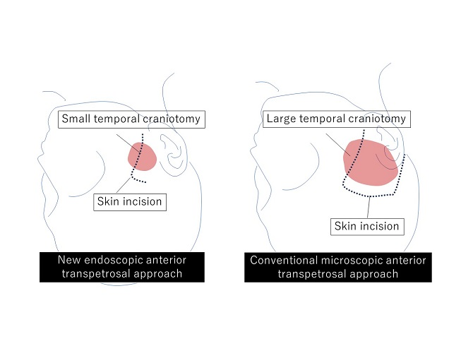
Diagram of skin incision and extent of craniotomy. New endoscopic neurosurgery approach does not require a large craniotomy, so the result is a smaller scar. Credit: Osaka Metropolitan University
The endoscopic technique means a smaller area of the skull needs to be surgically opened compared to the microscopic approach, an average of only 11.2 cm² versus 33.9 cm². The risk of damage to the brain is also reduced.
The team performed 10 neurosurgeries using their method from 2022 to 2023 at Osaka Metropolitan University Hospital and compared the results to 13 surgeries using the microscopic ATPA from 2014 to 2021. In terms of operative time, the endoscopic approach reduced it noticeably, from an average of 410.9 minutes to 252.9 minutes. Similarly, blood loss lessened from a mean of 193 ml to 90 ml. The degree of tumour resection (surgical removal) was just as high as the microscopic method, while neurological functions were preserved at a rate equal to or higher than with the conventional approach.
“Comparison of the new endoscopic method and the conventional microscopic method showed no significant difference in tumour resection rate or in the ability to perform daily activities before and after surgery, with the new endoscopic approach resulting in shorter operative times and less blood loss,” Professor Goto stated. “The widespread use of this surgical procedure is expected to improve the treatment results of brain tumours in the base of the skull, not only in Japan but also worldwide.”
Dr Tim Forgan at the surgeon’s console of the da Vinci robotic system. (Photo: Biénne Huisman/Spotlight)
By Biénne Huisman
Within South Africa’s beleaguered public health sector – unsettled by budget cuts, understaffing, and divisive NHI legislation – cutting edge surgical robots that have been used to perform more than 600 surgeries at two Cape Town public hospitals are beacons of excellence that offer a glimmer of hope. Spotlight’s Biénne Huisman visited Dr Tim Forgan at Tygerberg Hospital to learn more.
Cutting edge robotic surgery might not immediately come to mind when one thinks of public hospitals, but in a first for public healthcare in South Africa, such systems are being used at two hospitals in the Western Cape.
The da Vinci Xi systems enable surgeons to control operations from a console – steering three arms with steel “hands” equipped with tiny surgical instruments; plus a fourth arm bearing a video camera (the laparoscope). The system translates a surgeon’s hand movements in real time, with enhanced precision, range and visuals, compared to manual surgery.
“It really is next level, it feels like you’re inside the patient,” says colorectal specialist Dr Tim Forgan, Tygerberg Hospital’s da Vinci robotics coordinator. “With this technology we can operate so much finer. You can see ten times better with this robot than with the naked eye; you can see tiny, tiny nerves you wouldn’t normally see. And you can manoeuvre surgical instruments so much better. Because of that, people have way better function after the procedure.”
He explains that the technology allows major surgery to be completed through small incisions – instead of larger cuts made by a doctor’s hand – leading to less bleeding and a faster recovery time.
Over 600 surgeries in two years
Lorraine Gys from Phillipstown in the Northern Cape can attest. On 22 February 2022, the 65-year-old pensioner became the first patient to undergo da Vinci robotic surgery in South Africa’s public sector. Forgan was behind the console, at Tygerberg Hospital.
Gys tells Spotlight: “The next day the sisters offered to wash me, I said to them ‘no, I’m not helpless.’ My recovery was very quick. I was up and about in no time, while the other patients had to be assisted. I was discharged on day four, and back at home I could even continue doing my own chores.”
Two years later, Gys is cancer free. The mother of three, who now lives in Eerste River, recalls how she made news headlines: “Before the operation, Dr Forgan explained everything to me. They asked my permission, saying that media will be there and the [provincial health] minister.”
Indeed, on the day Forgan operated on Gys, removing a cancerous rectal tumour, he was joined in theatre by several onlookers including former Western Cape MEC of Health and Wellness Nomafrench Mbombo.
“Yes it was a circus,” says Forgan, laughing. “A whole bunch of people watching me operate, quite bloody nerve-wracking. Fortunately I’m experienced at having lots of students around watching; plus performing surgery is just so immersive, everything else fades out.”
On that day, also in the operating room was colorectal surgeon Dr Roger Gerjy, keeping an eye. “He’s a very well-known robotic surgeon; a Swedish surgeon who works in Dubai,” says Forgan. “And if there was a problem, Roger would’ve taken over. He was also there to impart tips and tricks: move the instrument like this, shape it like a hockey stick; because with the robot it’s like having your whole arm inside [the body]. He’d give me advice on what to do with my extra floating arm – where to place it and how to manipulate it – because remember you’re controlling three arms at a time.”
Since 2022, the da Vinci robots installed at Cape Town’s two tertiary hospitals: Groote Schuur and Tygerberg, have enabled over 600 minimally invasive surgeries – including colorectal operations, prostatectomies, cystectomies (bladder removal surgery), and gynaecological procedures to treat endometriosis.
Groote Schuur Hospital has the other da Vinci Xi system run by Western Cape public healthcare
A spokesperson for the Western Cape Ministry of Health and Wellness, under former MEC Mbombo, Luke Albert explains: “We can see the immense impact it has for patients and the health system. For example, a traditional open cystectomy patient would require three days of ICU stay, as well as two weeks of hospital stay to recuperate. During this time, on average, 42% of patients require blood transfusions and almost 20% need total parenteral nutrition (when a patient is fed intravenously). A patient undergoing robotic surgery for a cystectomy requires no ICU stay and goes straight to a general ward for no more than six days on average, with no blood transfusions needed.”
Where the money came from
Asked how the department was able to afford R40 million per system for these machines in the context of severe budget cuts, Albert says: “The purchase was applicable to 2021/22 and not the current financial year; with all provincial health departments currently managing the effects of budget cuts.”
Asked the same question, Forgan explains the investment derived from surplus budget discovered within the throes of the COVID-19 pandemic: “There was a surplus because certain services just couldn’t be done. I mean, for us, we couldn’t do elective surgery. And how state funding works; if you don’t spend your [provincial] budget within the financial year, it goes back to central government.”
What it looks like
On a Friday afternoon at Tygerberg Hospital, Forgan is guiding Spotlight along corridors and up grey linoleum stairs, to the theatre where the da Vinci system is used. Dressed in black surgical scrubs bearing his name and a cap; on his feet Forgan is wearing bright pink crocs. In passing, he waves hello to fellow healthcare staff.
Inside the small blindingly white room, Forgan points out the three core components of the da Vinci system. There is a console with two control levers similar to refined joysticks – he demonstrates how to delicately hold them between forefingers and thumbs – a patient-side cart with four interactive metal arms (they are disposable; each arm can be used on twelve patients), and another trolley with a television screen. All connected by blue fibre optic cables.
As we speak, nurses arrive in the theatre, preparing it for upcoming gynaecology procedures scheduled for Monday. Forgan greets them, then continues to expand on his passion for colorectal surgery.
“With colorectal surgery, there’s a high rate of complications, but I really enjoy it, I really enjoy my job. When you have a successful outcome, saving a person from their cancer and prolonging their life through your intervention, that is the reward. Colorectal cancer is a very unpleasant disease, and operating like this can make one hell of a difference in a patient’s life.”
Colorectal cancer on the increase
Forgan adds that colorectal cancer is on the increase: “There aren’t many colorectal surgeons in South Africa, with a dire need for people to operate in this subspecialty. I mean, there are so few of us, we’re all on a WhatsApp group.”
Colorectal or colon cancer is the second most common cancer in South African men (following prostate cancer), and the third most common cancer in women (following breast and cervical cancer), according to the Cancer Association of South Africa.
Originally from Johannesburg, Forgan attended medical school at the University of the Witwatersrand. He qualified as a general surgeon at Stellenbosch University, sub-specialising in colorectal surgery at the University of Cape Town, before studying minimally invasive colorectal surgery at the Academic Medical Centre in Amsterdam.
He is also president of the South African Colorectal Society and runs a part-time private practise with his Tygerberg colleague, Dr Imraan Mia, at Cape Town’s Christiaan Barnard Hospital, where he has 32 all five-star Google reviews.
‘Early adopter’
Forgan considers himself an early adopter. But learning to use the da Vinci system did not happen overnight.
“We trained for ages,” he says. “On the surgical console there’s a simulator, so you spend hours and days and days doing procedures, over and over and over again. You have to get over 95% for each one of the procedures, before you can move on to the next skill.
“Then it’s how to use the machine, how to put it together, what to do if there’s an emergency; what if there’s a power failure and the machine stops working? How to safely remove it from the person. Then we went to the University of Lyon [in France] for two days of hands-on robotics training. And then a proctor – an international expert – comes to your theatre and does the procedures with you. So that was Dr Roger Gerjy, and that’s when we did Lorraine…”
First introduced by American biotechnology company Intuitive Surgical in 1999, the da Vinci Xi systems have sparked some liability lawsuits. An article from the Tampa Bay Times in February cites a lawsuit filed at the United States District Court in West Palm Beach, with a man claiming that a stray electrical arc from a surgical robot burned his wife’s small intestine during a colon cancer procedure, causing her death. The article quotes Intuitive Surgical’s 2023 financial report, which notes 8 606 da Vinci systems in use worldwide, having performed 2 286 000 procedures in 2023. The financial report mentions an undisclosed number of pending lawsuits, which the company disputes.
Nevertheless, Forgan remains an advocate.
Exiting via Tygerberg’s maze of corridors, he continues to reflect on his job. After our meeting, he is set to deliver a talk at the Cape Town International Convention Centre. His manner is earnest. Shrugging, he describes himself as a “glorified plumber”.
A 3D map of the islet density routes throughout the healthy human pancreas. Source: Wikimedia CC0
Northwestern University researchers have developed a new antioxidant biomaterial that someday could provide much-needed relief to people living with chronic pancreatitis. The study was published in the journal Science Advances.
Before surgeons remove the pancreas from patients with severe, painful chronic pancreatitis, they first harvest insulin-producing tissue clusters, called islets, and transplant them into the vasculature of the liver. The goal of the transplant is to preserve a patient’s ability to control their own blood-glucose levels without insulin injections.
Unfortunately, the process inadvertently destroys 50–80% of islets, and one-third of patients become diabetic after surgery. Three years post-surgery, 70% of patients require insulin injections, which are accompanied by a list of side effects, including weight gain, hypoglycaemia and fatigue.
In the new study, researchers transplanted islets from the pancreas to the omentum – the large, flat, fatty tissue that covers the intestines – instead of the liver. And, to create a healthier microenvironment for the islets, the researchers adhered the islets to the omentum with an inherently antioxidant and anti-inflammatory biomaterial, which rapidly transforms from a liquid to a gel when exposed to body temperature.
In studies with mouse and non-human primates, the gel successfully prevented oxidative stress and inflammatory reactions, significantly improving survival and preserving function of transplanted islets. It marks the first time a synthetic antioxidant gel has been used to preserve function of transplanted islets.
“Although islet transplantation has improved over the years, long-term outcomes remain poor,” said Northwestern’s Guillermo A. Ameer, who led the study. “There is clearly a need for alternative solutions. We have engineered a cutting-edge synthetic material that provides a supportive microenvironment for islet function. When tested in animals, we were successful. It kept islet function maximised and restored normal blood sugar levels. We also report a reduction in units of insulin that animals required.”
“With this new approach, we hope that patients will no longer have to choose between living with the physical pain of chronic pancreatitis or the complications of diabetes,” added Jacqueline Burke, a research assistant professor of biomedical engineering at Northwestern and the paper’s first author.
‘Compromised quality of life’
For patients living without a pancreas, side effects such as managing blood-sugar levels can be a lifelong struggle. By secreting insulin in response to glucose, islets help the body maintain glycaemic control. Without functioning islets, people must closely monitor their blood-sugar levels and frequently inject insulin.
“Living without functional islets places a great burden on patients,” Burke said. “They must learn to count carbs, dose insulin at the appropriate time and continuously monitor blood glucose. This consumes much of their time and mental energy. Even with great care, exogeneous insulin therapy is not as effective as islets for maintaining glucose control.”
“It’s a compromised quality of life,” Ameer said. “Instead of multiple insulin injections, we would love to collect and preserve as many islets as possible.”
But, unfortunately, the current standard of care for preserving islets often leads to poor outcomes. After the surgery to remove the pancreas, surgeons isolate islets from the pancreas and transplant them to the liver through portal vein infusion. This intraportal perfusion procedure has several common complications. Islets in direct contact with blood flow undergo an inflammatory response, more than half of the islets die, and transplanted islets can cause dangerous clots in the liver. For those reasons, physicians and researchers have been searching for an alternate transplantation site.
In previous clinical studies, researchers transplanted islets to the omentum instead of the liver in order to bypass issues with clotting. To secure the islets on the omentum, physicians used plasma from the patients’ own blood to form a biologic gel. While the omentum appeared to work better than the liver as a transplantation site, several issues, including clots and inflammation, remained.
“There’s been significant interest in the research and medical communities to find an alternate islet transplantation site,” Ameer said. “The results from the omentum study were encouraging, but outcomes were varied. We believe that’s because the use of the patients’ blood and the added components required to create the biologic gel can affect reproducibility among patients.”
A citrate solution
To protect the islets and improve outcomes, Ameer turned to the citrate-based biomaterials platform with inherent antioxidant properties developed in his laboratory. Used in products approved by U.S. Food and Drug Administration for musculoskeletal surgeries, citrate-based biomaterials have demonstrated the ability to control the body’s inflammatory responses. Ameer set out to investigate whether a version of these biomaterials with biodegradable and temperature-responsive phase-changing properties would provide a superior alternative to a biologic gel obtained from blood.
In cell cultures, both mouse and human islets stored within the citrate-based gel maintained viability much longer than islets in other solutions. When exposed to glucose, the islets secreted insulin, demonstrating normal functionality. Moving beyond cell cultures, Ameer’s team tested the gel in small and large animal models. Liquid at room temperature, the material turns into a gel at body temperature, so it’s simple to apply and easily stays in place.
In the animal studies, the gel effectively secured the islets onto the omentum of the animals. Compared to the current methods, more islets survived, and, over time, the animals restored normal blood glucose levels. According to Ameer, the success is partially due to the new material’s biocompatibility and antioxidant nature.
“Islets are very sensitive to oxygen,” Ameer said. “They are affected by both too little oxygen and too much oxygen. The material’s innate antioxidant properties protect the cells. Plasma from your own blood doesn’t offer the same level of protection.”
Integrating into tissues
After about three months, the body resorbed 80-90% of the biocompatible gel. But, at that point, it was no longer needed.
“What was fascinating is that the islets regenerated blood vessels,” Ameer said. “The body generated a network of new blood vessels to reconnect the islets with the body. That is a major breakthrough because the blood vessels keep the islets alive and healthy. Meanwhile, our gel is simply resorbed into the surrounding tissue, leaving little evidence behind.”
Next, Ameer aims to test his hydrogel in animal models over a longer period of time. He said the new hydrogel also could be used for various cell replacement therapies, including stem cell-derived beta cells for treating diabetes.
Researchers who had been using Fitbit data to help predict surgical outcomes have a new method to more accurately gauge how patients may recover from spine surgery.
Using machine learning techniques developed at the AI for Health Institute at Washington University in St. Louis, Chenyang Lu, the Fullgraf Professor in the university’s McKelvey School of Engineering, collaborated with Jacob Greenberg, MD, assistant professor of neurosurgery at the School of Medicine, to develop a way to predict recovery more accurately from lumbar spine surgery.
The results show that their model outperforms previous models to predict spine surgery outcomes. This is important because in lower back surgery and many other types of orthopaedic operations, the outcomes vary widely depending on the patient’s structural disease but also varying physical and mental health characteristics across patients. The study is published in Proceedings of the ACM on Interactive, Mobile, Wearable and Ubiquitous Technologies.
Surgical recovery is influenced by both preoperative physical and mental health. Some people may have catastrophising, or excessive worry, in the face of pain that can make pain and recovery worse. Others may suffer from physiological problems that cause worse pain. If physicians can get a heads-up on the various pitfalls for each patient, that will allow for better individualized treatment plans.
“By predicting the outcomes before the surgery, we can help establish some expectations and help with early interventions and identify high risk factors,” said Ziqi Xu, a PhD student in Lu’s lab and first author on the paper.
Previous work in predicting surgery outcomes typically used patient questionnaires given once or twice in clinics that capture only one static slice of time.
“It failed to capture the long-term dynamics of physical and psychological patterns of the patients,” Xu said. Prior work training machine learning algorithms focus on just one aspect of surgery outcome “but ignore the inherent multidimensional nature of surgery recovery,” she added.
Researchers have used mobile health data from Fitbit devices to monitor and measure recovery and compare activity levels over time but this research has shown that activity data, plus longitudinal assessment data, is more accurate in predicting how the patient will do after surgery, Greenberg said.
The current work offers a “proof of principle” showing, with the multimodal machine learning, doctors can see a much more accurate “big picture” of all the interrelated factors that affect recovery. Proceeding this work, the team first laid out the statistical methods and protocol to ensure they were feeding the AI the right balanced diet of data.
Prior to the current publication, the team published an initial proof of principle in Neurosurgery showing that patient-reported and objective wearable measurements improve predictions of early recovery compared to traditional patient assessments. In addition to Greenberg and Xu, Madelynn Frumkin, a PhD psychological and brain sciences student in Thomas Rodebaugh’s laboratory in Arts & Sciences, was co-first author on that work. Wilson “Zack” Ray, MD, the Henry G. and Edith R. Schwartz Professor of neurosurgery in the School of Medicine, was co-senior author, along with Rodebaugh and Lu. Rodebaugh is now at the University of North Carolina at Chapel Hill.
In that research, they show that Fitbit data can be correlated with multiple surveys that assess a person’s social and emotional state. They collected that data via “ecological momentary assessments” (EMAs) that employ smart phones to give patients frequent prompts to assess mood, pain levels and behaviour multiple times throughout day.
We combine wearables, EMA -and clinical records to capture a broad range of information about the patients, from physical activities to subjective reports of pain and mental health, and to clinical characteristics,” Lu said.
Greenberg added that state-of-the-art statistical tools that Rodebaugh and Frumkin have helped advance, such as “Dynamic Structural Equation Modeling,” were key in analyzing the complex, longitudinal EMA data.
For the most recent study they then took all those factors and developed a new machine learning technique of “Multi-Modal Multi-Task Learning (M3TL)” to effectively combine these different types of data to predict multiple recovery outcomes.
In this approach, the AI learns to weigh the relatedness among the outcomes while capturing their differences from the multimodal data, Lu adds.
This method takes shared information on interrelated tasks of predicting different outcomes and then leverages the shared information to help the model understand how to make an accurate prediction, according to Xu.
It all comes together in the final package producing a predicted change for each patient’s post-operative pain interference and physical function score.
Greenberg says the study is ongoing as they continue to fine tune their models so they can take these more detailed assessments, predict outcomes and, most notably, “understand what types of factors can potentially be modified to improve longer term outcomes.”