Newborns with heart complications can rely on their newly developed immune systems to regenerate cardiac tissues, but adults aren’t so lucky. After a heart attack, most adults struggle to regenerate healthy heart tissue, leading to scar-tissue buildup and, often, heart failure.
A new Northwestern Medicine study in experimental animals reveals a critical difference in how macrophages help repair the heart in newborns versus adults after a heart attack. The study highlights a fundamental difference in how the immune system drives healing based on age.
“Understanding why newborns can regenerate their hearts while adults cannot will open the door to developing treatments that could ‘reprogram’ adult macrophages,” said first and co-corresponding author Connor Lantz, lead scientist of the bioinformatics core at the Comprehensive Transplant Center at Northwestern University Feinberg School of Medicine.
In newborns, macrophages perform a process called efferocytosis, which recognizes and eats dying cells. This process triggers the production of a bioactive lipid called thromboxane, signaling nearby heart muscle cells to divide, and allowing the heart to regenerate damaged heart muscle, the study found. In adults, macrophages produce much less thromboxane, leading to a weaker repair signal.
“By mimicking the effects of thromboxane, we might one day improve tissue repair after a heart attack in adults,” Lantz said.
How the study worked
The study examined how the immune system responds to heart injury in mice of different ages, including newborn mice (one day old) and adult mice (eight weeks old). The researchers found the ability of macrophages to engulf dying cells was enhanced in newborn mice due to increased expression of MerTK, a receptor that recognizes dying cells. Therefore, when the scientists blocked this key receptor, newborn mice lost their ability to regenerate their hearts, resembling adult hearts after a heart attack.
Engulfment of dying cells by newborn macrophages triggered a chemical chain reaction that produced a molecule called thromboxane A2, which unexpectedly stimulated heart muscle cells to multiply and repair the damage, the study found. Additionally, nearby muscle heart cells in newborns are primed to respond to thromboxane A2, leading them to change their metabolism to support their growth and healing. But in adults, this process did not work the same way – after an injury, their macrophages did not produce enough thromboxane A2, limiting their ability to regenerate heart tissue.
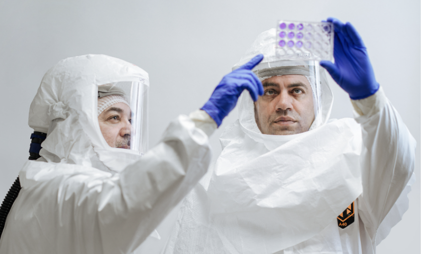
Professor Luis Martinez-Sobrido, Ph.D., (left) and Staff Scientist Ahmed Mostafa Elsayed, PhD, (right) review test results for the presence of bird flu while wearing protective equipment required for biosafety level-3 laboratories.
One of the earliest strains of bird flu isolated from a human in Texas shows a unique constellation of mutations that enable it to more easily replicate in human cells and cause more severe disease in mice compared to a strain found in dairy cattle, researchers from Texas Biomedical Research Institute (Texas Biomed) report in Emerging Microbes & Infections.
The finding highlights a key concern about the H5N1 strains of bird flu currently circulating in the U.S.: the speed at which the virus can mutate when introduced to a new host.
Naturally found in wild birds and lethal in chickens, H5N1 has spread to a wide variety of mammals and began infecting dairy cows for the first time in spring 2024. As of early 2025, the outbreak had spread through herds across multiple states in the U.S. and infected dozens of people, mostly farm workers. So far, most people infected experience mild illness and eye inflammation and the virus is not spreading between people. The first H5N1 death in the U.S. was reported in January 2025 following exposure to infected chickens.
“The clock is ticking for the virus to evolve to more easily infect and potentially transmit from human to human, which would be a concern,” said Texas Biomed Professor Luis Martinez-Sobrido, PhD, whose lab specialises in influenza viruses and has been studying H5N1 since the outbreak began last year. The team has developed specialised tools and animal models to test prophylactic vaccines and therapeutic antivirals.
Human vs bovine
In the recent study, they compared H5N1 strains isolated from a human patient and from dairy cattle in Texas.
“There are nine mutations in the human strain that were not present in the bovine strain, which suggests they occurred after human infection,” Dr Martinez-Sobrido said.
In mouse studies, they found that compared to the bovine strain, the human strain replicated more efficiently, caused more severe disease and was found in much higher quantities in brain tissue. They also tested several FDA-approved antiviral medications to see if they were effective against both virus strains in cells.
“Fortunately, the mutations did not affect the susceptibility to FDA-approved antivirals,” said Staff Scientist Ahmed Mostafa Elsayed, PhD, first author of the study.
Antivirals will be a key line of defence should a pandemic occur before vaccines are widely available, Dr Martinez-Sobrido said. This is especially true since humans have no preexisting immunity against H5N1 and seasonal flu vaccines appear to offer very limited protection, according to a separate study conducted in collaboration with Aitor Nogales, PhD, at the Center for Animal Health Research in Spain.
Dr Elsayed shows the host species of the four types of influenza viruses: A, B, C and D. Avian influenza is part of the influenza A group and has infected a wide range of species. Influenza A and B are responsible for seasonal flu in humans.
Next steps and recommendations
Texas Biomed is now exploring the human H5N1 mutations individually to determine which are responsible for increased pathogenicity and virulence. The team wants to figure out what allows H5N1 to infect such a wide range of mammal species; why H5N1 causes mild disease in cows but is lethal in cats; and why infections via cows are less harmful to people than infections from chickens.
In a third paper, Dr Elsayed and collaborators analysed the history of H5N1 in dairy cattle for the journal mBio and called for a One Health approach to protect both animals and people.
“A key priority will be to eradicate bird flu from dairy cows to minimise risk of mutations and transmission to people and other species,” Dr Elsayed said. “Steps that can be taken now include thorough decontamination of milking equipment and more stringent quarantine requirements, which will help eliminate the virus more quickly in cows.”
(Left) An image of a 3D-printed material implanted in vivo for 4 weeks. The photo was taken using a scanning electron microscope. Credit: Dr Zhidao Xia. (Right) A photo of coral. Credit: Jesus Cobaleda.
Researchers at Swansea University have developed a revolutionary bone graft substitute inspired by coral which not only promotes faster healing but dissolves naturally in the body after the repair is complete.
Bone defects caused by conditions like fractures, tumours, and non-healing injuries are one of the leading causes of disability worldwide. Traditionally, doctors use either a patient’s own bone (autograft) or donor bone (allograft) to fill these gaps. However, these methods come with challenges, including a limited supply, the risk of infection and ethical concerns.
By using advanced 3D-printing technology, the team have developed a biomimetic material that mimics the porous structure and chemical composition of coral-converted bone graft substitute, blending perfectly with human bone and offering several incredible benefits:
Rapid Healing – It helps new bone grow within just 2–4 weeks.
Complete Integration – The material naturally degrades within 6–12 months after enhanced regeneration, leaving behind only healthy bone.
Cost-Effective – Unlike natural coral or donor bone, this material is easy to produce in large quantities.
In preclinical in vivo studies, the material showed remarkable results: it fully repaired bone defects within 3–6 months and even triggered the formation of a new layer of strong, healthy cortical bone in 4 weeks.
Most synthetic bone graft substitutes currently on the market can’t match the performance of natural bone. They either take too long to dissolve, don’t integrate well, or cause side effects like inflammation. This new material overcomes these problems by closely mimicking natural bone in both structure and biological behaviour.
Dr Xia explained: “Our invention bridges the gap between synthetic substitutes and donor bone. We’ve shown that it’s possible to create a material that is safe, effective, and scalable to meet global demand. This could end the reliance on donor bone and tackle the ethical and supply issues in bone grafting.”
Innovations like this not only promise to improve patient quality of life but also reduce healthcare costs and provide new opportunities for the biomedical industry.
The Swansea University team is now looking to partner with companies and healthcare organisations to bring this life-changing technology to patients around the world.
Right side heart failure. Credit: Scientific Animations CC4.0
After severe heart failure, the ability of the heart to heal by forming new cells is very low. But now Karolinska Institutet researchers found that, after use of a supportive heart pump, the capacity of a damaged heart to repair itself with new cardiomyocytes becomes significantly higher – even greater than that of a healthy heart. This study is published in the journal Circulation.
The ability of the human heart to renew itself by regenerating its muscle cells, myocytes, is very limited. But what happens to this capability when the heart is damaged by severe heart failure has been unknown.
Researchers at Karolinska Institutet have now discovered that after an injury, the rate of cell renewal is even lower than in a healthy heart. Standard-of-care for patients with advanced heart failure is a surgically implanted pump that helps propel blood, a so-called left ventricular assist device (LVAD).
Kick-starting repair
Surprisingly, the researchers found that patients with such a heart pump, who have shown significant improvement in their heart function, can regenerate heart muscle cells at a rate more than six times higher than in healthy hearts.
“The results suggest that there might be a hidden key to kick-start the heart’s own repair mechanism”, says Olaf Bergmann, senior researcher at the Department of Cell and Molecular Biology at Karolinska Institutet and last author of the paper.
The mechanism behind the effect is still unknown and there is not yet any hypothesis to explain it.
“It is difficult to say. In the existing data we cannot find an explanation for the effect, but we will now continue to study this process at a cellular and molecular level,” says Olaf Bergmann.
The findings open the possibility of developing new therapies for patients with serious heart conditions that stimulate the heart’s ability to repair itself after damage. This way, patients wouldn’t have to rely only on heart transplants or other kinds of long-term mechanical support.
“This offers some hope that the recovery after a heart incident can somehow be boosted,” says Olaf Bergmann.
Atomic bombs enable cell age estimation
It is generally difficult to determine the age of cells in the human body and to decide which cells are new and which are old. However, by using a method earlier devised by Jonas Frisén, professor of stem cell research at Karolinska Institutet, the group has been able to count the rate of renewal of myocytes in the heart. The method is based upon the fact that the percentage of radioactive carbon in the atmosphere, and subsequently in our cells, has steadily decreased since the nuclear test ban in 1963. For every following year, there is a little less radioactivity in newly formed cells, which means that they can be ‘dated’.
As described in research published in the Biotechnology Journal, investigators have developed a novel patch that can help liver tissue regenerate. The patch is a combination of decellularised liver matrix, a liver growth factor, and an anticoagulant. In lab tests with liver cells, the patch helped liver cells regain function after exposure to a toxin.
In rats, patches attached to the liver and gut promoted recovery from liver fibrosis, with notable decreases in scarring and inflammation.
“The decellularised liver matrix–based hepatic patch has demonstrated the ability to restore liver function and inhibit inflammation in fibrotic livers,” said corresponding author Yung-Te Hou, PhD, of National Taiwan University. “This approach shows great potential for treating various liver-related diseases, ranging from mild conditions such as fatty liver to severe conditions like liver cirrhosis.”
Coup and contrecoup brain injury. Credit: Scientific Animations CC4.0
Birmingham scientists have shown light therapy delivered transcranially can aid tissue repair after mild traumatic brain injury (mTBI). Their research, published in Bioengineering & Translational Medicine, indicates that this novel method could result in a new treatment option in an area of medicine that currently has few, if any, treatment options.
Traumatic brain injury (mTBI) results when the initial trauma of head injury is magnified by a complex set of inflammatory changes that occur in the brain. These secondary processes, which take place from minutes to hours after head injury, can dramatically worsen outcomes for patients.
The method invented by scientists at the University of Birmingham, UK and patented by University of Birmingham Enterprise aims to protect against this secondary damage, and stimulate faster, and better recovery for patients.
We want to develop this method into a medical device that can be used to enhance recovery for patients with traumatic brain or spinal cord injury, with the aim of improving outcomes for patients.
Professor Zubair Ahmed, College of Medicine & Health
In the study, the Birmingham team, comprising researchers Professor Zubair Ahmed, Professor Will Palin, Dr Mohammed Hadis and surgeons Mr Andrew Stevens and Mr David Davies, examined the effect of two wavelengths of near infrared light (660nm and 810nm) on recovery following injury.
The study in preclinical models used daily two-minute bursts of infrared light, delivered by a laser, for three days post-injury.
The findings showed significant reductions in the activation of astrocytes and microglial cells, which are heavily implicated in the inflammatory processes in the brain that follow head trauma, and significant reductions in biochemical markers of apoptosis (cell death).
At four weeks, there were significant improvements in performance in functional tests involving balance and cognitive function. The red light therapy also accelerated recovery compared to controls, with superior outcomes for light with a wavelength of 810nm.
The study builds on research published earlier this year which showed near infrared light delivered directly to the site of spinal cord injury both improves survival of nerve cells and stimulates new nerve cell growth.
Professor Ahmed, who led the study, said: “We want to develop this method into a medical device that can be used to enhance recovery for patients with traumatic brain or spinal cord injury, with the aim of improving outcomes for patients.”
The researchers are seeking commercial partners to co-develop the device and take it to market.
Newly published research from the University of Houston College of Pharmacy identifies key mechanisms of skeletal muscle regeneration and growth of muscles following resistance exercise. The findings, published in EMBO Reports, open the door to the development of targeted therapies for various muscle disorders, like Muscular Dystrophy, which affect millions of people worldwide.
When it comes to muscles and muscle disorders, the importance of a discovery like this cannot be overstated.
Muscles’ regenerative powers
Skeletal muscles are formed during embryonic development by the fusion of hundreds of specialised cells called myoblasts. Adult skeletal muscles maintain regenerative capacity, which is attributed to the presence of muscle stem cells, named satellite cells.
After injury, satellite cells undergo several rounds of proliferation followed by their differentiation into myoblasts. These myoblasts once again fuse with each other and to injured myofibres to accomplish muscle regeneration.
In many muscular disorders, this intrinsic capacity of muscles to regenerate is diminished resulting in the loss of muscle mass and function.
Key signalling protein
UH researchers found that Inositol-requiring enzyme 1, a key signalling protein, is essential for myoblast fusion during muscle formation and growth.
“During muscle regeneration, IRE1 augments the activity of X-box binding protein 1 which in turn stimulates the gene expression of multiple transmembrane proteins required for myoblast fusion,” reports Ashok Kumar, professor of pharmacy in the Department of Pharmacological and Pharmaceutical Sciences.
According to researchers, increasing the levels of IRE1 or XBP1 in muscle stem cells outside the body, followed by their injection in patients’ muscle tissues will improve muscle repair and reduce the severity of disease.
“We also found that augmenting the levels of IRE1α or XBP1 in myoblasts leads to the formation of myotubes (muscle cells) having an increased diameter,” said Kumar.
That increase in diameter can be significant.
“Size is very important for muscle. Muscle grows only in size, not in number,” said Aniket Joshi, a graduate student in Kumar’s lab and first author on the article. “Muscular people have larger muscle cells. Larger muscles generally work better- can lift more weight, run and walk faster, and improve overall metabolism of the body and prevent various diseases, such as type II diabetes.”
Flexing their muscles
This new research is not the first flex for Kumar’s team. In 2021, research from Kumar’s lab published in the ELife journaldescribed the role of the IRE1α/XBP1 signaling axis in regeneration of healthy skeletal muscle after acute injury and in models of Duchenne Muscular Dystrophy. In this study, they found that IRE1α/XBP1 signaling axis also plays an important cell autonomous role in satellite cells.
Fat tissue holds the key to 3D printing layered living skin and potentially hair follicles, according to researchers who recently harnessed fat cells and supporting structures from clinically procured human tissue to precisely correct injuries in rats. The advancement could have implications for reconstructive facial surgery and even hair growth treatments for humans.
The team’s findings published inBioactive Materials, and the team received a patent in February for the bioprinting technology it developed and used in this study.
“Reconstructive surgery to correct trauma to the face or head from injury or disease is usually imperfect, resulting in scarring or permanent hair loss,” said Ibrahim T. Ozbolat, professor of engineering science and mechanics, of biomedical engineering and of neurosurgery at Penn State, who led the international collaboration that conducted the work. “With this work, we demonstrate bioprinted, full thickness skin with the potential to grow hair in rats. That’s a step closer to being able to achieve more natural-looking and aesthetically pleasing head and face reconstruction in humans.”
While scientists have previously 3D bioprinted thin layers of skin, Ozbolat and his team are the first to intraoperatively print a full, living system of multiple skin layers, including the bottom-most layer or hypodermis. Intraoperatively refers to the ability to print the tissue during surgery, meaning the approach may be used to more immediately and seamlessly repair damaged skin, the researchers said. The top layer — the epidermis that serves as visible skin — forms with support from the middle layer on its own, so it doesn’t require printing. The hypodermis, made of connective tissue and fat, provides structure and support over the skull.
“The hypodermis is directly involved in the process by which stem cells become fat,” Ozbolat said. “This process is critical to several vital processes, including wound-healing. It also has a role in hair follicle cycling, specifically in facilitating hair growth.”
The researchers started with human adipose, or fat, tissue obtained from patients undergoing surgery at Penn State Health Milton S. Hershey Medical Center. Collaborator Dino J. Ravnic, associate professor of surgery in the Division of Plastic Surgery at Penn State College of Medicine, led his lab in obtaining the fat for extraction of the extracellular matrix to make one component of the bioink.
Ravnic’s team also obtained stem cells, which have the potential to mature into several different cell types if provided the correct environment, from the adipose tissue to make another bioink component. Each component was loaded into one of three compartments in the bioprinter. The third compartment was filled with a clotting solution that helps the other components properly bind onto the injured site.
“The three compartments allow us to co-print the matrix-fibrinogen mixture along with the stem cells with precise control,” Ozbolat said. “We printed directly into the injury site with the target of forming the hypodermis, which helps with wound healing, hair follicle generation, temperature regulation and more.”
They achieved both the hypodermis and dermis layers, with the epidermis forming within two weeks by itself.
“We conducted three sets of studies in rats to better understand the role of the adipose matrix, and we found the co-delivery of the matrix and stem cells was crucial to hypodermal formation,” Ozbolat said. “It doesn’t work effectively with just the cells or just the matrix – it has to be at the same time.”
They also found that the hypodermis contained downgrowths, the initial stage of early hair follicle formation. According to the researchers, while fat cells do not directly contribute to the cellular structure of hair follicles, they are involved in their regulation and maintenance.
“In our experiments, the fat cells may have altered the extracellular matrix to be more supportive for downgrowth formation,” Ozbolat said. “We are working to advance this, to mature the hair follicles with controlled density, directionality and growth.”
According to Ozbolat, the ability to precisely grow hair in injured or diseased sites of trauma can limit how natural reconstructive surgery may appear. He said that this work offers a “hopeful path forward,” especially in combination with other projects from his lab involving printing bone and investigating how to match pigmentation across a range of skin tones.
A small molecule previously shown to enhance strength in injured or old laboratory mice does so by restoring lost connections between nerves and muscle fibres, Stanford Medicine researchers have found.
The molecule blocks the activity of an aging-associated enzyme, or gerozyme, called 15-PGDH that naturally increases in muscles as they age. The study, which was published in Science Translational Medicine, showed that levels of the gerozyme increase in muscles after nerve damage and that it is prevalent in muscle fibres of people with neuromuscular diseases.
The research is the first to show that damaged motor neurons can be induced to regenerate in response to a drug treatment and that lost strength and muscle mass can be at least partially regained. It suggests that, if similar results are seen in humans, the drug may one day be used to prevent muscle loss of muscle strength due to aging or disease or to hasten recovery from injury.
It’s estimated that sarcopenia, or debilitating muscle frailty, affects about 30% of people over 80 and costs the United States around $380 billion each year.
“There is an urgent, unmet need for drug treatments that can increase muscle strength due to aging, injury or disease,” said Helen Blau, PhD, professor of microbiology and immunology. “This is the first time a drug treatment has been shown to affect both muscle fibres and the motor neurons that stimulate them to contract in order to speed healing and restore strength and muscle mass. It’s unique.”
Blau, the Donald E. and Delia B. Baxter Foundation Professor and director of the Baxter Laboratory for Stem Cell Biology, is the senior author of the study. Postdoctoral scholar Mohsen Bakooshli, PhD, and former postdoctoral scholar Yu Xin Wang, PhD, are the lead authors of the study. Wang is now an assistant professor at the Sanford Burnham Prebys Medical Discovery Institute in San Diego.
Addressing loss of strength
The finding is the latest from the Blau laboratory focused on understanding how muscles weaken from aging or disease, and whether it’s possible to combat this decline. In 2021, the group showed that blocking the activity of 15-PGDH in 24-month-old laboratory mice significantly enhances the animals’ leg strength and endurance when running on a treadmill. (Laboratory mice typically live about 26 to 30 months.) But it wasn’t clear exactly how.
The new research shows that the effect is due to the restoration of lost connections between the nerves and the muscle. These connections, called neuromuscular junctions, are how the brain signals muscles to contract. In aging, some of these connections are lost, causing muscle contractions to become less powerful and muscles to atrophy. People typically lose muscle mass and strength, up to 10% per decade, after the age of 50.
Conditions other than aging can also destabilise these connections, including the disuse of muscles due to bedrest after illness or injury, or muscle-wasting diseases like spinal muscular atrophy or amyotrophic lateral sclerosis (also known as ALS).
Blau’s previous research showed that a molecule called PGE2 is critical to the function of stem cells in muscle fibres that repair damage – including the microtears from exercise that lead to an increase in muscle mass and strength. They subsequently showed that levels of 15-PGDH, which breaks down PGE2, increase in the muscles with age and that the loss of strength with aging could be overcome by inhibiting the activity of this PGE2-degrading enzyme.
“PGE2 is part of the body’s natural healing mechanism, and its levels increase in muscle after injury,” Blau said. “We wanted to learn how age triggers an increase in 15-PGDH, and therefore the degradation and loss of PGE2.”
A lack of nerves
The researchers knew that muscles become less innervated, or infiltrated with nerves, as people and animals age. They wondered if that loss could be what triggers the rising levels of 15-PGDH.
“We found that when you cut the nerve that innervates the leg muscles of mice, the amount of 15-PGDH in the muscle increases rapidly and dramatically,” Blau said. “This was an exciting new insight. But what surprised us most was that when these mice are treated with a drug that inhibits 15-PGDH activity, the nerve grows back and makes contact with the muscle more quickly than in control animals, and that this leads to a faster recovery of strength and function.”
Additional experiments showed that treatment with the drug restored neuromuscular junctions lost during aging and increased muscle strength and function in old laboratory mice. The researchers also identified discrete clumps of 15-PGDH in the muscle fibres of people with several types of neuromuscular disorders suggesting that the gerozyme may have a role in causing these human disorders.
Blau and her colleagues plan to investigate at a molecular level how neural growth is stimulated by blocking 15-PGDH activity. Blau has also co-founded a company, Epirium Bio, to develop similar drugs for use in humans. Although her lab is still conducting animal studies, the company hopes to launch a clinical trial within the next year or so.
“Our next steps will be to examine whether blocking 15-PGDH function in people with spinal muscular atrophy can increase lost muscle strength in combination with gene therapy or other treatments,” Blau said. “We are also looking at ALS to see if something like this might help these patients. It’s really exciting that we are able to affect both muscle function and motor neuron growth.”
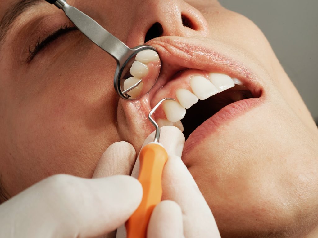
Tissue regeneration might one day replace the pain and discomfort of a root canal for most people. ADA Forsyth scientists are testing a novel technology to treat endodontic diseases (diseases of the soft tissue or pulp of the teeth) more effectively. The technology may also even be applicable to other parts of the body, such as helping to regrow bones.
The study, published in The Journal of Dental Research, demonstrates regenerative properties of resolvins, specifically Resolvin E1 (RvE1), when applied to dental pulp. Resolvins are part of a greater class of Specialised Proresolving Mediators (SPMs). This class of molecule is naturally produced by the body and is exquisitely effective in the control of excess inflammation associated with disease.
“Pulpitis (inflammation of dental pulp) is a very common oral health disease that can become a serious health condition if not treated properly,” said Dr Thomas Van Dyke, Vice President at the Center for Clinical and Translational Research at ADA Forsyth, and a senior scientist leading the study.
“Root canal therapy (RCT) is effective, but it does have some problems since you are removing significant portions of dentin, and the tooth dries out leading to a greater risk of fracture down the road. Our goal is to come up with a method for regenerating the pulp, instead of filling the root canal with inert material.”
Inflammation of this tissue is usually caused by damage to the tooth through injury, cavities or cracking, and the resulting infection can quickly kill the pulp and cause secondary problems if not treated.
The study applied RvE1 to different levels of infected and damaged pulp to explore its regenerative and anti-inflammatory capacities.
There were two major findings. First, they showed RvE1 is very effective at promoting pulp regeneration when used in direct pulp-capping of vital or living pulp (replicating conditions of reversible pulpitis). They were also able to identify the specific mechanism supporting tissue regeneration.
Second, the scientists found that placing RvE1 on exposed and severely infected and necrotic pulp did not facilitate regeneration.
However, this treatment did effectively slow down the rate of infection and treat the inflammation, preventing the periapical lesions (abscesses) that typically occur with this type of infection.
Previous publications have shown that if the infected root canal is cleaned before RvE1 treatment, regeneration of the pulp does occur.
While this study focused on this technology in treating endodontic disease, the potential therapeutic impact is far reaching.
Dr Van Dyke explained, “because application of RvE1 to dental pulp promotes formation of the type of stem cells that can differentiate into dentin (tooth), bone, cartilage or fat, this technology has huge potential for the field of regenerative medicine beyond the tissues in the teeth. It could be used to grow bones in other parts of the body, for instance.”