Category: Diet and Nutrition

Long-term High-fat Diets Shown to Worsen Cognitive Abilities

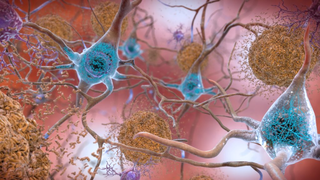
Plaques and neurons. Source: NIAH

A study published in Metabolic Brain Disease has established a clear link between mice being fed a high-fat diet for 30 weeks, resulting in diabetes, and a subsequent worsening of their cognitive abilities. This included developing anxiety, depression and worsening Alzheimer’s disease.

Mice with impaired cognitive function were also more likely to gain excessive weight due to poor metabolism caused by brain changes.

Neuroscientist and biochemist Associate Professor Larisa Bobrovskaya, who co-led the study said that the research adds to the growing body of evidence linking chronic obesity and diabetes with Alzheimer’s disease.

“Obesity and diabetes impair the central nervous system, exacerbating psychiatric disorders and cognitive decline. We demonstrated this in our study with mice,” said Associate Prof Bobrovskaya.

In the study, mice were randomised to a standard diet or a high-fat diet for 30 weeks, starting at eight weeks of age. Food intake, body weight and glucose levels were monitored at different intervals, along with glucose and insulin tolerance tests and cognitive dysfunction.

The mice on the high-fat diet gained significant weight, developed insulin resistance and started behaving abnormally compared to those fed a standard diet.

Genetically modified Alzheimer’s disease mice showed a significant deterioration of cognition and pathological changes in the brain while fed the high fat diet.

“Obese individuals have about a 55 per cent increased risk of developing depression, and diabetes will double that risk,” Assoc Prof Bobrovskaya said.

“Our findings underline the importance of addressing the global obesity epidemic. A combination of obesity, age and diabetes is very likely to lead to a decline in cognitive abilities, Alzheimer’s disease and other mental health disorders.”

Source: University of South Australia

Vitamin D ‘Overdoses’ are Possible – and Dangerous

Vitamin D pills
Photo by Michele Blackwell on Unsplash

Doctors have warned that it it is possible to ‘overdose’ on Vitamin D, with harmful effects, after they treated a man hospitalised for excessive Vitamin D intake, a case which was detailed in BMJ Case Reports.

They further stress that ‘hypervitaminosis D’ is on the increase, and linked to a wide range of potentially serious health issues.

The case concerns a middle-aged man who was referred to hospital by his family GP after complaining of recurrent vomiting, nausea, abdominal pain, leg cramps, tinnitus, dry mouth, increased thirst, diarrhoea, and weight loss (12.7 kg). 

The patient had been experiencing these symptoms had for almost three months, which had started around one month after he began an intensive vitamin supplement regimen on the advice of a nutritional therapist.

The man had had a number of health issues, including tuberculosis; a left vestibular schwannoma, resulting in deafness in that ear, hydrocephalus; bacterial meningitis; and chronic sinusitis.  

He had been taking high doses of more than 20 over the counter supplements every day containing: vitamin D 50 000mg – the daily requirement is 600mg or 400IU; vitamin K2 100mg (daily requirement 100–300 μg); vitamin C, vitamin B9 (folate) 1000mg (daily requirement 400 μg); vitamin B2 (riboflavin), vitamin B6, omega-3 2000mg twice daily (daily requirement 200–500 mg), plus several other vitamin, mineral, nutrient, and probiotic supplements.

Once he developed symptoms, he stopped taking his daily supplement cocktail, but his symptoms didn’t go away.

Blood test results from his family doctor revealed that he had very high levels of calcium and slightly raised levels of magnesium. And his vitamin D level was seven times over the level required for sufficiency.

The tests also indicated acute kidney injury. The results of various x-rays and scans to check for cancer were normal. 

The man was hospitalised for 8 days, during which time he was given intravenous fluids to flush out his system and treated with bisphosphonates – ordinarily used to treat osteoporosis or hypercalcaemia.

Two months post-discharge, his calcium level had returned to normal, but his vitamin D level was still abnormally high.

“Globally, there is a growing trend of hypervitaminosis D, a clinical condition characterised by elevated serum vitamin D3 levels,” with women, children and surgical patients most likely to be affected, the authors noted. 

Recommended vitamin D levels can be obtained through diet, sunlight exposure to sunlight, and supplements.

“Given its slow turnover (half-life of approximately two months), during which vitamin D toxicity develops, symptoms can last for several weeks,” the authors warned.

Hypervitaminosis D has many and varied symptoms, they point out, and are mostly caused by hypercalcaemia. They include drowsiness, confusion, apathy, psychosis, depression, stupor, coma, anorexia, abdominal pain, vomiting, constipation, peptic ulcers, pancreatitis, hypertension, abnormal heart rhythm, and kidney abnormalities, including renal failure. 

Other associated features, such as keratopathy (inflammatory eye disease), joint stiffness (arthralgia), and hearing loss or deafness, have also been reported, they add.

This is just one case, and while hypervitaminosis D is on the rise, it is still relatively uncommon, the authors cautioned.

Nevertheless, complementary therapy, including the use of dietary supplements, is popular, and people may not realise that it’s possible to overdose on vitamin D, or the potential consequences of doing so, they say.

“This case report further highlights the potential toxicity of supplements that are largely considered safe until taken in unsafe amounts or in unsafe combinations,” they concluded.

Source: The BMJ

Multivitamins and Dietary Supplements are a ‘Waste of Time’ for Most

Vitamin C pills and orange
Photo by Diana Polekhina on Unsplash

For those who aren’t pregnant, vitamins are a waste of money because the evidence for cardiovascular disease or cancer prevention is lacking, according to researchers at Northwestern University Feinberg School of Medicine.

“Patients ask all the time, ‘What supplements should I be taking?’”

Dr Jeffrey Linder, Northwestern University

The researchers penned an editorial in JAMA that supports new recommendations from the United States Preventive Services Task Force (USPSTF), a national panel which makes evidence-based recommendations on clinical prevention. 

Based on a systematic review of 84 studies, the USPSTF’s new guidelines state there was “insufficient evidence” that taking multivitamins, paired supplements or single supplements can help prevent cardiovascular disease and cancer in otherwise healthy, non-pregnant adults. 

“Patients ask all the time, ‘What supplements should I be taking?’ They’re wasting money and focus thinking there has to be a magic set of pills that will keep them healthy when we should all be following the evidence-based practices of eating healthy and exercising,” said Dr Jeffrey Linder, one of the editorial’s authors.

“The task force is not saying ‘don’t take multivitamins,’ but there’s this idea that if these were really good for you, we’d know by now,” Dr Linder added. 

The task force is specifically recommending against taking beta-carotene supplements because of a possible increased risk of lung cancer, and is recommending against taking vitamin E supplements because it has no net benefit in reducing mortality, cardiovascular disease or cancer.

“The harm is that talking with patients about supplements during the very limited time we get to see them, we’re missing out on counselling about how to really reduce cardiovascular risks, like through exercise or smoking cessation,” Dr Linder said.

No substitute for actual fruits and vegetables

Eating fruits and vegetables is associated with decreased cardiovascular disease and cancer risk, they said, so it is reasonable to think those key vitamins and minerals in pills could prevent disease. But, they explain, whole fruits and vegetables contain a mixture of vitamins, phytochemicals, fibre and other nutrients that probably act synergistically to deliver health benefits. Micronutrients on their own may also have a different effect than when consumed with others in foods.

Dr Linder noted that individuals with vitamin deficiency can still benefit from taking dietary supplements, such as calcium and vitamin D, which have been shown to prevent fractures and perhaps falls in older adults. 

New guidelines do not apply to those who are pregnant

The new USPSTF guidelines do not apply to people who are pregnant or trying to get pregnant, said JAMA editorial co-author Dr Natalie Cameron, a physician at Northwestern. 

“Pregnant individuals should keep in mind that these guidelines don’t apply to them,” said Dr Cameron. “More data is needed to understand how specific vitamin supplementation may modify risk of adverse pregnancy outcomes and cardiovascular complications during pregnancy.” 

Source: Northwestern University

A Way to Prepare Potatoes for a Lower Glycaemic Index

Photo by Clark Douglas on Unsplash

Researchers have tested a new potato processing technique designed to slow down the digestion of potato starch. Their experiments show that the approach prevents certain digestive enzymes from reaching the potato starch as quickly, leading to a more controlled release of dietary glucose.

Foods with lower glycaemic index have a variety of health benefits, such as reducing the risk of cardiovascular disease.

“There is a perception that potato foods are unhealthy because eating a large amount of some potato foods can cause a rapid increase in blood sugar, which is a risk for people with diabetes or those who want to control body weight,” said Amy Lin, PhD, the study’s principal investigator at A*STAR. “Our team revealed that toggling the accessibility of two digestion enzymes – α-amylase and mucosal α-glucosidase – in the small intestine is a successful strategy to make dietary glucose slowly and continuously release from potatoes.”

The findings are being presented online at NUTRITION 2022 LIVE ONLINE, the flagship annual meeting of the American Society for Nutrition.

The researchers cut potatoes into cubes and blanched them in hot water with a food grade ingredient for 30 minutes., though they did not disclose what the ingredient was.

An enzyme barrier of pectin

This process causes a reaction with pectin, a water-soluble fibre in potatoes, creating a gelling structure which makes a porous barrier between starch granules and digestive enzymes. The size of the barrier’s pores can can be determined by the processing method to moderate how quickly α-amylase is able to penetrate the potato parenchyma cells and degrade starch to small molecules. Converting starch molecules to glucose relies on mucosal α-glucosidase, which is too big to penetrate those pores. Therefore, the elevation of dietary glucose of processed potatoes depends on the how quickly small starch molecules leach out of parenchyma cells and are digested by mucosal α-glucosidase.

“Without our treatment, enzymes move freely in and out of cells, and starch is quickly degraded by both enzymes and rapidly converted to glucose,” said Dr Lin. “The treatment allows the starch to be slowly degraded to prevent a spike in glycemia and then fully converted to glucose to meet our energy and nutritional needs.”

The technique is not designed to prevent the potato from being digested, but rather to slow digestion to avoid a rapid increase in blood sugar. Researchers say the modification could also help consumers feel full for a longer period after eating the treated potatoes, helping to avoid overeating.

Researchers report that the method performed well in tests with a simulated digestion process in the laboratory. Treatment increased the fraction of the starch that is considered slowly digestible from 10% to 35% and significantly reduced the ability for the enzyme a-amylase to access starch within the cell walls.

But how do they taste?

Since the process essentially pre-cooks the potatoes, treated potatoes are not shelf-stable but could be frozen and then cooked or further processed for dishes such as roasted potatoes, hash browns, soups or stir-fry, researchers say. Initial taste tests had good results in terms of digestibility and texture.

As a next step, the researchers are preparing to further test impacts on digestibility in a clinical trial. They also plan to study whether a similar approach could be used to improve other staple foods.

Source: American Society for Nutrition

Humans Naturally Moderate their Intake of Energy-rich Meals

A hamburger
Photo by Ilya Mashkov on Unsplash

A new study has shown that, instead of overeating, humans moderate the size of energy-rich meals they consume, suggesting people are smarter eaters than previously thought.

The findings, published in The American Journal of Clinical Nutrition, revisit the long-held belief that humans don’t notice the energy content of the foods they consume, making them prone to eating the same quantity of food by weight, regardless of it being energy-rich or energy-poor.

The study, led by the University of Bristol, challenges a common view among researchers that people tend to overconsume high-energy foods.

Previous studies manipulated the energy content of foods or meals to create low- and high-energy versions. In those studies, people were not informed of which version they ate, and findings showed they tended to eat meals of the same weight, resulting in greater calorie intake with the high-energy version.

“For years we’ve believed that humans mindlessly overeat energy-rich meals. Remarkably, this study indicates a degree of nutritional intelligence whereby humans manage to adjust the amount they consume of high-energy density options,” said lead author Annika Flynn, Doctoral Researcher in Nutrition and Behaviour at the University of Bristol.

Rather than artificially manipulating the calories in single foods, this study looked at data from a trial using a normal, everyday meals with different energy densities, such as a chicken salad sandwich with fig roll biscuits or porridge with blueberries and almonds. The trial involved 20 healthy adults who temporarily lived in a hospital ward where they were served a variety of meals for four weeks.

The international team of researchers calculated the calories, grams, and energy density (calories per gram) for every meal each participant consumed. They found that meal calorie intake increased with energy density in energy-poor meals as previous observations with artificially manipulated foods also found. Surprisingly, with greater energy density a turning point was observed whereby people start to respond to increases in calories by reducing the size of the meals they consume. This suggests a previously unrecognised sensitivity to the energy content of the meals people were eating.

As this finding was based on data from a small, highly-controlled trial, the researchers next investigated whether the general population followed this pattern eating freely. Using data from the UK National Diet and Nutrition Survey, researchers again found meal calorie intake increased with energy density in meals which were energy-poor and then decreased in energy-rich meals. Importantly, for this turning point pattern to occur, participants would have needed to consume smaller meals, by weight, of the more energy-rich meals.

Annika said: “For instance, people ate smaller portions of a creamy cheese pasta dish, which is an energy-rich meal, than a salad with lots of different vegetables which is relatively energy-poor.”

This research sheds new light on human eating behaviour, specifically an apparent subtle sensitivity to calories in energy-rich meals.

Co-author Jeff Brunstrom, Professor of Experimental Psychology, said: “This research gives added weight to the idea humans aren’t passive overeaters after all, but show the discerning ability to moderate how much of an energy-rich meal they consume.

“This work is particularly exciting as it reveals a hidden complexity to how humans interact with modern energy-rich foods, something we’ve been referring to as ‘nutritional intelligence’. What this tells us is we don’t seem to passively overconsume these foods and so the reason why they are associated with obesity is more nuanced than previously thought. For now, at least this offers a new perspective on a longstanding issue and it opens the door to a range of important new questions and avenues for future research.”

Source: University of Bristol

Changes in Brain Structures Found in Patients with Anorexia Nervosa

Anorexia photo created by freepik – www.freepik.com

A major study published in the journal Biological Psychiatry has revealed key differences in brain structure between people with and without anorexia nervosa.

Anorexia nervosa is an eating disorder defined by restriction of energy intake relative to requirements, leading to a significantly low body weight. Patients will have an intense fear of gaining weight and distorted body image and are unable to recognise the seriousness of their significantly low body weight.

Little is known about why some people develop anorexia whilst others do not, although biological factors are widely recognised. The findings from the study, which was coordinated by neuroscientists at the University of Bath with international partners, draws on extensive analyses of brain scans taken from patients around the world and goes some way to answering the question.

They reveal that people with anorexia demonstrate ‘sizeable reductions’ in three critical measures of the brain: cortical thickness, subcortical volumes and cortical surface area. Brain size reductions are significant due the implied loss of brain cells or the connections between them.

The results are some of the clearest yet to show links between structural changes in the brain and eating disorders. The team says that the effect sizes in their study for anorexia are in fact the largest of any psychiatric disorder investigated to date.

This means that people with anorexia showed reductions in brain size and shape two to four times greater than people with conditions such as depression, ADHD, or OCD. The changes observed in brain size for anorexia may be attributable to reductions in body mass index (BMI).

The team emphasised the importance of early treatment to help people with anorexia avoid long-term, structural brain changes. Existing treatment typically involves forms of cognitive behavioural therapy and, critically, weight gain. Many people with anorexia are successfully treated and these results show the positive impact such treatment has on brain structure.

Their study pooled nearly 2000 pre-existing brain scans for people with anorexia, including people in recovery and ‘healthy controls’ (people neither with anorexia nor in recovery). For people in recovery from anorexia, the study found that reductions in brain structure were less severe, suggesting that, with appropriate early treatment and support, brain self-repair is possible.

Lead researcher, Dr Esther Walton of the Department of Psychology at the University of Bath explained: “For this study, we worked intensively over several years with research teams across the world. Being able to combine thousands of brain scans from people with anorexia allowed us to study the brain changes that might characterise this disorder in much greater detail.

“We found that the large reductions in brain structure, which we observed in patients, were less noticeable in patients already on the path to recovery. This is a good sign, because it indicates that these changes might not be permanent. With the right treatment, the brain might be able to bounce back.”

“The international scale of this work is extraordinary,” said Paul Thompson, a professor of neurology and lead scientist for the ENIGMA Consortium, an international effort to understand the link between brain structure, function and mental health. “Scientists from 22 centres worldwide pooled their brain scans to create the most detailed picture to date of how anorexia affects the brain. The brain changes in anorexia were more severe than in other any psychiatric condition we have studied. Effects of treatments and interventions can now be evaluated, using these new brain maps as a reference.”

He added: “This study is novel in term of the thousands of brain scans analysed, revealing that anorexia affects the brain more profoundly than any other psychiatric condition. This really is a wake-up call, showing the need for early interventions for people with eating disorders.”

Source: University of Bath

Dietary Fibre Shown to Protect Against Atopic Dermatitis

Research suggests that the gut-skin axis may have an influence on skin conditions. Photo by Romina Farias on Unsplash

A study published in Mucosal Immunology into the emerging gut-skin axis has found that microbial fermentation of dietary fibre in the gut can protect against atopic dermatitis. The research could potentially lead to novel treatments to prevent or treat allergies.

The Monash University led by Professor Ben Marsland showed that fermentation of fibre in the gut by bacteria and subsequent production of short chain fatty acids (SCFAs), in particular butyrate, protected against atopic dermatitis in mice.

Previous work had found that dietary fibre was connected to protection against flu through SFCAs activating cytotoxic T cells. SCFAs are also often found in sources including root vegetables such as chicory roots or the skins of citrus fruits

While it is well established that the gut microbiome shapes the immune system, the influence it has on the skin is less explored.

“Previous work from our group, and others, has focused on the local health benefits of SCFAs in the gut as well as at distal sites such as the lung and cardiovascular system,” Professor Marsland said. “We wondered if this might also extend to the skin, which is an area that has not really been investigated.

“People speculate that diet can influence skin health, but there is not a great deal of science behind this.”

The researchers fed mice a diet high in fermentable fibre or gave them purified SCFAs. “This treatment was profoundly protective against allergic skin inflammation,” Professor Marsland said.

They labelled the butyrate with isotopes and tracked it in the body, taking only minutes to reach the skin where it enhanced the metabolism of keratinocytes, priming them to mature and produce the key structural components required for a healthy skin barrier.

“The upshot of this was that the skin barrier was fortified against allergens – we were using house dust mite allergens – that would normally penetrate the skin barrier, activate the immune system and start an allergic reaction in these models,” he said.

“It turns out the immune system was secondary to this skin barrier function.”

Actively improving the skin barrier could have protective effects against environmental exposures that cause allergies and perhaps even other skin diseases which are underpinned by a damaged or weak skin barrier. SCFAs could be administered orally or directly on the skin as a cream, bypassing the gut, he said.

“The fact that short chain fatty acids can be given topically and are well-tolerated opens up possibilities for development of preventative strategies or disease-modifying interventions – that represents the most significant translational potential of our research.”

One possibility to explore is whether this could help children who are at risk of developing skin allergies that cascade towards food allergies and asthma, the so-called ‘Atopic March’.

Source: Monash University

An Egg a Day Keeps the Cardiologist Away

Photo by Annie Spratt on Unsplash

Research published in eLife has shown how moderate egg consumption can increase the amount of heart-healthy metabolites in the blood. The findings suggest that eating up to one egg per day may help lower the risk of developing cardiovascular disease.

A rich source of dietary cholesterol, eggs also contain a variety of essential nutrients. Eggs have long had a bad rap when it comes to cardiovascular health, with conflicting evidence as to whether egg consumption is beneficial or harmful to heart health. A large study in China showed that those who ate one egg a day had a lower cardiovascular disease risk than those who ate eggs occasionally. To explore this, researchers carried out a population-based study exploring how egg consumption affects markers of cardiovascular health in the blood.

“Few studies have looked at the role that plasma cholesterol metabolism plays in the association between egg consumption and the risk of cardiovascular diseases, so we wanted to help address this gap,” explained first author Lang Pan, MSc at the Department of Epidemiology and Biostatistics, Peking University, Beijing, China.

Pan and the team selected 4778 participants from the China Kadoorie Biobank, 3401 of whom had a cardiovascular disease and 1377 did not. Measuring 225 metabolites in plasma samples taken from the participants’ blood, they identified 24 that were associated with self-reported levels of egg consumption.

Their analyses showed that individuals who ate a moderate amount of eggs had higher levels of a protein in their blood called apolipoprotein A1- a building-block of high-density lipoprotein (HDL). These individuals especially had more large, protective HDL molecules in their blood.

The researchers further identified 14 metabolites linked to heart disease, and participants who ate fewer eggs had lower levels of beneficial metabolites and higher levels of harmful ones in their blood, compared to regular egg eaters.

“Together, our results provide a potential explanation for how eating a moderate amount of eggs can help protect against heart disease,” says author Canqing Yu, Associate Professor at the Department of Epidemiology and Biostatistics, Peking University. “More studies are needed to verify the causal roles that lipid metabolites play in the association between egg consumption and the risk of cardiovascular disease.”

“This study may also have implications for Chinese national dietary guidelines,” adds senior author Liming Li, Boya Distinguished Professor at the Department of Epidemiology and Biostatistics, Peking University. “Current health guidelines in China suggest eating one egg a day, but data indicate that the average consumption is lower than this. Our work highlights the need for more strategies to encourage moderate egg consumption among the population, to help lower the overall risk of cardiovascular disease.”

Source: eLife

High-fat Diets Increase Nitric Oxide, a Cancer Promoter

A hamburger
Photo by Ilya Mashkov on Unsplash

Research which appears in ACS Central Science has shown that a direct link exists between the amount of fat in the diet and bodily levels of nitric oxide, a naturally occurring signalling molecule that is related to inflammation and cancer development.

Nitric oxide (NO) is one of the critical components of the vasculature, regulating key signalling pathways in health. In macrovessels, NO functions to suppress cell inflammation as well as adhesion. In this way, it inhibits thrombosis and promotes blood flow. It also functions to limit vessel constriction and vessel wall remodelling. In microvessels and particularly capillaries, NO, along with growth factors, is important in promoting new vessel formation, a process termed angiogenesis. With age and cardiovascular disease, animal and human studies confirm that NO is dysregulated at multiple levels including decreased production, decreased tissue half-life, and decreased potency.

“We are trying to understand how subtle changes in the tumour microenvironment affect cancer progression at the molecular level. Cancer is a very complicated disease,” said Anuj Yadav, a senior research associate and the study’s lead coauthor.

Cancer is more than just a few tumour cells, rather the entire microenvironment of the tumour supporting the cells, Yadav explained.

“Inflammation can play a significant role in this environment. Certain inflammatory response comes from highly processed foods, which are high in calories and high in fat. We wanted to understand the links between food, inflammation, and tumors at a molecular level, so we had to develop advanced probes to be able to visualise these changes,” he said.

Yadav and coauthors are familiar with existing research linking increased NO levels to inflammation, and inflammation to cancer. Proving the connection between high-fat diets and NO levels on a molecular level required developing a highly sensitive molecular probe capable of deep-tissue imaging.

The team designed a molecular probe which is the first of its kind to be used in bioluminescence imaging of NO in cancer.

“Our group specialises in making designer molecules, which allows us to look at molecular features that are invisible to the naked eye,” said Jefferson Chan, an associate professor of chemistry at the University of Illinois Urbana-Champaign and the study’s principal investigator. “We design these custom-made molecules to discover things that weren’t previously known.”

With the probe, the researchers compared the tumourigenicity of the breast-cancer-carrying mice on a high-fat diet (60% of calories coming from fat) with mice on a low-fat diet (10% of calories coming from fat) by measuring the NO levels in both groups.

“As a result of the high-fat diet, we saw an increase in nitric oxide in the tumor microenvironment,” said Michael Lee, a student researcher in the Chan lab and a lead coauthor on this study. “The implication of this is that the tumor microenvironment is a very complex system, and we really need to understand it to understand how cancer progression works. A lot of factors can go into this from diet to exercise – external factors that we don’t really take into account that we should when we consider cancer treatments.”

The authors emphasised the importance of proving a direct link between a high-fat diet, NO levels, and cancer development. With this association now known, new implications exist for cancer diagnosis and treatment.

“Without this technology, you wouldn’t see this missing molecular link,” said Chan. “Now that we know that this is happening, how do we prevent it, and how do we improve the situation?”

Source: Beckman Institute

Dentures Could Worsen Nutritional Intake

Dentist checking teeth
Image by Caroline LM on Unsplash

Dentures could potentially worsen a person’s nutritional intake, according to new research in the Journal of Prosthodontics. The research team examined electronic dental and health records to gain a better understanding of how oral health treatments affect individuals’ overall health over time.

This is first known study to link lab values of nutritional biomarkers to dental records.

Thankam Thyvalikakath, DMD, MDS, PhD, senior author, explained the impact of dentures. “Dentures are a significant change for a person. They do not provide the same chewing efficiency, which may alter eating habits. Dentists need to be aware of this and provide advice or a referral for nutrition counselling. These patients need support during the transition and possible continued monitoring.”

The researchers matched the dental records of more than 10 000 patients which included lab test data, including malnutrition markers. The data included complete blood count, basic metabolic profile and lipid and thyroid panel tests. Comparing the lab results from two years before a patient received dentures to the two years after, the researchers found that people with dentures had a significant decline in certain nutrition markers over those two years.

People who did not wear dentures did not experience this decline. While marker levels were still within normal range, but the levels could potentially fall as more time passes, and the researchers urged dentists to be aware of this possibility.

Future research will look at other factors that may influence nutrition, including insurance status and dental clinic characteristics.

Source: Regenstrief Institute