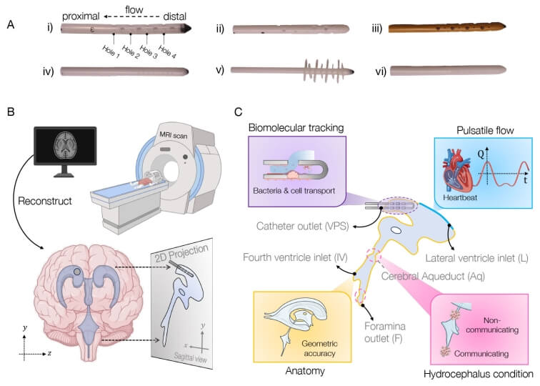
Schematic of approach to simulating brain shunt fluid dynamics. Credit: Harvard SEAS
Millions of people worldwide suffer from hydrocephalus, a condition which recently received greater attention when Billy Joel announced his diagnosis. Treatment usually involves surgical placement of shunts to divert cerebrospinal fluid away, but this procedure often leads to complications, infections, and multiple re-treatments.
Bioengineers in the Harvard John A. Paulson School of Engineering and Applied Sciences (SEAS) have now developed a new computational model to aid the creation of shunts tailored to individual patients’ anatomy and needs. The model combines brain anatomy, fluid flow, and biomolecular transport dynamics to simulate shunt performance with pinpoint accuracy.
The work was supported by federal funding from the National Science Foundation and published in Proceedings of the National Academy of Sciences. It was led by SEAS postdoctoral fellow Haritosh Patel, who works in the labs of Joanna Aizenberg, Professor of Materials Science at SEAS and Professor of Chemistry and Chemical Biology; and Venkatesh Murthy, Professor of Molecular and Cellular Biology and Director of the Center for Brain Science.
Repeat surgeries due to infection or obstruction
Tens of thousands of shunt procedures are performed annually in the U.S. — many of which are repeat surgeries due to the inserted devices becoming blocked or obstructed, or the patient suffering an infection.
“Some elderly patients told me they had had over 10 surgeries — one every two to three years,” Patel said. “We really wanted to understand why this was happening, and we realised that many of these obstructions and infections were tied to shunt designs that didn’t fully consider fluid dynamics as a fundamental part of their geometry. We noticed that the tubing geometry used in shunts closely resembles the kind of piping we rely on in household plumbing. While that simplicity has its advantages, we saw an opportunity to explore more creative, biomimetic solutions that better suit the complexity of the brain’s environment.”
Pursuing the problem from both a material and design perspective, the team quickly realized there was no universally accepted fluid flow model for the brain ventricle space to guide them. “Okay, well, we can’t test our devices in a model, so why don’t we first make a better model?” Patel said.
Computational tool simulates fluid flow in brain
The result is their computational tool, called BrainFlow, which combines detailed anatomical and physiological features of the brain to simulate the flow of cerebrospinal fluid flow in the presence of shunt implants.
The model incorporates patient-specific medical imaging data along with pulse-induced flow to mimic a patient’s cerebrospinal fluid dynamics, all to offer insight into optimal shunt design, placement, and even choice of materials.
“We believe that our model, combined with novel geometries and materials improvements such as anti-biofouling coatings developed in my lab, could lead to smoother integration of optimized, patient-specific medical devices into patients’ brains, with less likelihood of complications, and a better quality of life,” Aizenberg said.
The Harvard team is currently conducting studies that use the model to test different designs of shunts and calculate their efficacy.
Neurotransmitters at a synapse. Credit: Scientific Animations CC4.0
A new study from the University of Colorado Anschutz Medical Campus has upended decades of neuroscience dogma by revealing that the neurotransmitter dopamine communicates in the brain with extraordinary precision – not broad diffusion as previously believed. This groundbreaking research offers fresh hope for millions of people living with dopamine-related disorders, marking a significant advance in the quest for precision-based neuroscience and medicine.
For years, scientists thought of dopamine as a kind of chemical “broadcast system,” flooding large areas of the brain to influence behaviour. But new research, published in Science, found that dopamine acts more like a finely-tuned postal service, delivering highly localised messages to specific nerve cell branches at exact moments in time.
“Our current research found that dopamine signaling and transmission in the brain is much more complex than we thought,” said Christopher Ford, PhD, professor at the University of Colorado School of Medicine and lead author. “We knew that dopamine plays a role in many different behaviours, and our work gives the beginning of a framework for understanding how all those different behaviours could all be regulated by dopamine.”
‘We are really only at the tip of the iceberg in trying to understand how dysfunctions in dopamine contribute to diseases like Parkinson’s disease, schizophrenia or addiction.’
– Christopher Ford, PhD
Using advanced microscopy techniques, researchers found that dopamine is released in concentrated hotspots which enable targeted, rapid responses in nearby brain cells, while broader signals activate slower, widespread effects. This dual signaling system allows dopamine to simultaneously fine-tune individual neural connections and orchestrate complex behaviours like movement, decision-making, and learning.
The implications are far-reaching: dopamine system dysfunction plays a central role in a wide range of brain disorders, including Parkinson’s disease, addiction, schizophrenia, ADHD and depression. Current treatments largely focus on restoring overall dopamine levels – but this research suggests that the precision of dopamine signalling may be just as crucial.
“We are really only at the tip of the iceberg in trying to understand how dysfunctions in dopamine contribute to diseases like Parkinson’s disease, schizophrenia or addiction,” said Ford. “More work is needed to grasp how these specific changes in dopamine signalling are affected in these different neurological and psychiatric diseases. The goal, of course, would then be to build on those findings to come up with new and improved treatments for those disorders.”
Weizmann Institute scientists have discovered hundreds of molecules that promote nerve regeneration in mice – and may even encourage growth in brain neurons
Top: Overexpression of genes from the B2-SINE family in retinal ganglion neurons led to accelerated growth after injury. Bottom: Ganglion cells after injury without B2-SINE overexpression. Credit: Weizmann Institute of Science
Unlike the brain and spinal cord, peripheral nerve cells, whose long extensions reach the skin and internal organs, are capable of regenerating after injury. This is why injuries to the central nervous system are considered irreversible, while damage to peripheral nerves can, in some cases, heal, even if it takes months or years. Despite decades of research, the mechanisms behind peripheral nerve regeneration remain only partially understood.
In a new study published in Cell, researchers from Prof Michael (Mike) Fainzilber’s lab at the Weizmann Institute of Science discovered that a family of hundreds of RNA molecules with no known physiological function is essential to nerve regeneration. Remarkably, the study showed that these molecules can stimulate growth not only in the peripheral nervous system of mice but also in their central nervous system. These findings could pave the way for new treatments for a variety of nerve injuries and neurodegenerative diseases.
For a peripheral nerve to regenerate, it must maintain communication between the neuron’s cell body and its long extension – the axon – which in humans can reach more than a meter in length. In a series of studies over the past two decades, Fainzilber’s lab has revealed key components of this communication: proteins that act like postal couriers, delivering instructions for the production of growth-controlling factors and other proteins, from the cell body to the axon. These molecular couriers also help assess the distance between the cell body and the axon tip, allowing the neuron to modulate its growth accordingly. Yet one central issue remained: What triggers the regenerative growth after injury, and why does this not happen in central nervous system cells?
“While the growth acceleration observed in our study is not yet sufficient to address clinical paralysis, it is definitely significant”
In the new study, Dr Indrek Koppel of Fainzilber’s lab, in collaboration with Dr Riki Kawaguchi of the University of California, Los Angeles (UCLA), examined a specific kind of gene expression in the peripheral nerves of mice following injury. The researchers were surprised to find that one day after damage, the neurons increased the expression of an entire family of short genetic sequences called B2-SINEs, whose role was previously unknown. These sequences do not encode any proteins, and because they are known for “jumping” around the genome, meaning that they can appear at the wrong place or time, they have a bad reputation. But the researchers found that after injury, the neurons began expressing many B2-SINE RNA transcripts, in parallel with other processes preparing the cell for regeneration and repair.
However, B2-SINE is an enormous family, comprising some 150 000 sequences scattered throughout the mouse genome. The initial analysis could not determine which of these were responsible for promoting growth. Dr. Eitan Erez Zahavi, also of Fainzilber’s lab, who led the new study alongside Koppel, used bioinformatics tools to identify 453 B2-SINE sequences that are highly expressed after injury, promoting nerve growth. Collaborating with international research teams, the scientists showed that this overexpression after injury is unique to peripheral nerve cells and does not occur in the central nervous system.
The periphery leads, the center follows
The researchers then tested whether B2-SINEs from peripheral nerve cells could also stimulate neuronal growth in the central nervous system. They induced retinal neurons in mice to overexpress RNA molecules of the B2-SINE type and observed faster regeneration after injury. A similar experiment in the mouse motor cortex – the brain region that controls muscle movement via long axons projecting to the spinal cord – showed that neurons expressing high levels of B2-SINE also regenerated faster than control neurons.
“There are still no effective treatments to accelerate nerve cell growth and regeneration,” Fainzilber notes. “While the growth acceleration observed in our study is not yet sufficient to address clinical paralysis, it is definitely significant. Of course, the path from basic research to clinical application is long, and we must make sure that enhancing growth mechanisms does not, for example, increase the risk of cancer.”
One final mystery remained: How do B2-SINE RNA molecules actually promote regeneration? With help from Prof Alma L. Burlingame’s group at the University of California, San Francisco, the researchers discovered that these RNAs promote a physical link between the molecular “couriers” carrying instructions for producing growth-associated proteins and the ribosomes that read these instructions and carry them out. This means that production of the critical factors takes place closer to the cell body rather than to the tip of the axon. The researchers believe that this signals to the neuron that it is “too small,” triggering a growth response.
“There are over a million sequences called Alu elements in the human genome, the human equivalent of B2-SINEs in mice,” says Fainzilber. “These molecules had been previously shown to bind to ribosomes and mail couriers, but why this happens was unknown. We’re now trying to determine whether Alu or other noncoding RNA elements are involved in nerve regeneration in humans.”
“Recovery from peripheral nerve injuries, or from systemic diseases like diabetes that affect these nerves, can be very slow,” he adds. “That’s why we’re now testing a therapy that might speed up regeneration by mimicking B2-SINE activity. This therapy involves small molecules that connect the couriers to ribosomes while keeping them close to the nerve cell body, promoting faster growth. We are conducting this research in collaboration with Weizmann’s Bina unit for early-stage research with applicative potential.”
Beyond promoting peripheral nerve regeneration, the new study also hints at an even broader prospect: regeneration in the central nervous system. “We are currently working with UCLA on a study showing that the mechanism we discovered plays a role in recovery from stroke in mouse models,” Fainzilber says. “Additionally, we’re collaborating with Tel Aviv University, Hebrew University and Sheba Medical Center to study its possible role in ALS, a progressive neurodegenerative disease. Neurodegenerative conditions affect many millions of people worldwide. While the road ahead is long, I truly hope we’ll one day be able to harness our newly discovered regeneration mechanism to treat them.”
Science Numbers
After injury, the axon of a peripheral nerve cell regrows at a rate of around 1 millimetre a day.
In a new study, investigators from McLean Hospital (a member of Mass General Brigham), Harvard Medical School, and the National Institute on Drug Abuse Intramural Research Program (NIDA-IRP) discovered that the tendency of people’s arousal to wane over the course of brain scans has been distorting the brain connection maps produced by functional magnetic resonance imaging (fMRI).
The team found that as people’s arousal levels dwindle during an fMRI, such as if they become more relaxed and sleepy, changes in breathing and heart rates alter blood oxygen levels in the brain—which are then falsely detected on the scan as neuronal activity.
“You’re laying down in a snug scanner for quite some time, often with only a low-engagement button press task to attend to or nothing to do at all, as the scanner monotonously hums and vibrates around you,” said first author Cole Korponay, PhD, MPA, a research fellow at the McLean Hospital Imaging Center. “These arousal-dampening conditions create the illusion that people’s brain connection strengths continuously inflate throughout the scan.”
fMRI scans are commonly used to non-invasively map brain connectivity, but the technique relies on changes in brain blood oxygen to indirectly measure neuronal activity. It is therefore vulnerable to “noise” from other processes that can affect blood oxygen – such as changes in breathing and heart rates.
And because breathing and heart rate patterns are closely tied to arousal levels, changes in arousal can introduce significant noise into fMRI data. Problematically, the conditions of an fMRI scan tend to progressively lull people into lower arousal states.
In the present study, the research team identified a specific blood flow signal that seemed to track both the decline in subject arousal levels and the illusory inflation of functional brain connection strengths.
This non-neuronal, physiological noise signal, termed the “systemic low frequency oscillation” (sLFO) signal, grew over time during scanning, in a spatial and temporal pattern that closely followed the pattern of the connection strength increases.
The researchers then demonstrated that a method called RIPTiDe, developed by co-senior author Blaise Frederick, PhD, an associate biophysicist at the McLean Imaging Center, to remove the sLFO signal from fMRI data, was able to eliminate the illusory connection strength increases.
“By adopting this sLFO denoising procedure, future studies can mitigate the distortive effects of arousal changes during brain scans and enhance the validity and reliability of fMRI findings,” said Korponay.
This research was supported by the National Institute on Drug Abuse, the National Institute of Mental Health, and the National Institute on Aging, all part of the National Institutes of Health.
A blood-test analysis developed at Stanford Medicine can determine the “biological ages” of 11 separate organ systems in individuals’ bodies and predict the health consequences.
Beside our chronological age, research has shown that we also have what’s called a “biological age,” a cryptic but more accurate measure of our physiological condition and likelihood of developing aging-associated disorders from heart trouble to Alzheimer’s disease.
How old someone’s internal organs are is a challenge to determine compared to looking at wrinkles and greying hair. Internal organs are ageing at different speeds, too, according to a new study by Stanford Medicine investigators.
“We’ve developed a blood-based indicator of the age of your organs,” said Tony Wyss-Coray, PhD, professor of neurology and neurological sciences and director of the Knight Initiative for Brain Resilience at the Wu Tsai Neurosciences Institute. “With this indicator, we can assess the age of an organ today and predict the odds of your getting a disease associated with that organ 10 years later.”
They can even predict who is most likely to die from medical conditions associated with one or more of the 11 separate organ systems the researchers looked at: brain, muscle, heart, lung, arteries, liver, kidneys, pancreas, immune system, intestine and fat.
The brain is the gatekeeper of longevity. If you’ve got an old brain, you have an increased likelihood of mortality. If you’ve got a young brain, you’re probably going to live longer.”
The biological age of one organ, the brain, plays an outsized role in determining how long you have left to live, Wyss-Coray said.
“The brain is the gatekeeper of longevity,” he said. “If you’ve got an old brain, you have an increased likelihood of mortality. If you’ve got a young brain, you’re probably going to live longer.”
Wyss-Coray is the senior author of the study, published online July 9 in Nature Medicine. The lead author is Hamilton Oh, PhD, a former graduate student in Wyss-Coray’s group.
Eleven organ systems, 3000 proteins, 45 000 people
The scientists used 44 498 randomly selected participants, ages 40 to 70, who were drawn from the UK Biobank. This ongoing effort has collected multiple blood samples and updated medical reports from some 600 000 individuals over several years. These participants were monitored for up to 17 years for changes in their health status.
Wyss-Coray’s team made use of an advanced commercially available laboratory technology that counted the amounts of nearly 3000 proteins in each participant’s blood. Some 15% of these proteins can be traced to single-organ origins, and many of the others to multiple-organ generation.
The researchers fed everybody’s blood-borne protein levels into a computer and determined the average levels of each of those organ-specific proteins in the blood of those people’s bodies, adjusted for age. From this, the scientists generated an algorithm that found how much the composite protein “signature” for each organ being assessed differed from the overall average for people of that age.
Based on the differences between individuals’ and age-adjusted average organ-assigned protein levels, the algorithm assigned a biological age to each of the 11 distinct organs or organ systems assessed for each subject. And it measured how far each organ’s multiprotein signature in any given individual deviated in either direction from the average for people of the same chronological age. These protein signatures served as proxies for individual organs’ relative biological condition. A greater than 1.5 standard deviation from the average put a person’s organ in the “extremely aged” or “extremely youthful” category.
One-third of the individuals in the study had at least one organ with a 1.5-or-greater standard deviation from the average, with the investigators designating any such organ as “extremely aged” or “extremely youthful.” One in four participants had multiple extremely aged or youthful organs.
For the brain, “extremely aged” translated to being among the 6% to 7% of study participants’ brains whose protein signatures fell at one end of the biological-age distribution. “Extremely youthful” brains fell into the 6% to 7% at the opposite end.
Health outcomes foretold
The algorithm also predicted people’s future health, organ by organ, based on their current organs’ biological age. Wyss-Coray and his colleagues checked for associations between extremely aged organs and any of 15 different disorders including Alzheimer’s and Parkinson’s diseases, chronic liver or kidney disease, Type 2 diabetes, two different heart conditions and two different lung diseases, rheumatoid arthritis and osteoarthritis, and more.
Risks for several of those diseases were affected by numerous different organs’ biological age. But the strongest associations were between an individual’s biologically aged organ and the chance that this individual would develop a disease associated with that organ. For example, having an extremely aged heart predicted higher risk of atrial fibrillation or heart failure, having aged lungs predicted heightened chronic obstructive pulmonary disease (COPD) risk, and having an old brain predicted higher risk for Alzheimer’s disease.
The association between having an extremely aged brain and developing Alzheimer’s disease was particularly powerful: 3.1 times that of a person with a normally aging brain. Meanwhile, having an extremely youthful brain was especially protective against Alzheimer’s – barely one-fourth that of a person with a normally aged brain.
In addition, Wyss-Coray said, brain age was the best single predictor of overall mortality. Having an extremely aged brain increased subjects’ risk of dying by 182% over a roughly 15-year period, while individuals with extremely youthful brains had an overall 40% reduction in their risk of dying over the same duration.
Predicting the disease, then preventing it
“This approach could lead to human experiments testing new longevity interventions for their effects on the biological ages of individual organs in individual people,” Wyss-Coray said.
Medical researchers may, for example, be able to use extreme brain age as a proxy for impending Alzheimer’s disease and intervene before the onset of outward symptoms, when there’s still time to arrest it, he said.
Careful collection of lifestyle, diet and prescribed- or supplemental-substance intake in clinical trials, combined with organ-age assessments, could throw light on the medical value of those factors’ contributions to the aging of various organs, as well as on whether existing, approved drugs can restore organ youth before people develop a disease for which an organ’s advanced biological age puts them at high risk, Wyss-Coray added.
If commercialised, the test could be available in the next two to three years, Wyss-Coray said. “The cost will come down as we focus on fewer key organs, such as the brain, heart and immune system, to get more resolution and stronger links to specific diseases.”
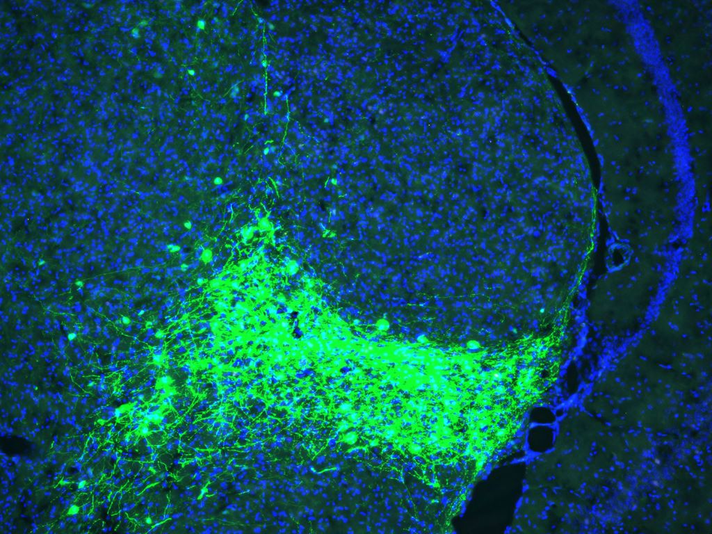
Salk scientists uncover a key neural circuit in mice that gives pain its emotional punch, opening new doors for treating fibromyalgia, migraine, and PTSD
CGRP-expressing neurons (green) in the parvocellular subparafascicular nucleus (SPFp) of the thalamus. Credit: Salk Institute
More than just a physical sensation, pain also carries emotional weight. That distress, anguish, and anxiety can turn a fleeting injury into long-term suffering.
Salk Institute researchers have now identified a brain circuit that gives physical pain its emotional tone, revealing a new potential target for treating chronic and affective pain conditions such as fibromyalgia, migraine, and post-traumatic stress disorder (PTSD).
Published in PNAS, the study identifies a group of neurons in a central brain area called the thalamus that appears to mediate the emotional or affective side of pain in mice. This new pathway challenges the textbook understanding of how pain is processed in the brain and body.
“For decades, the prevailing view was that the brain processes sensory and emotional aspects of pain through separate pathways,” says senior author Sung Han, associate professor and holder of the Pioneer Fund Developmental Chair at Salk. “But there’s been debate about whether the sensory pain pathway might also contribute to the emotional side of pain. Our study provides strong evidence that a branch of the sensory pain pathway directly mediates the affective experience of pain.”
The physical sensation of pain allows immediate detection, assessment of its intensity, and identification of its source. The affective part of pain is what makes it so unpleasant – the emotional discomfort motivates avoidance.
This is a critical distinction. Most people start to perceive pain at the same stimulus intensities, meaning the sensory side of pain is processed similarly. But the ability to tolerate pain varies greatly. The degree of suffering or feeling threatened by pain is determined by affective processing, and if that becomes too sensitive or lasts too long, it can result in a pain disorder. This makes it important to understand which parts of the brain control these different dimensions of pain.
Sensory pain was thought to be mediated by the spinothalamic tract, a pathway that sends pain signals from the spinal cord to the thalamus, which then relays them to sensory processing areas across the brain.
Affective pain was generally thought to be mediated by a second pathway called the spinoparabrachial tract, which sends pain information from the spinal cord into the brainstem.
However, previous studies using older research methods have suggested the circuitry of pain may be more complex. This long-standing debate inspired Han and his team to revisit the question with modern research tools.
Using advanced techniques to manipulate the activity of specific brain cells, the researchers discovered a new spinothalamic pathway in mice. In this circuit, pain signals are sent from the spinal cord into a different part of the thalamus, which has connections to the amygdala, the brain’s emotional processing center. This particular group of neurons in the thalamus can be identified by their expression of CGRP (calcitonin gene-related peptide), a neuropeptide originally discovered in Professor Ronald Evans’ lab at Salk.
When the researchers “turned off” (genetically silenced) these CGRP neurons, the mice still reacted to mild pain stimuli, such as heat or pressure, indicating their sensory processing was intact. However, they didn’t seem to associate lasting negative feelings with these situations, failing to show any learned fear or avoidance behaviors in future trials. On the other hand, when these same neurons were “turned on” (optogenetically activated), the mice showed clear signs of distress and learned to avoid that area, even when no pain stimuli had been used.
“Pain processing is not just about nerves detecting pain; it’s about the brain deciding how much that pain matters,” says first author Sukjae Kang, a senior research associate in Han’s lab. “Understanding the biology behind these two distinct processes will help us find treatments for the kinds of pain that don’t respond to traditional drugs.”
Many chronic pain conditions—such as fibromyalgia and migraine—involve long, intense, unpleasant experiences of pain, often without a clear physical source or injury. Some patients also report extreme sensitivity to ordinary stimuli like light, sound, or touch, which others would not perceive as painful.
Han says overactivation of the CGRP spinothalamic pathway may contribute to these conditions by making the brain misinterpret or overreact to sensory inputs. In fact, transcriptomic analysis of the CGRP neurons showed that they express many of the genes associated with migraine and other pain disorders.
Notably, several CGRP blockers are already being used to treat migraines. This study may help explain why these medications work and could inspire new nonaddictive treatments for affective pain disorders.
Han also sees potential relevance for psychiatric conditions that involve heightened threat perception, such as PTSD. Growing evidence from his lab suggests that the CGRP affective pain pathway acts as part of the brain’s broader alarm system, detecting and responding to not only pain but a wide range of unpleasant sensations. Quieting this pathway with CGRP blockers could offer a new approach to easing fear, avoidance, and hypervigilance in trauma-related disorders.
Importantly, the relationship between the CGRP pathway and the psychological pain associated with social experiences like grief, loneliness, and heartbreak remains unclear and requires further study.
“Our discovery of the CGRP affective pain pathway gives us a molecular and circuit-level explanation for the difference between detecting physical pain and suffering from it,” says Han. “We’re excited to continue exploring this pathway and enabling future therapies that can reduce this suffering.”
A study in the journal Science presents compelling new evidence that neurons in the brain’s memory centre, the hippocampus, continue to form well into late adulthood. The research from Karolinska Institutet provides answers to a fundamental and long-debated question about the human brain’s adaptability.
The hippocampus is a brain region that is essential for learning and memory and involved in emotion regulation. Back in 2013, Jonas Frisén’s research group at Karolinska Institutet showed in a high-profile study that new neurons can form in the hippocampus of adult humans. The researchers then measured carbon-14 levels in DNA from brain tissue, which made it possible to determine when the cells were formed.
Identifying cells of origin
However, the extent and significance of this formation of new neurons (neurogenesis) are still debated. There has been no clear evidence that the cells that precede new neurons, known as neural progenitor cells, actually exist and divide in adult humans.
“We have now been able to identify these cells of origin, which confirms that there is an ongoing formation of neurons in the hippocampus of the adult brain,” says lead researcher Jonas Frisén, professor of stem cell research at the Department of Cell and Molecular Biology.
In the new study, the researchers combined several advanced methods to examine brain tissue from people aged 0 to 78 years from several international biobanks. They used a method called single-nucleus RNA sequencing, which analyses gene activity in individual cell nuclei, and flow cytometry to study cell properties.
By combining this with machine learning, they were able to identify different stages of neuronal development, from stem cells to immature neurons, many of which were in the division phase.
To localise these cells, the researchers used two techniques that show where in the tissue different genes are active: RNAscope and Xenium. These methods confirmed that the newly formed cells were located in a specific area of the hippocampus called the dentate gyrus. This area is important for memory formation, learning and cognitive flexibility.
Hope for new treatments
The results show that the progenitors of adult neurons are similar to those of mice, pigs and monkeys, but that there are some differences in which genes are active. There were also large variations between individuals – some adult humans had many neural progenitor cells, others hardly any at all.
“This gives us an important piece of the puzzle in understanding how the human brain works and changes during life,” explains Jonas Frisén. “Our research may also have implications for the development of regenerative treatments that stimulate neurogenesis in neurodegenerative and psychiatric disorders.”
A team of scientists at the University of California, Riverside, explains in a paper published in PLoS Pathogens how the microscopic parasite Toxoplasma gondii can significantly disrupt brain function, even when it infects only a small number of neurons. The team found the parasite interferes with essential communication between brain cells — research that can offer new ways to detect and treat chronic brain infections.
Toxoplasma gondii can infect nearly any warm-blooded animal and prefers to live inside brain cells, forming cysts in neurons that can persist for life. The researchers report that they found infected neurons release fewer extracellular vesicles (EVs) — tiny, membrane-bound packets used by cells to exchange information.
“We found this disruption in EV signalling can interfere with how neurons and glial cells, especially astrocytes, maintain a healthy brain environment,” said Emma H. Wilson, a professor of biomedical sciences in the UC Riverside School of Medicine who led the research team. “Even a handful of infected neurons can shift the brain’s neurochemical balance. This suggests that communication between neurons and supporting glial cells is not only critical, but also vulnerable to hijacking by parasites.”
Approximately 10–30% of people in the United States are infected with Toxoplasma gondii, often without knowing it. The parasite is typically contracted through undercooked meat or exposure to cat feces. Although the immune system typically keeps the infection in check, the parasite can lie dormant in the brain for decades. In individuals with weakened immunity, it can reactivate and cause serious illness.
Current diagnostic tools can only detect whether someone has been exposed to Toxoplasma gondii by identifying antibodies. The tools cannot confirm whether the parasite is still present in the brain or how it may be affecting brain function.
“Our research opens the door to using EVs as biomarkers, which can be isolated from blood,” Wilson said.
The study was conducted using mouse models and human cells in a laboratory setting.
Wilson explained that in healthy mouse brains astrocytes regulate neurotransmitters like glutamate, ensuring that neurons do not become overexcited. But when neurons infected with Toxoplasma gondii stop sending the right EV signals, this regulation breaks down. The result is elevated glutamate levels, which can lead to seizures, neural damage, or altered brain connectivity.
“The parasite may play a larger role in neurological and behavioural conditions than we previously thought,” she said.
Wilson’s research team is now working to analyse samples from human blood banks to look for EVs linked to Toxoplasma gondii brain infection. The team also hopes to better understand how glial cells detect and respond to parasite proteins — insights that could one day lead to new therapies or even vaccines.
“Our brains have built-in defences that may recognise and respond to neurons infected by Toxoplasma gondii,” Wilson said. “If we can learn how to support or enhance that process, we may be able to better protect people, especially the most vulnerable.”
Despite its potential impact, Toxoplasma gondii is often misunderstood, Wilson added.
“There’s no need to avoid someone who is infected; most people live their entire lives without symptoms,” she said. “Pregnant individuals should be cautious as the parasite can cause serious birth defects if contracted for the first time during pregnancy. The most effective prevention is proper food handling and hygiene. Cook meat thoroughly, wash vegetables, and always wash your hands after handling cat litter, especially from young cats, which are more likely to shed the parasite.”
A drug commonly used to treat type 2 diabetes may reduce excess fluid in the brains of patients with hydrocephalus, which could help treat the disease less invasively than current treatments, according to a Northwestern Medicine study published in theJournal of Clinical Investigation.
Normal pressure hydrocephalus occurs when excess cerebrospinal fluid builds up inside the skull and puts pressure on the brain. The cause of the condition is elusive and affects up to three percent of individuals over the age of 65, with symptoms including cognitive decline, difficulty walking and bladder problems.
Patients are typically treated with permanent ventriculoperitoneal shunts, which are surgically implanted in the front or back of the skull and are connected to a valve that diverts excess cerebrospinal fluid away from the brain and into the abdomen where it is absorbed. The procedure has been shown to dramatically improve mobility, bladder control and cognitive functioning in patients with hydrocephalus, according to senior study author Stephen Magill, MD, PhD.
“It’s a great procedure because it’s one of the few things you can do that actually reverses these symptoms,” said Magill, who is assistant professor of Neurological Surgery.
There is, however, no pharmacological treatment currently approved to treat hydrocephalus. Additionally, nearly 20% of patients with normal pressure hydrocephalus also have type 2 diabetes and take sodium/glucose cotransporter 2 (SGLT2) inhibitors to manage their blood sugar, cardiovascular and kidney function, and weight loss.
Magill recently observed a reduction in the brain ventricle size in a patient with hydrocephalus who had a ventriculoperitoneal shunt surgically implanted and then began taking SGLT2 inhibitors to treat their type 2 diabetes. This phenomenon prompted Magill to further investigate the impact of SGLT2 inhibitors on ventricular size in patients with hydrocephalus.
“The medication inhibits a receptor found in the kidneys, which is where it works for diabetes. However, that receptor is also expressed in the choroid plexus, which is the structure in the brain that secretes the spinal fluid. Although this was known from animal studies, the clinical aspects of this biology have not been fully appreciated,” Magill said.
In the current study, three patients with hydrocephalus underwent CT scans both before and after surgery for ventriculoperitoneal shunts. After surgery, each patient began taking SGLT2 inhibitors for a medical indication and then underwent additional CT scans.
From analyzing these scans, Magill’s team discovered that all three patients showed a reduction in ventricle size as well as structural changes in their brains after starting SGLT2 therapy. One patient demonstrated dramatic ventricle size reduction due to ventricular collapse and required a shunt valve adjustment to reduce cerebrospinal fluid drainage.
“It’s a really interesting clinical observation because it raises the possibility that these medications could be used to treat normal pressure hydrocephalus in the future, which would normally require surgery,” Magill said.
Magill said the findings have sparked a new line of research in studying how SGLT2 inhibitors could help prevent hydrocephalus, adding that his team is now studying SGLT2 knockout mouse models to better understand the drug’s impact on ventricular size.
Their findings could ultimately inform new therapeutic strategies for treating normal pressure hydrocephalus as well as post-traumatic hydrocephalus, or the buildup of cerebrospinal fluid after traumatic brain injury, according to Magill.
“This sparks a new line of research on how normal pressured hydrocephalus develops, what causes it, how this protein works in creating and secreting spinal fluid, and has direct translational implications,” Magill said. “There’s a whole new avenue of potentially treating this disease that might save a patient from having surgery, and there’s always risks with surgery. It will also evolve our understanding of how these drugs work.”
Researchers at Karolinska Institutet and Lund University have identified a new treatment strategy for neuroblastoma, an aggressive form of childhood cancer. By combining two antioxidant enzyme inhibitors, they have converted cancer cells in mice into healthy nerve cells. The study is published in the journal Proceedings of the National Academy of Sciences (PNAS).
Neuroblastoma is a type of childhood cancer that affects the nervous system and is the leading cause of cancer-related death in young children. Some patients have a good prognosis, but those with metastatic tumours often cannot be cured despite modern combinations of surgery, radiation, chemotherapy and immunotherapy.
“The children who survive often have lifelong cognitive difficulties due to the harsh treatment, so there is a great need for new forms of therapies for children with neuroblastoma,” says Marie Arsenian Henriksson, professor at the Department of Microbiology, Tumour and Cell Biology at Karolinska Institutet.
Transform cancer cells
Differentiation therapy is a treatment method used in neuroblastoma that aims to transform cancer cells into more mature and healthy cells. The problem with the current retinoic acid differentiation therapy is that many patients do not respond to treatment, and about half develop resistance.
In collaboration with researchers at Lund University, Marie Arsenian Henriksson’s research team has shown that inhibition of two specific enzymes, PRDX6 and GSTP1, could be an alternative to retinoic acid treatment.
Mature into healthy neurons
Neuroblastoma is characterised by high oxidative stress due to the active metabolism in the cancer cells. Tumours are therefore dependent on antioxidant enzymes such as PRDX6 and GSTP1 to manage the stress and avoid cell death. High levels of these enzymes are associated with a poorer prognosis.
“When we inhibit these enzymes in cell cultures as well as in mouse models, some of the tumour cells die while others mature into active, healthy neurons, impairing tumour growth,” says Judit Liaño-Pons, researcher at the Department of Microbiology, Tumour and Cell Biology.
Needs to be tested in children
In the next step, the treatment will need to be tested in a clinical trial to investigate its safety and efficacy in children. One of the inhibitors has received orphan drug designation from the US Food and Drug Administration for the treatment of a different diagnosis in adults, making it a particularly promising drug candidate, according to the scientists.