Scientists Discover the Neurological Basis of Food Cravings in Pregnancy

By examining mice, which get pregnancy cravings similar to humans, scientists have identified the neurological basis of food craving during pregnancy.
During pregnancy, the mother’s body undergoes a series of physiological and behavioural changes to create an environment facilitating the embryo’s development. Frequent consumption of tasty, high calorie foods driven by the cravings contributes to weight gain and obesity in pregnancy, with possible negative consequences for the baby’s health.
“There are many myths and popular beliefs regarding these cravings, although the neuronal mechanisms that cause them are not widely known,” noted study leader March Claret, at the University of Barcelona and leader of the study published in the journal Nature Metabolism.
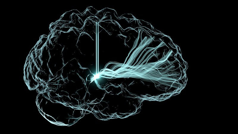
The researchers found that the brains of pregnant female mice undergoes changes in the functional connections of the brain reward circuits, as well as the taste and sensorimotor centres. Mice, like pregnant women, are also more sensitive to sweet food, and develop binge-eating behaviours towards high calorie foods. “The alteration of these structures made us explore the mesolimbic pathway, one of the signal transmission pathways of dopaminergic neurons. Dopamine is a key neurotransmitter in motivational behaviours,” notes Claret, member of the Department of Medicine of the UB and the Diabetes and Associated Metabolic Diseases Networking Biomedical Research Centre (CIBERDEM).
The team saw that dopamine levels and dopamine receptor (D2R) activity increased in the nucleus accumbens, a brain region involved in the reward circuit. “This finding suggests that the pregnancy induces a full reorganisation of the mesolimbic neural circuits through the D2R neurons,” noted study leader Roberta Haddad-Tóvolli. “These neuronal cells – and their alteration – would be responsible for the cravings, since food anxiety, typical during pregnancy, disappeared after blocking their activity.”
The team demonstrated that persistent cravings have consequences for the offspring, affecting the metabolism and development of neural circuits that regulate food intake, leading to weight gain, anxiety and eating disorders. “These results are shocking, since many of the studies are focused on the analysis of how the mother’s permanent habits – such as obesity, malnutrition, or chronic stress – affect the health of the baby. However, this study indicates that short but recurrent behaviours, such as cravings, are enough to increase the psychological and metabolic vulnerability of the offspring,” concluded Claret.
The conclusions of the study could contribute to the improvement of nutritional guidelines for pregnant women in order to ensure a proper prenatal nutrition and prevent the development of diseases.
Source: University of Barcelona