Category: Neurology

Experiments to Test Consciousness All Fall Short
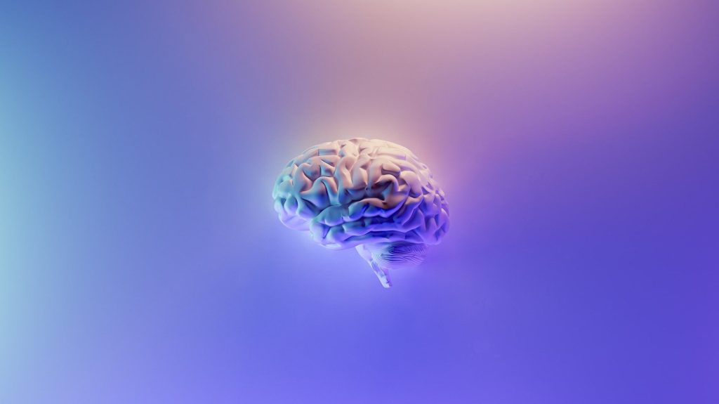
Depiction of a human brain
Image by Fakurian Design on Unsplash

A study examining various experiments each designed to prove one of four conflicting theories of consciousness has discovered that they are all flawed: predetermined to prove the theory they are designed to test. The surprising conclusion is that the nature of the experiment largely determines its result.

In neuroscience, there are currently four leading theories trying to explain how the experience of consciousness emerges from neural activity. In this unique study, researchers re-examined hundreds of experiments that support contradictory theories.

The study, published in Nature Human Behaviour, shows that the inconsistencies in the experiments’ findings are mainly due to methodological differences or the methodological choices made by the researchers, predetermines their results.

Employing artificial intelligence, researchers re-examined 412 experiments, and found that scientists’ methodological choices actually determined the result of the experiment – so much so that an algorithm could predict which theory they were designed to support with 80% success.

Professor Liad Mudrik led the study. He explained: “The big question is how consciousness is born out of activity in the brain, or what distinguishes between conscious processing and unconscious processing,” Prof Mudrik explained. “For example, if I see a red rose, my visual system processes the information and reports that there is a red stimulus in front of me. But what allows me – unlike a computer for example – to experience this colour? To know how it feels? In recent years, a number of neuroscientific theories have been proposed to explain how conscious experience arises from neural activity. And although the theories provide utterly different explanations, each of them was able to gather empirical evidence to justify itself, based on multiple experiments that were conducted. We re-examined all these experiments, and showed that the parameters of the experiment actually determine its results. The artificial intelligence we used knew how to predict with an 80% success rate which theory the experiment would support, based solely on the researchers’ methodological choices.”

The study of consciousness has four leading theories, with contradicting predictions about the neural underpinnings of conscious experience. The Global Neuronal Workspace Theory maintains that there is a central neural network, and when information enters it, it is being broadcasted throughout the brain, becoming conscious. The Higher Order Thought Theory claims that there is a higher order neural state that ‘points’ at activity in lower-level areas, marking this content as conscious. A third theory, called Recurrent Processing Theory, claims that information that is reprocessed within the sensory areas themselves, in the form of recurrent processing, becomes conscious. And finally, a fourth theory – Integrated Information Theory – defines consciousness as integrated information in the brain, claiming that the posterior regions are the physical substrates of consciousness.

“Each of these theories offers convincing experiments to support them, so the field is polarized, with no agreed-upon neuroscientific account of consciousness,” said Prof Mudrik.

In-depth analysis of all of the 412 experiments designed to test the four leading theories showed that they were constructed differently. For example, some experiments focused on different states of consciousness, such as a coma or a dream, and others studied changes in the content of consciousness of healthy subjects. Some experiments tested connectivity metrics were tested, and others did not. “Researchers make a series of decisions as they build their experiment, and we demonstrated that these decisions alone – without even knowing the results of the experiments – already predict which theory these experiments will support. That is, these theories were tested in different manners, though they try to explain the same phenomenon,” Prof Mudrik said.

“Another one of our findings was that the vast majority of the experiments we analysed supported the theories, rather than challenging them. There appears to be a built-in confirmation bias in our scientific praxis, though the philosopher of science Karl Popper said that science advances by refuting theories, not by confirming them,” added Prof. Mudrik. “Moreover, when you put together all of the findings that were reported in these experiments, it seems like almost the entire brain is involved in creating the conscious experience, which is not consistent with any of the theories. In other words, it would appear that the real picture is larger and more complex than any of the existing theories suggest. It would seem that none of them is consistent with the data, when aggregated across studies, and that the truth lies somewhere in the middle.”

Source: EurekAlert!

Antiepileptics in Comatose Cardiac Arrest Survivors are Ineffective, Study Shows

Image by Falkurian Design on Unsplash

A large scale study of comatose intensive care (ICU) patients admitted after cardiac arrest and resuscitation has shown that antiepileptics to treat epilepsy-like brain activity has no effect, and may even prolong ICU stay.

Following a cardiac arrest and resuscitation, patients may need an ICU stay, and are in a coma. By that stage, the cardiac arrest may have damaged the brain to such an extent that half of the patients will not recover from coma. The other half will also have permanent damage, for example of memory functions. It is extremely difficult to predict if a patient will awaken and what their prognosis is, so clinicians make use of EEG (electroencephalography).

In 10–20% of the patients admitted to the ICU after cardiac arrest and resuscitation, there are signs of brain activity that appear similar to epilepsy: unlike an attack this activity is continuous. For a long time, it was unclear if anti-epileptic medication could help better recovery. As a result, some patients received this medication and some did not.

Now, a large-scale study done between 2014 and 2021 on 172 patients has proven that this medication is ineffective: it does not help recovery, even necessitating a longer ICU stay. The researchers, led by Professor Jeannette Hofmeijer of the University of Twente and Rijnstate Hospital in Arnhem, published their findings in the New England Journal of Medicine.

The conclusion from this study is that anti-epileptic medication does not lead to an improved recovery. The findings show that patients may need to stay longer at the ICU: for the patient an undesired situation, and it puts extra pressure on the health care system. 

Aside from patients who show continuous epileptic signals, a small group of patients show signs of a typical epileptic seizure: a short and heavy attack. In these situations, anti-epileptics could help, but this still needs further research.

“Although the outcome of the trial may be disappointing in terms of chances of recovery, it also takes away uncertainties from the family. The signals point at serious brain damage that would lead to a much longer stay at the ICU,” said Prof Hofmeijer.

Source: University of Twente

Scientists Unravel Neurological Origins of the Placebo Effect

Researchers at Massachusetts General Hospital (MGH) have discovered a network of brain regions activated by the placebo effect overlaps with several regions targeted by brain-stimulation therapy for depression.

The findings of this study, published in Molecular Psychiatry, will help in understanding the neurobiology of placebo effects and could inform how brain stimulation trial results are interpreted. In addition, this could provide insights on how to harness placebo effects for the treatment of a variety of conditions.

The placebo effect occurs when a patient’s symptoms improve because they expect a therapy to help (due to a variety of factors), but not from the specific effects of the treatment itself. Recent research indicates that there is a neurological basis for the placebo effect, with imaging studies identifying a pattern of changes that happen in certain brain regions when a person experiences this phenomenon.

The use of brain-stimulation techniques for patients with depression that doesn’t respond adequately to medication or psychotherapy has gained wider use in recent years. Transcranial magnetic stimulation (TMS) delivers electromagnetic pulses to the brain, and its effect on brain activity has been established over the last three decades in animal and human research studies, with several TMS devices approved by the Food and Drug Administration for treating depression. In addition, for treatment depression, deep brain stimulation (DBS, which requires an implanted device) has shown some promise.

Senior author Emiliano Santarnecchi, PhD, saw studies of brain stimulation as a unique opportunity to learn more about the neurobiology of the placebo effect. Santarnecchi and his co-investigators conducted a meta-analysis and review of neuroimaging studies involving healthy subjects and patients to create a “map” of brain regions activated by the placebo effect. They also analysed studies of people treated with TMS and DBS for depression to identify brain regions targeted by the therapies. The team found that several sites in the brain that are activated by the placebo effect overlap with brain regions targeted by TMS and DBS.

Dr Santarnecchi and his colleagues believe that this overlap has critical importance in interpreting the results of research on brain stimulation for conditions such as depression. In clinical trials, a significant portion of depression patients receiving brain stimulation improve — but so do many patients receiving placebo (sham) treatment, in which no stimulation is administered, which has led to confusion over the therapy’s benefits.

A possible explanation is “that there is a significant placebo effect when you do any form of brain stimulation intervention,” said Dr Santarnecchi. TMS involves a clinical setting, with loud clicks as the pulse is delivered. “So the patient thinks, ‘Wow, they are really activating my brain’, so you get a lot of expectation,” said Dr Santarnecchi.

Elevated placebo effects associated with brain stimulation may create problems when studying the intervention, said first author Matthew Burke, MD, a cognitive neurologist. If brain stimulation and the placebo effect overlap in activating the same brain regions, then those circuits could be maximally activated by placebo effects, which could make it difficult to show any additional benefit from TMS or DBS, said Dr Burke. If so, this could explain the disparity of results in neurostimulation treatment of depression. Screening out placebo from brain stimulation’s direct impact on brain activity will help in designing studies where the real potential of techniques such as TMS will be more easily quantified, thus improving the effect of treatment protocols.

The findings from this study also suggest broad applications for the placebo effect, said Dr Santarnecchi. “We think this is an important starting point for understanding the placebo effect in general, and learning how to modulate and harness it, including using it as a potential therapeutic tool by intentionally activating brain regions of the placebo network to elicit positive effects on symptoms,” he said.

Dr Santarnecchi and his colleagues are currently designing trials that they hope will “disentangle” the effects of brain stimulation from placebo effects and offer insights about how they can be leveraged in clinical settings.

Source: Massachusetts General Hospital

Reduced Antiepileptic Drug Effectiveness in Pregnancy Uncovered

Photo by Shvets Productions on Pexels

Blood levels of many commonly used antiepileptic drugs drop dramatically with the onset of pregnancy, which can result in ‘breakthrough seizures’ according to a study published in JAMA Neurology.

The findings, collected as part of the multicentre study Maternal Outcomes and Neurodevelopmental Effects of Antiepileptic Drugs (MONEAD), explain why many people with epilepsy start experiencing breakthrough seizures after conception, underscoring the need to increase antiseizure medication doses and closely monitor blood levels over the course of pregnancy.

A fine-tuned medication regime is critical in epilepsy. “Some people mistakenly believe that changes in the drugs’ blood concentration won’t occur until after 20 weeks of pregnancy, but our study shows how important it is to start monitoring and adjusting patients’ medication dosages early on,” said lead author Dr Page Pennelll. “Nearly half of all pregnancies in the United States are unplanned, so it is important to ensure that doctors have a clear picture of each patient’s baseline drug level even if they are not trying to conceive.”

A life-altering neurological condition, two-thirds of epilepsy cases do not have a known cause. In people with epilepsy, nerve cells in the brain are hyper-reactive, causing them to change the pattern of their electrical activity and become spontaneously active. That synchronous activation is manifested in seizures.

Epilepsy has a fraught history of diagnosis and management; people with epilepsy go undiagnosed or under-treated. First-generation drugs to control it had many dangerous side effects and were contraindicated for people who are trying to conceive.

Since then, safer medications have entered the U.S. market and become widely available, but clinicians started noticing a new problem – patients whose epilepsy was successfully managed with medications started having seizures soon after becoming pregnant.

“Identifying which antiseizure medications may have changes in concentrations and at what point in pregnancy those changes occur is important for determining which patients may need to be monitored more closely during pregnancy and after delivery,” said senior author Professor Angela Birnbaum at the University of Minnesota.

To solve the mystery, the researchers embarked on a study to analyse blood concentrations of 10 commonly used antiseizure drugs and compare them across different stages of pregnancy and after childbirth.

The study found that blood levels of seven out of 10 of the medications they examined dropped dramatically — from 29.7% for lacosamide, a commonly prescribed anticonvulsant, and up to 56.4% for lamotrigine.

In addition, the researchers noted that the drop in drug levels occurred mere days after conception.

Source: University of Pittsburgh

Body Fat Linked to Risk of Reduced Cognitive Function

Photo by Andres Ayrton from Pexels

A new study published in JAMA Network Open has found that greater body fat is a risk factor for reduced cognitive function, such as processing speed, in adults.

Even when accounting for cardiovascular risk factors or vascular brain injury, the association between body fat and lower cognitive scores persisted. This suggests other, currently unknown, mechanisms linking excess body fat to reduced cognitive function.

For the study, 9166 participants were measured by bioelectrical impedance analysis to assess their total body fat. In addition, 6733 underwent magnetic resonance imaging (MRI) to measure abdominal fat packed around the organs known as visceral fat, and the MRI measured vascular brain injury, including silent brain infarctions and high white matter hyperintensities. Cardiovascular risk factors were measured using health and lifestyle questions and physical measures, and cognitive assessment was measured by the Digital Symbol Substitution Test and the Montreal Cognitive Assessment.

Lead author Sonia Anand, lead author, professor of medicine of McMaster University said: “Our results suggest that strategies to prevent or reduce having too much body fat may preserve cognitive function.”

She added that “the effect of increased body fat persisted even after adjusting for its effect on increasing cardiovascular risk factors like diabetes and high blood pressure, as well as vascular brain injury, which should prompt researchers to investigate which other pathways may link excess fat to reduced cognitive function.”

Co-author Eric Smith, associate professor of clinical neurosciences at the University of Calgary, said that “preserving cognitive function is one of the best ways to prevent dementia in old age. This study suggests that one of the ways that good nutrition and physical activity prevent dementia may be by maintaining a healthy weight and body fat percentage.”

Participants had no existing cardiovascular diseases, and ranged in age from 30 to 75, with an average age of about 58 and 56% were women. Most were White European origin, with about 16% other ethnic backgrounds. 

Source: McMaster University

An Updated Look at the Link Between Alcohol and Epilepsy

Photo by Pavel Danilyuk on Pexels

A new meta-analysis has established an association between alcohol and epilepsy, in contrast to previous studies which reported conflicting results on the relationship.

Epilepsy is one of the most common neurological conditions, with an annual incidence of 40–70 per 100 000 people in industrialised countries. It is also a  disease that is highly stigmatised.

A number of studies have focused on how alcohol consumption leads to provoked seizures, commonly resulting from alcohol withdrawal, or heavy intoxication. Very few of these however focused on the link between alcohol consumption and unprovoked seizures. A 2010 meta-analysis found that alcohol users were more prone to developing unprovoked seizures – but data from recent cohort studies contradict these findings. A 2018 meta-analysis suggested that the relationship may only hold true for heavy drinkers.

Now, using more accurate diagnostic methods and recent data, a team of scientists from Pusan National University, South Korea, conducted an updated meta-analysis to conclusively clarify the relationship between alcohol consumption and unprovoked seizures and epilepsy.

For this meta-analysis, appearing in Drug and Alcohol Dependence, the researchers included a total of eight studies, of which five were case-control studies and three were cohort studies. They analysed the data to assess the dose-response relationship between alcohol intake and epilepsy. The results suggested that overall, compared to non-drinkers, alcohol drinkers were at a significantly higher risk of developing epilepsy, which increased with alcohol intake. These findings are consistent with previous meta-analyses.

An important finding was that cohort studies did not show a positive association between alcohol intake and epilepsy. In fact, 2 out of 3 cohort studies suggested that alcohol intake reduces the risk of epilepsy.

More large cohort studies are needed to prove a causal relationship between alcohol drinking and epilepsy, as well as a threshold of onset, said second author Professor Yun Hak Kim.

Source: EurekAlert!

Study Uncovers Mechanism Behind Visual Impairment in Traumatic Brain Injury

A healthy neuron.
A healthy neuron. Credit: NIH

Traumatic brain injury can lead to long-term visual impairment, which researchers have found is caused by a dramatic drop in the number of neurons in the visual cortex. Their findings were published in Communications Biology.

Traumatic brain injury (TBI) is associated with mechanical brain damage and a wide range of neuronal abnormalities.  Injuries to the posterior occipital cortex are common in humans, and can result in visual impairment. Up to 75% of current or former soldiers live with permanent visual dysfunction or cortical blindness. 

The human brain possesses surprising neuroplasticity, which allows other areas of the brain to take over the functions of a damaged area.

Such neuroplasticity is also characteristic of the sensory areas of the visual cortex, which is final component of the visual pathway, responsible for receiving and processing visual impressions. The primary visual cortex (V1) is reached by the nerve fibres of the optic radiation, which carry nerve impulses from the retinas of both eyes.

Until now, scientists knew little about the effects of TBI on long-term visual circuit function. Using mice, a team of researchers examined how neurons respond to visual stimuli two weeks and three months after mild injury to the primary visual cortex (V1). V1 neurons normally show sensitivity to different features of a visual stimulus, such as colour or direction of movement. The preprocessed data is transmitted to subsequent areas of the visual cortex. This study showed that although the primary visual cortex remained largely intact after the brain injury, there was a 35% reduction in the number of neurons. This loss largely affected inhibitory neurons rather than excitatory neurons, which inhibit or stimulate action in the target cells, respectively.

After TBI, fewer than half of the isolated neurons were sensitive to visual stimuli (32% at two weeks after injury; 49% at three months after the event), compared with 90% of V1 cells in the control group. Up to a threefold decrease in neuronal activity was seen after the brain injury, and the cells themselves had worse spatial orientation. The overall results mean that even minor, superficial brain injuries cause long-term impairment in the way visual stimuli are perceived, persisting several months after the event.

Such a deeper understanding of the functional impairments in damaged visual cortex could provide a basis for developing circuit-level therapies for visual cortex damage.

Source: Institute of Physical Chemistry of the Polish Academy of Sciences

Prompts During Sleep Boosts Recall of Names and Faces

Photo by Cottonbro on Pexels

Hearing names repeated during deep sleep may help bolster recall of names and faces, according to new research from Northwestern University.

The researchers found that people’s name recall improved significantly when memories of newly learned face-name associations were reactivated while they were napping. Uninterrupted deep sleep was key in this improvement.

“It’s a new and exciting finding about sleep, because it tells us that the way information is reactivated during sleep to improve memory storage is linked with high-quality sleep,” said lead author Nathan Whitmore, a PhD candidate in the Interdepartmental Neuroscience Program at Northwestern University.

The research is reported in the Nature partner journal npj Science of Learning.

The three main stages of the experiment of Whitmore et al. (2022). First, participants learned 80 face-name associations. Next, they slept while EEG was monitored to determine sleep stage, and 20 of the spoken names were presented softly over background music during slow-wave sleep. Finally, memory testing showed superior memory due to memory reactivation during sleep, but only when sleep was undisturbed by sound presentations. Credit: Nathan Whitmore, a Ph.D. candidate in the Interdepartmental Neuroscience Program at Northwestern University.

The results also highlighted the importance of adequate sleep: for study participants with EEG measurements that indicated disrupted sleep, the memory reactivation had no effect and may even be detrimental. But in those with uninterrupted sleep during the specific times of sound presentations, the reactivation helped participants recall just over 1.5 more names.

The study recruited 24 participants, aged 18-31 years old, who were asked to memorise the faces and names of 40 pupils from a hypothetical Latin American history class and another 40 from a Japanese history class. When each face was presented again, they were asked to recall the associated name. After the learning exercise, participants took a nap while the researchers carefully monitored brain activity using EEG measurements. When participants reached the N3 “deep sleep” state, some of the names were softly played on a speaker with music that was associated with one of the classes.

When participants awoke, they were again tested on recognising faces and recalling their names.

According to the researchers, the finding on the relationship between sleep disruption and memory accuracy is noteworthy for several reasons.

“We already know that some sleep disorders like apnoea can impair memory,” said Whitmore. “Our research suggests a potential explanation for this—frequent sleep interruptions at night might be degrading memory.”

The lab is currently exploring the reactivation of memories and deliberately disrupting sleep in order to learn more about the relevant brain mechanisms.

Source: EurekAlert!

A Brain ‘Breathalyser’ for THC Intoxication

Image by Falkurian Design on Unsplash

Scientists have developed a noninvasive brain imaging procedure to identify individuals whose performance has been impaired by THC, the psychoactive ingredient of cannabis. As reported in Neuropsychopharmacology, the technique uses functional near-infrared spectroscopy (fNIRS) to measure brain activation patterns linked to THC intoxication. The technology could have a great impact on road and workplace safety. 

The increasing legalisation of cannabis has driven the need for a portable brain imaging procedure that can distinguish between THC-caused impairment and mild intoxication. “Our research represents a novel direction for impairment testing in the field,” explained lead author Jodi Gilman, PhD. “Our goal was to determine if cannabis impairment could be detected from activity of the brain on an individual level. This is a critical issue because a ‘breathalyser’ type of approach will not work for detecting cannabis impairment, which makes it very difficult to objectively assess impairment from THC during a traffic stop.”

In previous studies, THC has been shown to impair cognitive and psychomotor performance essential to safe driving, a factor thought to at least double the risk of fatal motor vehicle accidents. However, concentration of THC in the body does not correspond well to functional impairment. Regular cannabis users often can have high levels of THC in the body and not be impaired. Metabolites of THC can remain in the bloodstream for weeks after the last cannabis use, well beyond the period of intoxication. Thus, there is a need for a different method to determine impairment from cannabis intoxication.

In the study, 169 cannabis users underwent fNIRS brain imaging before and after receiving either oral THC or a placebo. Participants who reported intoxication after being given oral THC showed an increased oxygenated haemoglobin concentration (HbO) – a type of neural activity signature from the prefrontal cortex region of the brain – compared to those who reported low or no intoxication.

“Identification of acute impairment from THC intoxication through portable brain imaging could be a vital tool in the hands of police officers in the field,” said senior author and principal investigator A. Eden Evins, MD, MPH, founding director of the Center for Addiction Medicine. “The accuracy of this method was confirmed by the fact impairment determined by machine learning models using only information from fNIRS matched self-report and clinical assessment of impairment 76% of the time.”

The study suggested the feasibility of inexpensive, lightweight, battery-powered fNIRS devices that could be incorporated into a headband or cap, and thus require minimal set-up time.

“Companies are developing breathalyser devices that only measure exposure to cannabis but not impairment from cannabis,” said Dr Gilman. “We need a method that won’t penalise medical marijuana users or others with insufficient amounts of cannabis in their system to impair their performance. While it requires further study, we believe brain-based testing could provide an objective, practical and much needed solution.”

Source: Massachusetts General Hospital

New Genetic Insights into Basal Ganglia Diseases

Source: Pixabay

A new study published in Developmental Medicine & Child Neurology uncovered a number of genetic causes of basal ganglia diseases.

Basal ganglia are deep grey matter structures in the brain involved in the control of posture and voluntary movements, cognition, behaviour, and motivational states. Several conditions are known to affect basal ganglia during childhood, but many questions remain.

In a study that included 62 children with basal ganglia diseases who were followed for two years, investigators identified multiple genetic aetiologies including mitochondrial diseases (57%), Aicardi–Goutières syndrome (20%), and single-gene causes of dystonia and/or epilepsy (17%) mimicking Leigh syndrome. Radiological abnormalities included T2-hyperintense lesions (n=26) and lesions caused by calcium or manganese mineralisation (n=9).

The researchers identified three clusters: the pallidal, neostriatal, and striatal, plus the last including mtDNA defects in the oxidative phosphorylation system with prominent brain atrophy. Mitochondrial biomarkers showed poor sensitivity and specificity in children with mitochondrial disease, whereas an interferon signature was observed in all patients with Aicardi–Goutières syndrome.

Radiological imaging tests also revealed several characteristics in patients that could help lead to an earlier diagnosis of basal ganglia diseases.

Source: Wiley