Over the past fifty years, there have been remarkable claims about the effects of Wolfgang Amadeus Mozart’s music. Reports about alleged symptom-alleviating effects of listening to Mozart’s Sonata KV448 in epilepsy attracted a lot of public attention. However, the empirical validity of the underlying scientific evidence has remained unclear. Now, University of Vienna psychologists Sandra Oberleiter and Jakob Pietschnig show in a new study published in the journal Nature Scientific Reports that there is no evidence for a positive effect of Mozart’s melody on epilepsy.
In the past, Mozart’s music has been associated with numerous ostensibly positive effects on humans, animals, and even microorganisms. For instance, listening to his sonata has been said to increase the intelligence of adults, children, or foetuses in the womb. Even cows were said to produce more milk, and bacteria in sewage treatment plants were said to work better when they heard Mozart’s composition.
However, most of these alleged effects have no scientific basis. The origin of these ideas can be traced back to the long-disproven observation of a temporary increase in spatial reasoning test performance among students after listening to the first movement allegro con spirito of Mozart’s sonata KV448 in D major.
More recently, the Mozart effect experienced a further variation: Some studies reported symptom relief in epilepsy patients after they had listened to KV448. However, a new comprehensive research synthesis by Sandra Oberleiter and Jakob Pietschnig from the University of Vienna, based on all available scientific literature on this topic, showed that there is no reliable evidence for such a beneficial effect of Mozart’s music on epilepsy. They found that this alleged Mozart effect can be mainly attributed to selective reporting, small sample sizes, and inadequate research practices in this corpus of literature. “Mozart’s music is beautiful, but unfortunately, we cannot expect relief from epilepsy symptoms from it” conclude the researchers.
Researchers at Massachusetts General Hospital have discovered a novel molecular mechanism behind the most common forms of acquired hydrocephalus – which could lead to the first non-surgical treatments for the life-threatening disease. Research in animal models uncovered a pathway through which infection or bleeding in the brain triggers inflammation, causing increased production of cerebrospinal fluid (CSF) by the choroid plexus and lead to swelling of the brain ventricles.
“Finding a nonsurgical treatment for hydrocephalus, given the fact neurosurgery is fraught with tremendous morbidity and complications, has been the holy grail for our field,” says Kristopher Kahle, MD, PhD, a paediatric neurosurgeon at MGH and senior author of the study in the journal Cell. “We’ve identified through a genome-wide analytical approach the mechanism that underlies the swelling of the ventricles which occurs after a brain bleed or brain infection in acquired hydrocephalus. We’re hopeful these findings will pave the way for approval of an anti-inflammatory drug to treat hydrocephalus, which could be a game-changer for populations in the US and around the world that don’t have access to surgery.”
Occurring in about 0.2% of births, acquired hydrocephalus is the most common cause of brain surgery in children, though it can affect people at any age. In underdeveloped regions where bacterial infection is the most prevalent form, hydrocephalus is often deadly for children due to the lack of surgical intervention. Brain surgery, where a shunt is implanted to drain fluid from the brain, is the only known treatment. But about half of all shunts in paediatric patients fail within two years of placement, according to the Hydrocephalus Association, requiring repeat neurosurgical operations and a lifetime of brain surgeries.
Pivotal to the process is the choroid plexus, the brain structure that routinely pumps cerebrospinal fluid into the four ventricles of the brain to keep the organ buoyant and injury-free within the skull. An infection or brain bleed, however, can create a dangerous neuroinflammatory response where the choroid plexus floods the ventricles with cerebral spinal fluid and immune cells from the periphery of the brain in a cytokine storm, swelling the brain ventricles.
“Scientists in the past thought that entirely different mechanisms were involved in hydrocephalus from infection and from haemorrhage in the brain,” explains co-author Bob Carter, MD, PhD, chair of the Department of Neurosurgery at MGH. “Dr Kahle’s lab found that the same pathway was involved in both types and that it can be targeted with immunomodulators like rapamycin, a drug that’s been approved by the US Food and Drug Administration for transplant patients who need to suppress their immune system to prevent organ rejection.”
MGH researchers are continuing to explore how rapamycin and other drugs which quell the inflammation seen in acquired hydrocephalus could be repurposed. “What has me most excited is that this noninvasive therapy could provide a way to help young patients who don’t have access to neurosurgeons or shunts,” says Kahle. “No longer would a diagnosis of hydrocephalus be fatal for these children.”
Typically developing infants perceive audio-video synchrony better than high-risk for autism infants, according to new research published in the European Journal of Pediatrics. The research from Rutgers University might enable far earlier autism diagnoses.
If follow-up research demonstrates that most infants who miss unmatched audio and video develop autism spectrum disorder (ASD), physicians may be able to diagnose the condition years earlier – a potentially important step as early treatment strongly predicts better outcomes.
“We’re a long way from validating this as a diagnostic tool, but the results definitely suggest it could be a diagnostic tool,” said senior author Michael Lewis, professor at Rutgers Robert Wood Johnson Medical School.
Lewis and other researchers have long known children with ASD struggle to perceive audio-visual speech as a unified event, and they’ve hypothesised that this difficulty may contribute to social impairments and language deficits in such children.
To study whether these difficulties arise before it’s currently possible to diagnose ASD, generally around age 3, the researchers assembled two groups of infants ages 4 to 24 months, one comprising children whose developmental delays indicate an elevated risk of ASD and the other comprising typically developing children.
The researchers showed that participants from both groups two types of videos with progressively longer time separation between image and sound. The first videos featured a ball making noises as it bounced against a wall. The second showed a woman talking.
When watching videos of the ball, the two groups performed similarly. When watching videos of the woman, however, the differences were stark. Typically, developing children perceive audio-visual gaps that are, on average, a tenth of a second smaller than those perceived by the kids with developmental delays.
Although this result confirmed the researchers’ initial hypothesis, some findings were surprising. The ability to perceive audio-visual mismatch wasn’t associated with vocabulary size in children old enough to have a vocabulary.
If a high percentage of the children who were slowest to identify mismatched audio and video go on to be diagnosed with autism – and the findings are repeated with far more children than the 88 who participated in this study – audio-visual tests might prove a revolutionary diagnostic tool for a condition that’s becoming far more common, Lewis said.
However, scientific validation is just the first step to adoption, he said. Insurers would need to pay for tests, and paediatricians would need to embrace them before they could be used to begin providing support services to children in need.
“Earlier diagnosis won’t allow us to cure ASD anytime soon, but it will allow for the earlier provision of support services that can help such children in areas of weakness and direct them toward areas of strength,” Lewis said. “The goal is to create happy people whose schooling and, eventually, careers are well suited to them, and that’s certainly an achievable goal for most.”
A “biocomputer” powered by human brain cells could be developed within our lifetime, according to an article in the journal Frontiers in Science. The Johns Hopkins University researchers expect such “organoid intelligence” technology to exponentially expand the capabilities of modern computing and create novel fields of study, as well as yielding insights into neurodegenerative diseases.
“Computing and artificial intelligence have been driving the technology revolution but they are reaching a ceiling,” said Thomas Hartung, a professor of environmental health sciences at the Johns Hopkins Bloomberg School of Public Health and Whiting School of Engineering who is spearheading the work. “Biocomputing is an enormous effort of compacting computational power and increasing its efficiency to push past our current technological limits.”
For nearly two decades scientists have used tiny organoids, lab-grown tissue resembling fully grown organs, to experiment on kidneys, lungs, and other organs without resorting to human or animal testing. More recently Hartung and colleagues at Johns Hopkins have been working with brain organoids, orbs the size of a pen dot with neurons and other features that promise to sustain basic functions like learning and remembering.
“This opens up research on how the human brain works,” Hartung said. “Because you can start manipulating the system, doing things you cannot ethically do with human brains.”
Hartung began to grow and assemble brain cells into functional organoids in 2012 using cells from human skin samples reprogrammed into an embryonic stem cell-like state. Each organoid contains about 50 000 cells, about the size of a fruit fly’s nervous system. He now envisions building a futuristic computer with such brain organoids.
Computers that run on this “biological hardware” could in the next decade begin to alleviate energy-consumption demands of supercomputing that are becoming increasingly unsustainable, Hartung said. Even though computers process calculations involving numbers and data faster than humans, brains are much smarter in making complex logical decisions, like telling a dog from a cat.
“The brain is still unmatched by modern computers,” Hartung said. “Frontier, the latest supercomputer in Kentucky, is a $600 million, 6,800-square-feet installation. Only in June of last year, it exceeded for the first time the computational capacity of a single human brain – but using a million times more energy.”
It might take decades before organoid intelligence can power a system as smart as a mouse, Hartung said. But by scaling up production of brain organoids and training them with artificial intelligence, he foresees a future where biocomputers support superior computing speed, processing power, data efficiency, and storage capabilities.
“It will take decades before we achieve the goal of something comparable to any type of computer,” Hartung said. “But if we don’t start creating funding programs for this, it will be much more difficult.”
Medical applications
Organoid intelligence could also revolutionise drug testing research for neurodevelopmental disorders and neurodegeneration, said Lena Smirnova, a Johns Hopkins assistant professor of environmental health and engineering who co-leads the investigations.
“We want to compare brain organoids from typically developed donors versus brain organoids from donors with autism,” Smirnova said. “The tools we are developing towards biological computing are the same tools that will allow us to understand changes in neuronal networks specific for autism, without having to use animals or to access patients, so we can understand the underlying mechanisms of why patients have these cognition issues and impairments.”
To assess the ethical implications of working with organoid intelligence, a diverse consortium of scientists, bioethicists, and members of the public have been embedded within the team.
New research published in Neurologymay explain why migraine attacks are more common during menstruation. The researchers found that, as oestrogen levels fluctuate, for female migraine sufferers, levels of the protein calcitonin gene-related peptide (CGRP) that plays a key role in starting the migraine process also fluctuate.
“This elevated level of CGRP following hormonal fluctuations could help to explain why migraine attacks are more likely during menstruation and why migraine attacks gradually decline after menopause,” said study author Bianca Raffaelli, MD, of Charité – Universitätsmedizin Berlin. “These results need to be confirmed with larger studies, but we’re hopeful that they will help us better understand the migraine process.”
The matched cohort study involved three groups of female participants with episodic migraine, all with least three days with migraine in the month before the study. The groups were those with a regular menstrual cycle, those taking oral contraceptives, and those who had gone through menopause. Each group had 30 people, for a total of 180, and were age-matched to women without migraine history.
Researchers collected blood and tear fluid to determine CGRP levels. In those with regular menstrual cycles, the samples were taken during menstruation when oestrogen levels are low and around the time of ovulation, when levels are the highest. In those taking oral contraceptives, samples were taken during the hormone-free time and the hormone-intake time. Samples were taken once from postmenopausal participants at a random time.
The study found that female participants with migraine and a regular menstrual cycle had higher CGRP concentrations during menstruation than those without migraine. Those with migraine had blood levels of 5.95 picograms per millilitre (pg/ml) compared to 4.61pg/ml for those without migraine. For tear fluid, those with migraine had 1.20ng/ml compared to 0.4ng/ml for those without migraine.
In contrast, those taking oral contraceptives or were postmenopausal had similar CGRP levels in the migraine and non-migraine groups.
“The study also suggests that measuring CGRP levels through tear fluid is feasible and warrants further investigation, as accurate measurement in the blood is challenging due to its very short half-life,” Raffaelli said. “This method is still exploratory, but it is non-invasive.”
Raffaelli noted that while hormone levels were taken around the time of ovulation, they may not have been taken exactly on the day of ovulation, so the fluctuations in oestrogen levels may not be fully reflected.
In patients with Huntington’s disease, neurons in a part of the brain called the striatum are some of the worst affected. Degeneration of these neurons contributes to patients’ loss of motor control, which is one of the major hallmarks of the disease.
Neuroscientists at MIT have now shown that two distinct cell populations in the striatum are affected differently by Huntington’s disease. Reporting their results in Nature Communication, they believe that neurodegeneration of one of these populations leads to motor impairments, while damage to the other population, located in structures called striosomes, may explain the mood disorders that are often see in the early stages of the disease.
“As many as 10 years ahead of the motor diagnosis, Huntington’s patients can experience mood disorders, and one possibility is that the striosomes might be involved in these,” says Ann Graybiel, an MIT Institute Professor and one of the senior authors of the study.
Using single-cell RNA sequencing to analyse the genes expressed in mouse models of Huntington’s disease and postmortem brain samples from Huntington’s patients, the researchers found that cells of the striosomes and another structure, the matrix, begin to lose their distinguishing features as the disease progresses. The researchers hope that their mapping of the striatum and how it is affected by Huntington’s could help lead to new treatments that target specific cells within the brain.
This kind of analysis could also shed light on other brain disorders that affect the striatum, such as Parkinson’s disease and autism spectrum disorder, the researchers say.
Neuron vulnerability
Huntington’s disease leads to degeneration of brain structures called the basal ganglia, which are responsible for control of movement and also play roles in other behaviors, as well as emotions. For many years, Graybiel has been studying the striatum, a part of the basal ganglia that is involved in making decisions that require evaluating the outcomes of a particular action.
Many years ago, Graybiel discovered that the striatum is divided into striosomes, which are clusters of neurons, and the matrix, which surrounds the striosomes. She has also shown that striosomes are necessary for making decisions that require an anxiety-provoking cost-benefit analysis.
In a 2007 study, Richard Faull of the University of Auckland discovered that in postmortem brain tissue from Huntington’s patients, the striosomes showed a great deal of degeneration. Faull also found that while those patients were alive, many of them had shown signs of mood disorders such as depression before their motor symptoms developed.
To further explore the connections between the striatum and the mood and motor effects of Huntington’s, Graybiel teamed up with Kellis and Heiman to study the gene expression patterns of striosomal and matrix cells. To do that, the researchers used single-cell RNA sequencing to analyze human brain samples and brain tissue from two mouse models of Huntington’s disease.
Within the striatum, neurons can be classified as either D1 or D2 neurons. D1 neurons are involved in the “go” pathway, which initiates an action, and D2 neurons are part of the “no-go” pathway, which suppresses an action. D1 and D2 neurons can both be found within either the striosomes and the matrix.
The analysis of RNA expression in each of these types of cells revealed that striosomal neurons are harder hit by Huntington’s than matrix neurons. Furthermore, within the striosomes, D2 neurons are more vulnerable than D1.
The researchers also found that these four major cell types begin to lose their identifying molecular identities and become more difficult to distinguish from one another in Huntington’s disease. “Overall, the distinction between striosomes and matrix becomes really blurry,” Graybiel says.
Striosomal disorders
The findings suggest that damage to the striosomes, which are known to be involved in regulating mood, may be responsible for the mood disorders that strike Huntington’s patients in the early stages of the disease. Later on, degeneration of the matrix neurons likely contributes to the decline of motor function, the researchers say.
In future work, the researchers hope to explore how degeneration or abnormal gene expression in the striosomes may contribute to other brain disorders.
Previous research has shown that overactivity of striosomes can lead to the development of repetitive behaviors such as those seen in autism, obsessive compulsive disorder, and Tourette’s syndrome. In this study, at least one of the genes that the researchers discovered was overexpressed in the striosomes of Huntington’s brains is also linked to autism.
Additionally, many striosome neurons project to the part of the brain that is most affected by Parkinson’s disease (the substantia nigra, which produces most of the brain’s dopamine).
“There are many, many disorders that probably involve the striatum, and now, partly through transcriptomics, we’re working to understand how all of this could fit together,” Graybiel says.
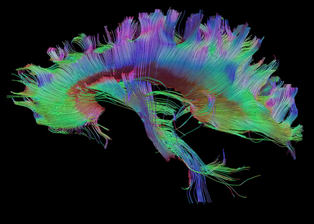
A study published in PLoS ONE has confirmed the role of the corpus callosum in language lateralisation, ie the distribution of language processing functions between the brain’s hemispheres. To get to this finding, the researchers applied advanced neuroimaging methods to study subjects performing an innovative language task for their study.
Functional asymmetry between the two cerebral hemispheres in performing higher-level cognitive functions is a major characteristic of the human brain. For example, the left hemisphere plays a leading role in language processing in most people. However, between 10% and 15% of people also use the right hemisphere to varying degrees for the same task.
Traditionally, language lateralisation to the right hemisphere was explained by handedness, as it is mainly found in left-handed and ambidextrous (using both hands equally well) individuals. But recent research has demonstrated a genetic difference in the way language is processed by left-handed and ambidextrous people. In addition to this, some right-handed people also involve their right hemisphere in language functions.
These findings prompted the scientists to consider alternative explanations, especially by looking at brain anatomy to find out why language functions can shift to the right hemisphere. Researchers at the HSE Centre for Language and Brain hypothesised that language lateralisation may have something to do with the anatomy of the corpus callosum, the largest commissural tract in the human brain connecting the two cerebral hemispheres.
Tractography of the corpus callosumDo Tromp et al. / brainimaging.waisman.wisc.edu
The researchers asked 50 study participants to perform a sentence completion task. The subjects were instructed to read aloud a visually presented Russian sentence and to complete it with an appropriate final word (eg ‘Teper’ ministr podpisyvaet vazhnoe…‘ – ‘Now the minister is signing an important …’). At the same time, the participants’ brain activity was recorded using functional magnetic resonance imaging (fMRI). Additionally, the volume of the corpus callosum was measured in each subject.
A comparison between the fMRI data and the corpus callosum measurements revealed that the larger the latter’s volume, the less lateralisation of the language function to the right hemisphere was observed.
When processing language, the brain tends to use the left hemisphere’s resources efficiently and the corpus callosum suppresses any additional involvement of the right hemisphere. The larger a person’s corpus callosum, the less involved their right hemisphere is in language processing (and vice versa). This finding is consistent with the inhibitory model suggesting that the corpus callosum inhibits the action of one hemisphere while the other is engaged in cognitive tasks.
The study’s innovative design and use of advanced neuroimaging have made this conclusion possible. Brain lateralisation in language processing is usually hard to measure accurately, as typical speech tasks used in earlier studies (eg image naming, selecting words that begin with a certain letter or listening to speech) tend to cause activation only in some parts of the brain responsible for language functions but not in others. Instead, we developed a unique speech task for fMRI: sentence completion, which reliably activates all language areas of the brain.
The researchers reconstructed the volume and properties of the corpus callosum from MRI data using an advanced tractography technique: constrained spherical deconvolution (CSD). This is more suitable than traditional diffusion tensor imaging for modelling crossing fibres in the smallest unit of volume, the voxel (3D pixel), and is therefore more reliable.
Researchers have found that compounds in the edible Lions’ Mane mushroom (Hericium erinaceus), already used in herbal medicine for stomach complaints, can promote nerve growth and boost memory. Their findings are published in The Journal of Neurochemistry.
The compounds are neurotrophins – a family of proteins associated with the growth, functioning and maintenance of neurons. In mammals, brain-derived neurotrophic factor (BDNF) is highly expressed in central nervous system neurons. Because BDNF pathway impairment is associated with several diseases, including schizophrenia, Alzheimer’s disease, Rett syndrome, and Huntington’s disease, experimental treatments for neurological and neurodegenerative disorders often target neutrophins.
So far, such treatments have run into problems with crossing the blood-brain barrier and off-target effects. H. erinaceus is high in neutrophins, though it has traditionally been used in herbal medicine to treat stomach complaints and cancer. It is also known for promoting peripheral nerve regeneration.
Two of the biologically active types of compounds, hericenones and erinacines, can successfully cross the the blood-brain barrier and confer neuroprotective effects.
The researchers tested crude and purified H. erinaceus extracts and found that they exhibited BDNF-like neurotrophic activity in both in vitro cultured hippocampal neurons and in mouse models of hippocampal memory. The extracts also promoted neurite outgrowth and improved memory.
They concluded that Hericene A acts through a novel signalling pathway, giving rise to improved cognitive performance. These findings however will need to be validated by future research.
When using psychedelic drugs to treat mental illness, it’s all down to location when rapidly rebuilding connections between nerve cells. In their paper published in Science, scientists show that engaging serotonin 2A receptors inside neurons promotes growth of new connections – but engaging the same receptor on the surface of nerve cells does not.
The findings will help guide efforts to discover new drugs for depression, PTSD and other disorders, according to senior author David E. Olson, associate professor at the University of California, Davis.
Drugs such as LSD, MDMA and psilocybin show great promise for treating a wide range of mental disorders that are characterised by a loss of neural connections. In laboratory studies, a single dose of these drugs can cause rapid growth of new dendrites from nerve cells, and formation of new spines on those dendrites.
Olson calls this group of drugs “psychoplastogens” because of their ability to regrow and remodel connections in the brain.
Earlier work from Olson’s and other labs showed that psychedelic drugs work by engaging the serotonin 2A receptor (5-HT2AR). But other drugs that engage the same receptor, including serotonin, do not have the same growth effects.
Maxemiliano Vargas, a graduate student in Olson’s lab, Olson and colleagues experimented with chemically tweaking drugs and using transporters to make it easier or harder for compounds to slip across cell membranes. Serotonin itself is polar, meaning it dissolves well in water but does not easily cross the lipid membranes that surround cells. The psychedelics, on the other hand, are much less polar and can easily enter the interior of a cell.
They found that the growth-promoting ability of compounds was correlated with the ability to cross cell membranes.
Drug receptors are usually thought of as being located on the cell membrane, facing out. But the researchers found that in nerve cells, serotonin 2A receptors were concentrated inside cells, mostly around a structure called the Golgi body, with some receptors on the cell surface. Other types of signalling receptors in the same class were on the surface.
The results show that there is a location bias in how these drugs work, Olson said. Engaging the serotonin 2A receptor when it is inside a cell produces a different effect from triggering it when it is on the outside.
“It gives us deeper mechanistic insight into how the receptor promotes plasticity, and allows us to design better drugs,” Olson said.
Most people remember emotional events, like their wedding day, very clearly, but researchers are not sure how the human brain prioritises emotional events in memory. In a study published in Nature Human Behaviour, Joshua Jacobs, associate professor of biomedical engineering at Columbia Engineering, and his team identified a specific neural mechanism in the human brain that tags information with emotional associations for enhanced memory.
The team demonstrated that high-frequency brain waves in the amygdala, a hub for emotional processes, and the hippocampus, a hub for memory processes, are critical to enhancing memory for emotional stimuli. Disruptions to this neural mechanism, brought on either by electrical brain stimulation or depression, impair memory specifically for emotional stimuli.
Rising prevalence of memory disorders
The rising prevalence of memory disorders such as dementia has highlighted the damaging effects that memory loss has on individuals and society. Disorders such as depression, anxiety, and post-traumatic stress disorder (PTSD) can also feature imbalanced memory processes, especially with the COVID pandemic. Understanding how the brain naturally regulates what information gets prioritised for storage and what fades away could provide critical insight for developing new therapeutic approaches to strengthening memory for those at risk of memory loss, or for normalising memory processes in those at risk of dysregulation.
“It’s easier to remember emotional events, like the birth of your child, than other events from around the same time,” says Salman E. Qasim, lead author of the study, who started this project during his PhD in Jacobs’ lab at Columbia Engineering. “The brain clearly has a natural mechanism for strengthening certain memories, and we wanted to identify it.”
The difficulty of studying neural mechanisms in humans
Most investigations into neural mechanisms take place in animals such as rats, because such studies require direct access to the brain to record brain activity and perform experiments that demonstrate causality, such as careful disruption of neural circuits. But it is difficult to observe or characterise a complex cognitive phenomenon like emotional memory enhancement in animal studies.
To study this process directly in humans. Qasim and Jacobs analysed data from memory experiments conducted with epilepsy patients undergoing direct, intracranial brain recording for seizure localisation and treatment. During these recordings, epilepsy patients memorised lists of words while the electrodes placed in their hippocampus and amygdala recorded the brain’s electrical activity.
Studying brain-wave patterns of emotional words
Qasim found that participants remembered more emotionally rated words, such as “dog” or “knife,” better than more neutral words, such as “chair.” Whenever participants successfully remembered emotional words, high-frequency neural activity (30-128 Hz) would become more prevalent in the amygdala-hippocampal circuit, a pattern which was absent when participants remembered more neutral words, or failed to remember a word altogether. Analysing 147 participant, they found a clear link between participants’ enhanced memory for emotional words and the prevalence in their brains of high-frequency brain waves across the amygdala-hippocampal circuit.
“Finding this pattern of brain activity linking emotions and memory was very exciting to us, because prior research has shown how important high-frequency activity in the hippocampus is to non-emotional memory,” said Jacobs. “It immediately cued us to think about the more general, causal implications – if we elicit high-frequency activity in this circuit, using therapeutic interventions, will we be able to strengthen memories at will?”
Electrical stimulation disrupts memory for emotional words
In order to establish whether this high-frequency activity actually reflected a causal mechanism, Jacobs and his team formulated a unique approach to replicate the kind of experimental disruptions typically reserved for animal research. First, they analysed a subset of these patients who had performed the memory task while direct electrical stimulation was applied to the hippocampus for half of the words that participants had to memorise. They found that electrical stimulation, which has a mixed history of either benefiting or diminishing memory depending on its usage, clearly and consistently impaired memory specifically for emotional words.
Uma Mohan, another PhD student in Jacobs’ lab at the time and co-author on the paper, noted that this stimulation also diminished high-frequency activity in the hippocampus. This provided causal evidence that, by knocking out the brain activity pattern correlating with emotional memory, stimulation was also selectively diminishing emotional memory.
Depression acts similarly to brain stimulation
Qasim further hypothesized that depression, which can involve dysregulated emotional memory, might act similarly to brain stimulation. He analyzed patients’ emotional memory in parallel with mood assessments the patients took to characterize their psychiatric state. And, in fact, in the subset of patients with depression, the team observed a concurrent decrease in emotion-mediated memory and high-frequency activity in the hippocampus and amygdala.
“By combining stimulation, recording, and psychometric assessment, they were able to demonstrate causality to a degree that you don’t always see in studies with human brain recordings,” said Bradley Lega, a neurosurgeon and scientist at the University of Texas Southwestern Medical Center and not an author on the paper. “We know high-frequency activity is associated with neuronal firing, so these findings open new avenues of research in humans and animals about how certain stimuli engage neurons in memory circuits.”
Next steps
Qasim is now investigating how individual neurons in the human brain fire during emotional memory processes. Qasim and Jacobs hope that their work might also inspire animal research exploring how this high-frequency activity is linked to norepinephrine, a neurotransmitter linked to attentional processes that they theorise might be behind the enhanced memory for emotional stimuli. They also hope that future research will target high-frequency activity in the amygdala-hippocampal circuit to protect memory.
“Our emotional memories are one of the most critical aspects of the human experience, informing everything from our decisions to our entire personality,” Qasim added. “Any steps we can take to mitigate their loss in memory disorders or prevent their hijacking in psychiatric disorders is hugely exciting.”