Category: Neurology

Neuron Cluster may Create a Little-understood Form of Chronic Pain

Source: Pixabay CC0

Stimulating a small cluster of neurons in the brain appears to create a response in mice that mimics nociplastic pain, a type of unexplained chronic pain, researchers at the University of Washington School of Medicine in Seattle have found. 

“When we stimulate these neurons, the mouse behaves as though gentle touch is very painful, which is one of the characteristics of nociplastic pain,” said Richard Palmiter, a professor of biochemistry and investigator of the Howard Hughes Medical Institute. Dr Logan Condon, who spearheaded this research as a PhD student at UW, was lead author on the paper, which was published in Cell Reports

Chronic pain can arise from ongoing injury or persistent damage to the nervous system. Pain caused by injury is called nociceptive from the Latin nocere “to harm.” Pain due to nerve damage is called neuropathic. But these categories do not explain a common form of chronic pain the persists even after an injury has fully healed and there is no evidence of neurological damage. This led the International Association for the Study of Pain to define a new category called nociplastic pain, meaning “able to be moulded.” 

Although the cause of nociplastic pain is unknown, scientists think it involves changes in pain circuits in the spinal cord and brain. These changes result in the perception of pain even when no nerve injury exists. 

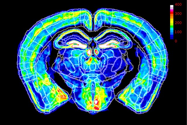
In the new study, researchers demonstrated that stimulating a cluster of cells in the brain’s parabrachial nucleus can generate chronic pain behaviour typical of nociplastic pain. They also showed that inhibiting these cells can prevent pain from nerve injury. 

The parabrachial nucleus is in an area of the brain known as the pons. It acts as a hub that relays aversive sensory information from the body to different parts of the brain. The parabrachial neurons found to create nociplastic pain are called Calca neurons, named for a defining gene for these cells. 

“You can think of these Calca neurons as a warning system for the brain,” said Palmiter. “They respond to any aversive event you can think of – a pinch, a visual threat, a bad odour, a loud noise – and they tell your brain that something bad is happening in the environment and you’d better do something about it.”

It is possible to manipulate genetically defined neurons using viral techniques to express molecules that activate, or inhibit, those neurons. 

The researchers also found that the nociplastic pain behaviour continues even after the Calca-neuron activation has stopped. This suggests signals from the stimulated Calca neurons cause persistent effects – a sign of plasticity – in the nerve circuits leading to the spinal cord. 

They also showed that it was possible to create nociplastic behaviours in the mice by exposing them to unpleasant, aversive experiences like nausea, chemotherapy drugs or migraine-like conditions. 

Palmiter’s team is currently focusing on the neural circuits and plasticity that arises when parabrachial Calca neurons are activated.

“The brain is somehow sending signals to the spinal cord,” he said. “We want to figure out the pathway for those signals.”

Source: University of Washington

The First Half of a Night’s Sleep Resets Brain Connections

…but not the second half

Source: CC0

During a night’s sleep, the brain weakens the new connections between neurons that had been forged while awake – but only during the first half, according to a new study in fish by UCL scientists.

The researchers say that their findings, published in Nature, provide insight into the role of sleep, but still leave an open question around what function the latter half of a night’s sleep serves.

The researchers say the study supports the Synaptic Homeostasis Hypothesis, a key theory on the purpose of sleep which proposes that sleeping acts as a reset for the brain.

Lead author Professor Jason Rihel (UCL Cell & Developmental Biology) said: “When we are awake, the connections between brain cells get stronger and more complex. If this activity were to continue unabated, it would be energetically unsustainable. Too many active connections between brain cells could prevent new connections from being made the following day.

“While the function of sleep remains mysterious, it may be serving as an ‘off-line’ period when those connections can be weakened across the brain, in preparation for us to learn new things the following day.”

For the study, the scientists used optically translucent zebrafish, with genes enabling synapses to be easily imaged. The research team monitored the fish over several sleep-wake cycles.

The researchers found that brain cells gain more connections during waking hours, and then lose them during sleep. They found that this was dependent on how much sleep pressure (need for sleep) the animal had built up before being allowed to rest; if the scientists deprived the fish from sleeping for a few extra hours, the connections continued to increase until the animal was able to sleep.

Professor Rihel added: “If the patterns we observed hold true in humans, our findings suggest that this remodelling of synapses might be less effective during a mid-day nap, when sleep pressure is still low, rather than at night, when we really need the sleep.”

The researchers also found that these rearrangements of connections between neurons mostly happened in the first half of the animal’s nightly sleep. This mirrors the pattern of slow-wave activity, which is part of the sleep cycle that is strongest at the beginning of the night.

First author Dr Anya Suppermpool (UCL Cell & Developmental Biology and UCL Ear Institute) said: “Our findings add weight to the theory that sleep serves to dampen connections within the brain, preparing for more learning and new connections again the next day. But our study doesn’t tell us anything about what happens in the second half of the night. There are other theories around sleep being a time for clearance of waste in the brain, or repair for damaged cells – perhaps other functions kick in for the second half of the night.”

Source: University College London

X-chromosome Inactivation may Reduce Females’ Autism Risk

X-chromosome inactivation varies across different areas of brains. Here, fluorescent imaging data from a mouse reveal where the father’s X chromosome is most active (white) and least active (blue). Credit: Eric Szelenyi

A study using mice published in the journal Cell Reports suggests how chromosome inactivation may protect women from autism disorder inherited from their father’s X chromosome.

Because cells do not need two copies of the X chromosome, the cells inactivate one copy early in embryonic development, a well-studied process known as X chromosome inactivation. As a result of this inactivation, every female is made up of a mix of cells, some have an active X chromosome from her father and others from her mother, a phenomenon known as mosaicism. 

For many years, it has been thought that this was random and would result, on average, in a roughly 50/50 mix of cells, with 50% having an active paternal X chromosome and 50% an active maternal X chromosome.

Now a new study finds that, in the mouse brain at least, this is not the case. Instead, there appears to be a bias in the process that results in the paternal X chromosome being inactivated in 60% of the cells rather than the expected 50%.

When the X-linked mutation that is the most common cause of autism spectrum disorder is inherited from the father, the pattern of X-chromosome inactivation in the brain circuitry of females can prevent the effects of that mutation, the study found.

“This bias may be a way to reduce the risk of harmful mutations, which occur more frequently in male chromosomes,” said corresponding author Eric Szelenyi, acting assistant professor of biological structure at the University of Washington School of Medicine in Seattle.

The X-chromosome is of particular interest because it carries more genes involved in brain development than any other chromosome. Mutations in the chromosome are linked to more than 130 neurodevelopmental disorders, including fragile X syndrome and autism.

In the study, the researchers first determined the ratio of X chromosome inactivation in healthy mice by analyzing roughly 40 million brain cells per mouse. The scientists did this by using high-throughput volumetric imaging and automated counting. This analysis revealed a systematic 60:40 ratio across all possible anatomical regions.

They then examined what would happen if they genetically added a mouse model for fragile X syndrome. This syndrome is the most common form of inherited intellectual and developmental disability in humans.

They first tested the mice for behaviors thought to be analogous to those impaired in people with fragile X syndrome. These tests evaluate such things as their sensorimotor function, spatial memory and tendencies towards anxiety and sociability.

They found that the mice who inherited the mutation on their mother’s X chromosome, which are less likely to be inactivated in the 60:40 ratio, were more likely to exhibit behaviour analogous to fragile X syndrome. They exhibited more signs of anxiety, less sociability, poor performance in spatial learning, and deficits in sensorimotor function. 

But mice that inherited the mutation from one their father’s X chromosomes, which were more likely to be inactivated, did not appear impaired. 

“What was most interesting is that using each animal’s behavioural performance was most accurately predicted by X chromosome inactivation in brain circuits, rather than just looking at the brain as a whole, or single brain regions,” said Szelenyi. “This suggests that having more mutant X-active cells due to maternal inheritance increases overall disease risk, but specific mosaic pattern within brain circuitry ultimately decides which behaviors are impacted the most.”

“This suggests that the 20% difference in mutant X-active cells created by the bias can be protective against X mutations from the father, which occur more commonly,” he said.

The findings may also explain why symptoms of X-linked syndromes, like X-linked autism spectrum disorder, vary more in females than males.

Source: University of Washington

Study Reveals ‘Profound’ Link between Dietary Choices and Brain Health

Photo by Fakurian Design on Unsplash

New research published in Nature has shown that a healthy, balanced diet was linked to superior brain health, cognitive function and mental wellbeing. The study, involving researchers at the University of Warwick, sheds light on how food preferences influence more than just physical health, and also significantly impact brain health.

With the help of machine learning, the researchers analysed a large sample of 181 990 participants from the UK Biobank, comparing their dietary choices against a range of physical evaluations, including cognitive function, blood metabolic biomarkers, brain imaging, and genetics.

The food preferences of each participant were collected via an online questionnaire, which the team categorised into 10 groups (eg, alcohol, fruits and meats).

A balanced diet was associated with better mental health, superior cognitive functions and even higher amounts of grey matter in the brain – linked to intelligence – compared with those with a less varied diet.

The study also highlighted the need for gradual dietary modifications, particularly for individuals accustomed to highly palatable but nutritionally deficient foods. By slowly reducing sugar and fat intake over time, individuals may find themselves naturally gravitating towards healthier food choices.

Genetic factors may also contribute to the association between diet and brain health, the scientists believe, showing how a combination of genetic predispositions and lifestyle choices shape wellbeing.

Lead Author Professor Jianfeng Feng, University of Warwick, emphasised the importance of establishing healthy food preferences early in life. He said: “Developing a healthy balanced diet from an early age is crucial for healthy growth. To foster the development of a healthy balanced diet, both families and schools should offer a diverse range of nutritious meals and cultivate an environment that supports their physical and mental health.”

Source: University of Warwick

How Spinal Cords can ‘Learn’ without Brain Involvement

In this study, spinal cords that associated limb position with an unpleasant experience learned to reposition the limb after only 10 minutes, and retained a memory the next day. Spinal cords that received random unpleasantness did not learn. Credit: RIKEN

Researchers in Japan have discovered the neural circuitry in the spinal cord that allows brain-independent motor learning. This study by Aya Takeoka at the RIKEN Center for Brain Science and colleagues found two critical groups of spinal cord neurons, one necessary for new adaptive learning, and another for recalling adaptations once they have been learned. The findings, published in Science, could help scientists develop ways to assist motor recovery after spinal cord injury.

It has been long been known that motor output from the spinal cord can be adjusted through practice even without a brain. This has been shown most dramatically in headless insects, whose legs can still be trained to avoid external cues. Until now, no one has figured out exactly how this is possible, and without this understanding, the phenomenon is not much more than a quirky fact. As Takeoka explains, “Gaining insights into the underlying mechanism is essential if we want to understand the foundations of movement automaticity in healthy people and use this knowledge to improve recovery after spinal cord injury.”

Before jumping into the neural circuitry, the researchers first developed an experimental setup that allowed them to study mouse spinal cord adaptation, both learning and recall, without input from the brain. Each test had an experimental mouse and a control mouse whose hindlegs dangled freely. If the experimental mouse’s hindleg drooped down too much it was electrically stimulated, emulating something a mouse would want to avoid. The control mouse received the same stimulation at the same time, but not linked to its own hindleg position.

After just 10 minutes, they observed motor learning only in the experimental mice; their legs remained high up, avoiding any electrical stimulation. This result showed that the spinal cord can associate an unpleasant feeling with leg position and adapt its motor output so that the leg avoids the unpleasant feeling, all without any need for a brain. Twenty-four hours later, they repeated the 10-minute test but reversed the experimental and control mice. The original experimental mice still kept their legs up, indicating that the spinal cord retained a memory of the past experience, which interfered with new learning.

Having thus established both immediate learning, as well as memory, in the spinal cord, the team then set out to examine the neural circuitry that makes both possible. They used six types of transgenic mice, each with a different set of spinal neurons disabled, and tested them for motor learning and learning reversal. They found that mice hindlimbs did not adapt to avoid the electrical shocks after neurons toward the top of the spinal cord were disabled, particularly those that express the gene Ptf1a.

When they examined the mice during learning reversal, they found that silencing the Ptf1a-expressing neurons had no effect. Instead, a group of neurons in the ventral part of the spinal cord that express the En1 gene was critical. When these neurons were silenced the day after learning avoidance, the spinal cords acted as if they had never learned anything. The researchers also assessed memory recall on the second day by repeating the initial learning conditions. They found that in wildtype mice, hindlimbs stabilised to reach the avoidance position faster than they did on the first day, indicating recall. Exciting the En1 neurons during recall increased this speed by 80%, indicating enhanced motor recall.

“Not only do these results challenge the prevailing notion that motor learning and memory are solely confined to brain circuits,” says Takeoka, “but we showed that we could manipulate spinal cord motor recall, which has implications for therapies designed to improve recovery after spinal cord damage.”

Source: RIKEN

Spinal Surgeons can Now Monitor their Procedure’s Effects Mid-surgery

Photo by Natanael Melchor on Unsplash

With technology developed at UC Riverside, scientists can, for the first time, make high resolution images of the human spinal cord during surgery. The advancement could help bring real relief to millions suffering chronic back pain.

The technology, known as fUSI or functional ultrasound imaging, not only enables clinicians to see the spinal cord, but also enables them to map the cord’s response to various treatments in real time. A paper published today in the journal Neuron details how fUSI worked for six people undergoing electrical stimulation for chronic back pain treatment.

“The fUSI scanner is freely mobile across various settings and eliminates the requirement for the extensive infrastructure associated with classical neuroimaging techniques, such as functional magnetic resonance imaging (fMRI),” said Vasileios Christopoulos, assistant professor of bioengineering at UCR who helped develop the technology. “Additionally, it offers ten times the sensitivity for detecting neuroactivation compared to fMRI.”

Until now, it has been difficult to evaluate whether a back pain treatment is working since patients are under general anaesthesia, unable provide verbal feedback on their pain levels during treatment. “With ultrasound, we can monitor blood flow changes in the spinal cord induced by the electrical stimulation. This can be an indication that the treatment is working,” Christopoulos said.

The spinal cord is an “unfriendly area” for traditional imaging techniques due to significant motion artifacts, such as heart pulsation and breathing. “These movements introduce unwanted noise into the signal, making the spinal cord an unfavorable target for traditional neuroimaging techniques,” Christopoulos said.

By contrast, fUSI is less sensitive to motion artifacts, using echoes from red blood cells in the area of interest to generate a clear image. “It’s like submarine sonar, which uses sound to navigate and detect objects underwater,” Christopoulos said. “Based on the strength and speed of the echo, they can learn a lot about the objects nearby.”

Christopoulos partnered with the USC Neurorestoration Center at Keck Hospital to test the technology on six patients with chronic low back pain. These patients were already scheduled for the last-ditch pain surgery, as no other treatments, including drugs, had helped to ease their suffering. For this surgery, clinicians stimulated the spinal cord with electrodes, in the hopes that the voltage would alleviate the patient’s discomfort and improve their quality of life.

“If you bump your hand, instinctively, you rub it. Rubbing increases blood flow, stimulates sensory nerves, and sends a signal to your brain that masks the pain,” Christopoulos said. “We believe spinal cord stimulation may work the same way, but we needed a way to view the activation of the spinal cord induced by the stimulation.”

The Neuron paper details how fUSI can detect blood flow changes at unprecedented levels of less than 1mm/s. For comparison, fMRI is only able to detect changes of 2cm/s.

“We have big arteries and smaller branches, the capillaries. They are extremely thin, penetrating your brain and spinal cord, and bringing oxygen places so they can survive,” Christopoulos said. “With fUSI, we can measure these tiny but critical changes in blood flow.”

Generally, this type of surgery has a 50% success rate, which Christopoulos hopes will be dramatically increased with improved monitoring of the blood flow changes. “We needed to know how fast the blood is flowing, how strong, and how long it takes for blood flow to get back to baseline after spinal stimulation. Now, we will have these answers,” Christopoulos said.

Moving forward, the researchers are also hoping to show that fUSI can help optimise treatments for patients who have lost bladder control due to spinal cord injury or age. “We may be able to modulate the spinal cord neurons to improve bladder control,” Christopoulos said.

“With less risk of damage than older methods, fUSI will enable more effective pain treatments that are optimised for individual patients,” Christopoulos said. “It is a very exciting development.”

Source: University of California Riverside

Essential Tremor Increases Cognitive Impairment Risks over Time

Photo by Matthias Zomer on Pexels

Essential tremor, a nervous system disorder that causes rhythmic shaking, is one of the most common movement disorders. A new study published in the Annals of Neurology reveals details on the increased risk of mild cognitive impairment (MCI) and dementia that individuals with essential tremor may face.

The research represents the longest available longitudinal prospective study of rates of MCI and dementia in people with essential tremor. The study enrolled 222 patients, 177 of whom participated in periodic evaluations over an average follow-up of 5 years.

Investigators observed a cumulative prevalence of 26.6% and 18.5% for MCI and dementia, respectively. They also noted a cumulative incidence of 18.2% and 11.2% for MCI and dementia, respectively. Each year, 3.9% of patients with normal cognition “converted” to having MCI, and 12.2% of those with MCI “converted” to having dementia.

“We know from related research that the presence of cognitive impairment in patients with essential tremor has meaningful clinical consequences. For example, patients with essential tremor who are diagnosed with dementia are more likely to need to use a walker or wheelchair, to employ a home health aide, and to reside in non-independent living arrangements than are patients with essential tremor without dementia,” said corresponding author Elan D. Louis, MD, MS, of the University of Texas Southwestern Medical Center. “With this in mind, the findings of the present study highlight the importance of cognitive screening and monitoring in patients with essential tremor. Early detection of impairment may provide opportunities for interventions that may slow further cognitive decline and improve the quality of life of patients and their families.”

Source: Wiley

A Single Gene Variant that Gave Rise to Humans’ Unique Skull Base

Source: CC0

One of the unique features that Homo sapiens have compared with other closely related hominin species and primates is the shape of the base of the skull, which enabled larger brains to evolve. Now, in a study recently published in the American Journal of Human Genetics, a team from Tokyo Medical and Dental University (TMDU), the University of Helsinki, and the University of Barcelona has analysed a genomic variant responsible for this unique human skull base morphology.

Most of the genomic changes that occurred during human evolution did not occur directly to genes themselves, but in regions responsible for controlling and regulating the expression of genes. Variants in these same regions are often involved in genetic conditions, causing aberrant gene expression throughout development. Identifying and characterising such genomic changes is therefore crucial for understanding human development and disease.

The development of the basicranial region, the base of the skull where it joins the vertebra, was key in the evolution of Homo sapiens, as we developed a highly flexed skull base that allowed our increased brain size. Therefore, variants that affect the development of this region are likely to have been highly significant in our evolution.

First, the team searched for variants in just a single letter of the DNA code, called single nucleotide polymorphisms (SNPs), that caused different regulation of genes in the basicranial region in Homo sapiens compared with other extinct hominins. One of these SNPs stood out, located in a gene called TBX1.

They then used cell lines to show that the SNP, called “rs41298798,” is located in a region that regulates the expression levels of the TBX1 gene, and that the “ancestral” form of the SNP, found in extinct hominins, is associated with lower TBX1 expression, while the form found in Homo sapiens gives us higher levels of TBX1.

“We then employed a mouse model with lower TBX1 expression,” explains lead author Noriko Funato, “which resulted in distinct alterations to the morphology at the base of the skull and premature hardening of a cartilage joint where the bones fuse together, restricting the growth ability of the skull.” The changes in the Tbx1-knockout mice were reminiscent of the known basicranial morphology of Neanderthals.

These morphological changes are also reflected in human genetic conditions associated with lower TBX1 gene dosage, such as DiGeorge syndrome and velocardiofacial syndrome, further indicating the significance of this genetic variant in the evolution of our unique skull base morphology.

The identification of this genomic variant sheds light on human evolution, as well as providing insight into common genetic conditions associated with lower expression of the TBX1 gene, paving the way for greater understanding and management of these conditions.

Source: Tokyo Medical and Dental University

How the Brain’s Working Memory… Works

Photo by Alex Green on Unsplash

Cedars-Sinai investigators have discovered how brain cells responsible for working memory – which holds onto things like phone numbers while we use them – coordinate intentional focus and short-term storage of information. Their discovery, which confirms the involvement of the hippocampus, is published in the journal Nature.

“We have identified for the first time a group of neurons, influenced by two types of brain waves, that coordinate cognitive control and the storage of sensory information in working memory,” said Jonathan Daume, PhD, a postdoctoral scholar in the Rutishauser Lab at Cedars-Sinai and first author of the study. “These neurons don’t contain or store information, but are crucial to the storage of short-term memories.”

Working memory, which requires the brain to store information for only seconds, is fragile and requires continued focus to be maintained, explained senior study author Ueli Rutishauser, PhD, director of the Center for Neural Science and Medicine at Cedars-Sinai. It can be affected by different diseases and conditions.

“In disorders such as Alzheimer’s disease or attention-deficit hyperactivity disorder, it is often not memory storage, but rather the ability to focus on and retain a memory once it is formed that is the problem,” said Rutishauser, who is a professor of Neurosurgery, Neurology and Biomedical Sciences at Cedars-Sinai. “We believe that understanding the control aspect of working memory will be fundamental for developing new treatments for these and other neurological conditions.”

To explore how working memory functions, investigators recorded the brain activity of 36 hospitalised patients who had electrodes surgically implanted in their brains as part of an epilepsy diagnosis procedure. The team recorded the activity of individual brain cells and brain waves while the patients performed a task that required use of working memory.

On a computer screen, patients were shown either a single photo or a series of three photos of various people, animals, objects or landscapes. Next, the screen went blank for just under three seconds, requiring patients to remember the photos they just saw. They were then shown another photo and asked to decide whether it was the one (or one of the three) they had seen before.

When patients performing the working memory task were able to respond quickly and accurately, investigators noted the firing of two groups of neurons: “category” neurons that fire in response to one of the categories shown in the photos, such as animals, and “phase-amplitude coupling,” or PAC, neurons.

PAC neurons, newly identified in this study, don’t hold any content, but use a process called phase-amplitude coupling to ensure the category neurons focus and store the content they have acquired. PAC neurons fire in time with the brain’s theta waves, which are associated with focus and control, as well as to gamma waves, which are linked to information processing. This allows them to coordinate their activity with category neurons, which also fire in time to the brain’s gamma waves, enhancing patients’ ability to recall information stored in working memory.

“Imagine when the patient sees a photo of a dog, their category neurons start firing ‘dog, dog, dog’ while the PAC neurons are firing ‘focus/remember,'” Rutishauser said. “Through phase-amplitude coupling, the two groups of neurons create a harmony superimposing their messages, resulting in ‘remember dog.’ It is a situation where the whole is greater than the sum of its parts, like hearing the musicians in an orchestra play together. The conductor, much like the PAC neurons, coordinates the various players to act in harmony.”

PAC neurons do this work in the hippocampus, a part of the brain that has long been known to be important for long-term memory. This study offers the first confirmation that the hippocampus also plays a role in controlling working memory, Rutishauser said.

Source: Cedars-Sinai Medical Center

Do More Mentally Challenging Jobs Protect against Cognitive Decline?

Source: Unsplash CC0

The harder your brain works at your job, the less likely you may be to have memory and thinking problems later in life, according to a new study published in Neurology®, the medical journal of the American Academy of Neurology. This study does not prove that stimulating work prevents mild cognitive impairment. It only shows an association.

“We examined the demands of various jobs and found that cognitive stimulation at work during different stages in life – during your 30s, 40s, 50s and 60s – was linked to a reduced risk of mild cognitive impairment after the age of 70,” said study author Trine Holt Edwin, MD, PhD, of Oslo University Hospital in Norway.

“Our findings highlight the value of having a job that requires more complex thinking as a way to possibly maintain memory and thinking in old age.”

The study looked at 7000 people and 305 occupations in Norway. Researchers measured the degree of cognitive stimulation that participants experienced while on the job. They measured the degree of routine manual, routine cognitive, non-routine analytical, and non-routine interpersonal tasks, which are skill sets that different jobs demand.

Routine manual tasks demand speed, control over equipment, and often involve repetitive motions, typical of factory work. Routine cognitive tasks demand precision and accuracy of repetitive tasks, such as in bookkeeping and filing.

Non-routine analytical tasks involve analysing information, engaging in creative thinking and interpreting information for others. Non-routine interpersonal tasks include establishing and maintaining personal relationships, motivating others and coaching. Non-routine cognitive jobs include public relations and computer programming.

Researchers divided participants into four groups based on the degree of cognitive stimulation that they experienced in their jobs. The most common job for the group with the highest cognitive demands was teaching. The most common jobs for the group with the lowest cognitive demands were mail carriers and custodians.

After age 70, participants completed memory and thinking tests to assess whether they had mild cognitive impairment. Of those with the lowest cognitive demands, 42% were diagnosed with mild cognitive impairment, compared to 27% for those with the highest cognitive demands.

After adjustment for age, sex, education, income and lifestyle factors, the group with the lowest cognitive demands at work had a 66% higher risk of mild cognitive impairment compared to the group with the highest cognitive demands at work.

“These results indicate that both education and doing work that challenges your brain during your career play a crucial role in lowering the risk of cognitive impairment later in life,” Edwin said. “Further research is required to pinpoint the specific cognitively challenging occupational tasks that are most beneficial for maintaining thinking and memory skills.”

A limitation of the study was that even within identical job titles, individuals might perform different tasks and experience different cognitive demands.

Source: American Academy of Neurology