Category: Neurology

New Insights into Sleep Uncover Mechanism for Enhancing Cognitive Function

Photo by Cottonbro on Pexels

While it’s well known that sleep enhances cognitive performance, the underlying neural mechanisms, particularly those related to nonrapid eye movement (NREM) sleep, remain largely unexplored. A new study by a team of researchers coordinated by Rice University’s Valentin Dragoi, has nonetheless uncovered a key mechanism by which sleep enhances neuronal and behavioural performance, potentially changing our fundamental understanding of how sleep boosts brainpower.

The research, published in Science, reveals how NREM sleep – such as in a nap – fosters brain synchronisation and enhances information encoding, shedding new light on this sleep stage. The researchers replicated these effects through invasive stimulation, suggesting promising possibilities for future neuromodulation therapies in humans. The implications of this discovery potentially pave the way for innovative treatments for sleep disorders and even methods to enhance cognitive and behavioural performance.

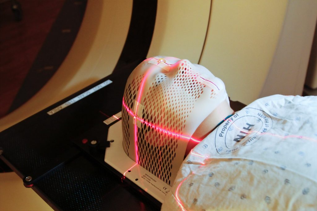
The investigation involved an examination of the neural activity in multiple brain areas in macaques while the animals performed a visual discrimination task before and after a 30-minute period of NREM sleep. Using multielectrode arrays, the researchers recorded the activity of thousands of neurons across three brain areas: the primary and midlevel visual cortices and the dorsolateral prefrontal cortex, which are associated with visual processing and executive functions. To confirm that the macaques were in NREM sleep, researchers used polysomnography to monitor their brain and muscle activity alongside video analysis to ensure their eyes were closed and their bodies relaxed.

The findings demonstrated that sleep improved the animals’ performance in the visual task with enhanced accuracy in distinguishing rotated images. Meanwhile, the macaques that experienced quiet wakefulness without falling asleep did not show the same performance boost.

“During sleep, we observed an increase in low-frequency delta wave activity and synchronised firing among neurons across different cortical regions,” said first author Dr Natasha Kharas. “After sleep, however, neuronal activity became more desynchronised compared to before sleep, allowing neurons to fire more independently. This shift led to improved accuracy in information processing and performance in the visual tasks.”

The researchers also simulated the neural effects of sleep through low-frequency electrical stimulation of the visual cortex. They applied a 4-Hz stimulation to mimic the delta frequency observed during NREM sleep while the animals were awake. This artificial stimulation reproduced the desynchronization effect seen after sleep and similarly enhanced the animals’ task performance, suggesting that specific patterns of electrical stimulation could potentially be used to emulate the cognitive benefits of sleep.

“This finding is significant because it suggests that some of the restorative and performance-enhancing effects of sleep might be achieved without the need for actual sleep,” said Dragoi, study co-author, professor of electrical and computer engineering at Rice and professor of neuroscience at Weill Cornell. “The ability to reproduce sleeplike neural desynchronisation in an awake state opens new possibilities for enhancing cognitive and perceptual performance in situations where sleep is not feasible – such as for individuals with sleep disorders or in extenuating circumstances such as space exploration.”

The researchers further investigated their findings by building a large neural network model. They found that during sleep, both excitatory and inhibitory connections in the brain become weaker, but they do so asymmetrically, making inhibitory connections weaker than excitatory connections, which causes an increase in excitation.

“We have uncovered a surprising solution that the brain employs after sleep whereby neural populations participating in the task reduce their level of synchrony after sleep despite receiving synchronizing inputs during sleep itself,” Dragoi said.

The idea that NREM sleep effectively “boosts” the brain in this way, and that this resetting can be mimicked artificially, offers potential for developing therapeutic brain stimulation techniques to improve cognitive function and memory.

“Our study not only deepens our mechanistic understanding of sleep’s role in cognitive function but also breaks new ground by showing that specific patterns of brain stimulation could substitute for some benefits of sleep, pointing toward a future where we might boost brain function independently of sleep itself,” Dragoi said.

Source: Rice University

Cannabis Disrupts Brain Activity in Young Adults Prone to Psychosis

Photo by Elsa Olofsson on Unsplash

Young adults at risk of psychosis show reduced brain connectivity, a deficit that cannabis use appears to worsen, a new study has found. The breakthrough paves the way for psychosis treatments targeting symptoms that current medications miss. In the first-of-its-kind study, McGill University researchers detected a marked decrease in synaptic density in individuals at risk of psychosis, compared to a healthy control group.

“Not every cannabis user will develop psychosis, but for some, the risks are high. Our research helps clarify why,” said Dr Romina Mizrahi, senior author of the study and professor in McGill’s Department of Psychiatry.

“Cannabis appears to disrupt the brain’s natural process of refining and pruning synapses, which is essential for healthy brain development.”

Hope for new treatments

Using advanced brain scanning technology, the team studied 49 participants aged 16 to 30, including individuals with recent psychotic symptoms and those considered at high risk. The results, published in JAMA Psychiatry, indicate that lower synaptic density is linked to social withdrawal and lack of motivation, symptoms the researchers say are difficult to treat.

“Current medications largely target hallucinations, but they don’t address symptoms that make it difficult to manage social relationships, work, or school,” said first author Belen Blasco, a PhD student at McGill’s Integrated Program in Neuroscience. “By focusing on synaptic density, we may eventually develop therapies that enhance social function and quality of life for those affected.”

While cannabis is a known risk factor for developing psychosis, which can progress to schizophrenia, this is the first time researchers have measured structural changes in the brains of a high-risk population in real time.

The team’s next research phase will explore whether these observed brain changes could predict psychosis development, potentially enabling earlier intervention.

Source: McGill University

Workouts – or Disturbed Sleep – Impact Brain Activity Weeks Later

Photo by Ketut Subiyanto on Pexels

In a rare, longitudinal study, researchers from Aalto University and the University of Oulu tracked one person’s brain and behavioural activity for five months using brain scans and data from wearable devices and smartphones. The results appear in PLOS Biology.

“We wanted to go beyond isolated events,” says research leader (and study participant) Ana Triana. “Our behaviour and mental states are constantly shaped by our environment and experiences. Yet, we know little about the response of brain functional connectivity to environmental, physiological, and behavioural changes on different timescales, from days to months.”

The study found that the brain does not respond to daily life in immediate, isolated bursts. Instead, brain activity evolves in response to sleep patterns, physical activity, mood, and respiration rate over many days. This suggests that even a workout or a restless night from last week could still affect the brain – and therefore attention, cognition and memory – well into next week.

The research also revealed a strong link between heart rate variability – a measure of the heart’s adaptability – and brain connectivity, particularly during rest. This suggests that impacts on the body’s relaxation response, like stress management techniques, could shape brain wiring even when not actively concentrating on a task. Physical activity was also found to positively influence the way brain regions interact, potentially impacting memory and cognitive flexibility. Even subtle shifts in mood and heart rate left lasting imprints for up to 15 days.

Study goes beyond a snapshot

The research is unusual in that few brain studies involve detailed monitoring over days and weeks. “The use of wearable technology was crucial,” says Triana. “Brain scans are useful tools, but a snapshot of someone lying still for half an hour can only show so much. Our brains do not work in isolation.”

Triana was herself the subject of the research, monitored as she went about her daily life. Her unique role as both lead author and study participant added complexity, but also brought firsthand insights into how best to maintain research integrity over several months of personalised data collection.  Data from the devices and twice-weekly brain scans were complemented by qualitative data from mood surveys. 

The researchers identified two distinct response patterns: a short-term wave lasting under seven days and a long-term wave up to 15 days. The former reflects rapid adaptations, like how focus is impacted by poor sleep, but it recovers quickly. The long wave suggests more gradual, lasting effects, particularly in areas tied to attention and memory. 

Single-subject studies offer opportunities for improving mental health care 

The researchers hope their innovative approach will inspire future studies that combine brain data with everyday life to help personalise mental health treatment. 

“We must bring data from daily life into the lab to see the full picture of how our habits shape the brain, but surveys can be tiring and inaccurate,” says study co-author, neuroscientist and physician Dr Nick Hayward. “Combining concurrent physiology with repeated brain scans in one person is crucial. Our approach gives context to neuroscience and delivers very fine detail to our understanding of the brain.”

The study is also a proof-of-concept for patient research. Tracking brain changes in real time could help detect neurological disorders early, especially mental health conditions where subtle signs might be missed.

“Linking brain activity with physiological and environmental data could revolutionise personalised healthcare, opening doors for earlier interventions and better outcomes,” says Triana.

Source: Aalto University

How Human Brain Functional Networks Emerge and Develop during the Birth Transition

Shedding light on the growth trajectory of global functional neural networks before and after birth

Photo by Christian Bowen on Unsplash

Brain-imaging data collected from foetuses and infants has revealed a rapid surge in functional connectivity between brain regions on a global scale at birth, possibly reflecting neural processes that support the brain’s ability to adapt to the external world, according to a study published November 19th, in the open-access journal PLOS Biology led by Lanxin Ji and Moriah Thomason from the New York University School of Medicine, USA.

Understanding the sequence and timing of brain functional network development at the beginning of human life is critical. Yet many questions remain regarding how human brain functional networks emerge and develop during the birth transition. To fill this knowledge gap, Thomason and colleagues leveraged a large functional magnetic resonance imaging dataset to model developmental trajectories of brain functional networks spanning 25 to 55 weeks of post-conceptual gestational age. The final sample included 126 foetal scans and 58 infant scans from 140 subjects.

The researchers observed distinct growth patterns in different regions, showing that neural changes accompanying the birth transition are not uniform across the brain. Some areas exhibited minimal changes in resting-state functional connectivity (RSFC) – correlations between blood oxygen level-dependent signals between brain regions when no explicit task is being performed. But other areas showed dramatic changes in RSFC at birth. The subcortical network, sensorimotor network, and superior frontal network stand out as regions that undergo rapid reorganisation during this developmental stage.

Additional analysis highlighted the subcortical network as the only region that exhibited a significant increase in communication efficiency within neighbouring nodes. The subcortical network represents a central hub, relaying nearly all incoming and outgoing information to and from the cortex and mediating communication between cortical areas. On the other hand, there was a gradual increase in global efficiency in sensorimotor and parietal-frontal regions throughout the foetal to neonatal period, possibly reflecting the establishment or strengthening of connections as well as the elimination of redundant connections.

According to the authors, this work unveils fundamental aspects of early brain development and lays the foundation for future research on the influence of environmental factors on this process. In particular, further studies could reveal how factors such as sex, prematurity, and prenatal adversity interact with the timing and growth patterns of children’s brain network development.

The authors add, “This study for the first time documents the significant change of brain functional networks over the birth transition. We observe that growth patterns are regionally specific, with some areas of the functional connectome showing minimal changes, while others exhibit a dramatic increase at birth.”

Provided by PLOS

New Minimally Invasive Neural Interface is a Revolutionary Development

The researchers’ experiments showed that the catheter electrodes could be successfully delivered and guided into the ventricular spaces and brain surface for electrical stimulation. Image courtesy of Rice University.

A team of researchers has developed a technique for diagnosing, managing and treating neurological disorders with minimal surgical risks. The team’s findings were published in Nature Biomedical Engineering.

While traditional approaches for interfacing with the nervous system often require creating a hole in the skull to interface with the brain, the researchers have developed an innovative method known as endocisternal interfaces (ECI), allowing for electrical recording and stimulation of neural structures, including the brain and spinal cord, through cerebral spinal fluid (CSF).

“Using ECI, we can access multiple brain and spinal cord structures simultaneously without ever opening up the skull, reducing the risk of complications associated with traditional surgical techniques,” said study leader Robinson Jacob Robinson, professor of electrical and computer engineering and bioengineering at Rice University.

ECI uses CSF, which surrounds the nervous system, as a pathway to deliver targeted devices. By performing a simple lumbar puncture in the lower back, researchers can navigate a flexible catheter to access the brain and spinal cord.

Using miniature magnetoelectric-powered bioelectronics, the entire wireless system can be deployed through a small percutaneous procedure. The flexible catheter electrodes can be navigated freely from the spinal subarachnoid space to the brain ventricles.

“This is the first reported technique that enables a neural interface to simultaneously access the brain and spinal cord through a simple and minimally invasive lumbar puncture,” said University of Texas Medical Branch’s Peter Kan, professor and Chair of Neurosurgery, who also led the study. “It introduces new possibilities for therapies in stroke rehabilitation, epilepsy monitoring and other neurological applications.”

To test the hypothesis, the research team characterised the endocisternal space and measured the width of the subarachnoid, or fluid-filled space, in human patients using MRI. The researchers then conducted experiments in large animal models, specifically sheep, to validate the feasibility of the new neural interface.

Their experiments showed that the catheter electrodes could be successfully delivered and guided into the ventricular spaces and brain surface for electrical stimulation. By using the magnetoelectric implant, the researchers were able to record electrophysiologic signals such as muscle activation and spinal cord potentials.

Preliminary safety results showed that the ECI remained functional with minimal damage up to 30 days after the electronic device was implanted chronically into the brain.

Moreover, the study revealed that unlike endovascular neural interfaces that require antithrombotic medication and are limited by the small size and location of blood vessels, ECI offers broader access to neural targets without the medication.

“This technology creates a new paradigm for minimally invasive neural interfaces and could lower the risk of implantable neurotechnologies, enabling access to wider patient populations,” said Josh Chen, lead author of the study.

Source: Rice University

A Multiple Sclerosis Drug may Help with Poor Working Memory

This is a pseudo-coloured image of high-resolution gradient-echo MRI scan of a fixed cerebral hemisphere from a person with multiple sclerosis. Credit: Govind Bhagavatheeshwaran, Daniel Reich, National Institute of Neurological Disorders and Stroke, National Institutes of Health

Fampridine is currently used to improve walking ability in multiple sclerosis. A new study shows that it could also help individuals with reduced working memory, as seen in mental health conditions like schizophrenia or depression.

Working memory allows a memory to be actively retained for a few seconds, for cognitive tasks such as remembering an email address to save it, or participating in a conversation. Certain conditions, such as schizophrenia or depression, as well as ADHD, impair working memory. Those affected lose track in conversations and struggle to organise their thoughts.

Fampridine is a drug that could help in such cases, as shown in a study led by Professors Andreas Papassotiropoulos and Dominique de Quervain at the University of Basel. The team has reported their findings in the journal Molecular Psychiatry.

Effective only if working memory is poor

In their study, the researchers tested the effectiveness of fampridine on working memory in 43 healthy adults. It was in those participants whose baseline working memory was at a low level that fampridine showed a more pronounced effect: after taking the active substance for three days, they scored better in the relevant tests than those who took the placebo. In contrast, in people who already had good baseline working memory, the drug showed no effect.

The researchers also observed that fampridine increased brain excitability in all participants, thus enabling faster processing of stimuli. The study was randomized and double-blind.

Established drug, new application

“Fampridine doesn’t improve working memory in everyone. But it could be a treatment option for those with reduced working memory,” explains Andreas Papassotiropoulos. Dominique de Quervain adds: “That’s why, together with researchers from the University Psychiatric Clinics Basel (UPK), we’re planning studies to test the efficacy of fampridine in schizophrenia and depression.”

The drug is currently used to improve walking ability in multiple sclerosis (MS). Particularly in capsule form, which releases the active ingredient slowly in the body, fampridine has shown effects on cognitive performance in MS patients: for some, it alleviates the mental fatigue that can accompany MS.

The researchers did not select the drug at random: this study followed comprehensive analyses of genome data in order to find starting points for repurposing established drugs. Fampridine acts on specific ion channels in nerve cells that, according to the researchers’ analyses, also play a role in mental disorders such as schizophrenia.

Source: University of Basel

Early Radiotherapy Treatment of Vestibular Schwannomas Prevents Problems

Photo by National Cancer Institute on Unsplash

Patients with a small cranial nerve tumour that can cause hearing loss, vertigo, imbalance and ringing in the ears have typically been watched rather than proactively treated, as the risks of early intervention were thought to outweigh the benefits. Now a study shows that even those patients benefit significantly from non-invasive stereotactic radiosurgery, led by UVA Health physicians has found. The findings were reported in Neurosurgery.

Doctors typically treat larger forms of the tumours, called vestibular schwannomas, while taking a “watch and wait” approach to smaller tumours that aren’t causing appreciable problems. But the new research, from UVA Health neurosurgeon Jason Sheehan, MD, PhD, and collaborators, could change how asymptomatic schwannomas are managed. Their findings demonstrated that stereotactic radiosurgery – a highly targeted form of radiation therapy – can prevent small tumours from growing over time while at the same time sparing patients from potentially irreversible problems in the future.

“This study and our recent Vestibular Schwannoma International Study of Active Surveillance versus Stereotactic Radiosurgery [VISAS] trial demonstrate that radiosurgery affords effective and durable tumour control while more often avoiding the neurological complications that come from watching a vestibular schwannoma,” Sheehan said. “Over time, Gamma Knife radiosurgery bends the curve of growth and problems that commonly arise from watching even the smallest of vestibular schwannomas.”

About vestibular schwannomas

Vestibular schwannomas are growths on a cranial nerve that connects the brain and inner ear. This nerve transmits information about head movements, helps us control our balance and allows us to hear. The growths, however, can disrupt the nerve’s important functions, causing hearing loss, unsteadiness, headaches, tinnitus (ringing in the ear), facial numbness/paralysis and other problems. 

Seeking to improve care for patients with these tumours, Sheehan and his team performed a trial through the International Radiosurgery Research Foundation looking at 261 adults with the smallest category of vestibular schwannomas. These were usually picked up early, and the patients often were high functioning and had the most to lose from tumour growth over time. Of the study participants, 182 received stereotactic radiosurgery, while 79 did not. 

The patients who underwent radiosurgery using the Gamma Knife system showed consistently better tumour control over time. In this group, 99% of the patients’ tumours either stayed the same size, grew very little (less than 25%) or shrank. This was true at 3 years, 5 years, and 8 years. Only one patient’s tumour significantly increased in size.

Tumour control was much worse among those who didn’t receive radiosurgery: 37% saw their tumours grow significantly at 3 years, 50% at five years, and 67% at eight years.

That difference was seen plainly in the symptoms the patients experienced. Radiosurgery was associated with a 54% lower rate of tinnitus, a 51% lower rate of cranial nerve deterioration and an 83% lower rate of vestibular dysfunction that causes dizziness and loss of balance. 

Even with Gamma Knife radiosurgery to treat the tumour arising from this very delicate neural structure, hearing was preserved similarly in both groups.

Sheehan, an expert in stereotactic radiosurgery and brain tumours, urges physicians to take note of the findings because tumour symptoms are often irreversible as the tumour grows. Acting early, before symptoms develop, could greatly improve patients’ long-term quality of life, he says.

“In brain surgery, particularly involving the hearing and balance nerve, our approach must be exceedingly refined,” he said. “This study shows that Gamma Knife radiosurgery substantially improves the future trajectory of vestibular schwannoma patients.”

Source: University of Virginia Health System

A Way to Make Glioblastoma Cells Visible to Immune Cells

MRI scan showing brain cancer. Credit: Michelle Monje, MD, PhD, Stanford University

Patients with glioblastoma typically survive less than two years after diagnosis, even with cutting-edge therapies. The latest immunotherapies have been unsuccessful, likely because glioblastoma cells have few, if any, natural targets for the immune system to attack.

In a cell-based study, scientists at Washington University School of Medicine have forced glioblastoma cells to display immune system targets, potentially making them visible to immune cells and newly vulnerable to immunotherapies. The strategy involves a combination of two drugs, each already FDA-approved to treat different cancers.

The study is online in the journal Nature Genetics.

“For patients whose tumours do not naturally produce targets for immunotherapy, we showed there is a way to induce their generation,” said co-senior author Ting Wang, PhD, professor of medicine and Department of Genetics head at WashU Medicine. “In other words, when there is no target, we can create one. This is a very new way of designing targeted and precision therapies for cancer. We are hopeful that in the near future we will be able to move into clinical trials, where immunotherapy can be combined with this strategy to provide new therapeutic approaches for patients with very hard-to-treat cancers.”

To create immune targets on cancer cells, Wang has focused on stretches of DNA in the genome known as transposable elements. In recent years, transposable elements have emerged as a double-edged sword in cancer, according to Wang. His work has shown that transposable elements play a role in causing tumours to develop even as they present vulnerabilities that could be exploited to create new cancer treatment strategies.

For this study, Wang’s team took advantage of the fact that transposable elements naturally can cause a tumour to churn out random proteins that are unique to the tumour and not present in normal cells. Called tumour antigens or neoantigens, these unusual proteins could be the targets for immunotherapies, such as checkpoint inhibitors, antibodies, vaccines and genetically engineered T cell therapies.

Even so, some tumours, including glioblastoma, have few immune targets produced naturally by transposable elements. To address this, Wang and his colleagues, including co-senior author Albert H. Kim, MD, PhD, neurosurgery professor, have demonstrated how to purposely force transposable elements to produce immune system targets on glioblastoma cells that normally lack them.

The researchers used a combination of two drugs that influence the epigenome, which controls gene activation. When treated with the two epigenetic therapy drugs, the tightly packed DNA molecules of the glioblastoma cells unfurled, triggering transposable elements to begin making the unusual proteins that could be used to target the cancer cells. The two drugs were decitabine, which is approved to treat myelodysplastic syndromes, a group of blood cancers; and panobinostat, which is approved for multiple myeloma, a cancer of white blood cells.

Before investigating this strategy in people, the researchers are seeking ways to target the epigenetic therapy so that only the tumour cells are induced to make neoantigens. In the new study, the researchers cautioned that normal cells also produced targets when exposed to the two drugs. Even though normal cells didn’t produce as many neoantigens as the glioblastoma cells did, Wang and Kim said there is a risk of unwanted side effects if normal cells create these targets as well.

In ongoing work, Wang and Kim are investigating how to use CRISPR molecular editing technology to induce specific parts of the genome in cancer cells to produce the same neoantigens from transposable elements that are common across the human population. Such a strategy could give many patients’ tumours – even different cancer types – the same targets that could respond to the same immunotherapy, while sparing healthy cells. There are then multiple possible ways to go after such a shared target, including checkpoint inhibitors, vaccines, engineered antibodies and engineered T cells.

Source: Washington University School of Medicine

Bilateral Magnetic Stimulation of the Brain Improves Symptoms of Depression

By BaburovOwn work, CC BY-SA 4.0, Link

A type of therapy that involves applying a magnetic field to both sides of the brain has been shown to be effective at rapidly treating depression in patients for whom standard treatments have been ineffective.

Our accelerated approach means we can do all of the sessions in just five days, rapidly reducing an individual’s symptoms of depression

Valerie Voon

The treatment – known as repetitive transcranial magnetic stimulation (TMS) – involves placing an electromagnetic coil against the scalp to relay a high-frequency magnetic field to the brain.

Around one in 20 adults is estimated to suffer from depression. Although treatments exist, such as anti-depressant medication and cognitive behavioural therapy (‘talking therapy’), they are ineffective for just under one in three patients.

One of the key characteristics of depression is under-activity of some regions (such as the dorsolateral prefrontal cortex) and over-activity of others (such as the orbitofrontal cortex (OFC)).

Repetitive transcranial magnetic stimulation applied to the left side of the dorsolateral prefrontal cortex (an area at the upper front area of the brain) is approved for treatment of depression in the UK by NICE and in the US by the FDA. It has previously been shown to lead to considerable improvements among patients after a course of 20 sessions, but because the sessions usually take place over 20-30 days, the treatment is not ideal for everyone, particularly in acute cases or where a person is suicidal.

In research published in Psychological Medicine, scientists from Cambridge, UK, and Guiyang, China, tested how effective an accelerated form of TMS is. In this approach, the treatment is given over 20 sessions, but with four sessions per day over a period of five consecutive days.

The researchers also tested a ‘dual’ approach, whereby a magnetic field was additionally applied to the right-hand side of the OFC (which sits below the dorsolateral prefrontal cortex).

Seventy-five patients were recruited to the trial from the Second People’s Hospital of Guizhou Province in China. The severity of their depression was measured on a scale known as the Hamilton Rating Scale of Depression.

Participants were split randomly into three groups: a ‘dual’ group receiving TMS applied first to the right- and then to the left-hand sides of the brain; a ‘single’ group receiving sham TMS to the right-side followed by active TMS applied to the left-side; and a control group receiving a sham treatment to both sides. Each session lasted in total 22 minutes.

There was a significant improvement in scores assessed immediately after the final treatment in the dual treatment group compared to the other two groups. When the researchers looked for clinically-relevant responses – that is, where an individual’s score fell by at least 50% – they found that almost half (48%) of the patients in the dual treatment group saw such a reduction, compared to just under one in five (18%) in the single treatment group and fewer than one in 20 (4%) in the control group.

Four weeks later, around six in 10 participants in both the dual and single treatment groups (61% and 59% respectively) showed clinically relevant responses, compared to just over one in five (22%) in the control group.

Professor Valerie Voon from the Department of Psychiatry at the University of Cambridge, who led the UK side of the study, said: “Our accelerated approach means we can do all of the sessions in just five days, rapidly reducing an individual’s symptoms of depression. This means it could be particularly useful in severe cases of depression, including when someone is experiencing suicidal thoughts. It may also help people be discharged from hospital more rapidly or even avoid admission in the first place.

“The treatment works faster because, by targeting two areas of the brain implicated in depression, we’re effectively correcting imbalances in two import processes, getting brain regions ‘talking’ to each other correctly.”

The treatment was most effective in those patients who at the start of the trial showed greater connectivity between the OFC and the thalamus (an area in the middle of the brain responsible for, among other things, regulation of consciousness, sleep, and alertness). The OFC is important for helping us make decisions, particularly in choosing rewards and avoiding punishment. Its over-activity in depression, particularly in relation to its role in anti-reward or punishment, might help explain why people with depression show a bias towards negative expectations and ruminations.

Dr Yanping Shu from the Guizhou Mental Health Centre, Guiyang, China, said: “This new treatment has demonstrated a more pronounced – and faster – improvement in response rates for patients with major depressive disorder. It represents a significant step forward in improving outcomes, enabling rapid discharge from hospitals for individuals with treatment-resistant depression, and we are hopeful it will lead to new possibilities in mental health care.”

Dr Hailun Cui from Fudan University, a PhD student in Professor Voon’s lab at the time of the study, added: “The management of treatment-resistant depression remains one of the most challenging areas in mental health care. These patients often fail to respond to standard treatments, including medication and psychotherapy, leaving them in a prolonged state of severe distress, functional impairment, and increased risk of suicide.

“This new TMS approach offers a beacon of hope in this difficult landscape. Patients frequently reported experiencing ‘lighter and brighter’ feelings as early as the second day of treatment. The rapid improvements, coupled with a higher response rate that could benefit a broader depressed population, mark a significant breakthrough in the field.”

Just under a half (48%) of participants in the dual treatment group reported local pain where the dual treatment was applied, compared to just under one in 10 (9%) of participants in the single treatment group. However, despite this, there were no dropouts.

For some individuals, this treatment may be sufficient, but for others ‘maintenance therapy’ may be necessary, with an additional day session if their symptoms appear to be worsening over time. It may also be possible to re-administer standard therapy as patients can then become more able to engage in psychotherapy. Other options include using transcranial direct current stimulation, a non-invasive form of stimulation using weak electrical impulses that can be delivered at home.

The researchers are now exploring exactly which part of the orbitofrontal cortex is most effective to target and for which types of depression.

The research was supported by in the UK by the Medical Research Council and by the National Institute for Health and Care Research Cambridge Biomedical Research Centre.*

Reference
Cui, H, Ding, H & Hu, L et al. A novel dual-site OFC-dlPFC accelerated repetitive transcranial magnetic stimulation for depression: a pilot randomized controlled study. Psychological Medicine; 23 Oct 2024; DOI: 10.1017/S0033291724002289

*A full list of funders is available in the journal paper.

Source: University of Cambridge. The original text of this story is licensed under a Creative Commons Attribution-NonCommercial-ShareAlike 4.0 International LicenseNote: Content may be edited for style and length.

Memories are Stored in Cells Outside the Brain, Too

It’s common knowledge that the neurons in the brain store memories. But a team of scientists has discovered that cells from other parts of the body also perform a memory function, opening new pathways for understanding how memory works and creating the potential to enhance learning and to treat memory-related afflictions. 

“Learning and memory are generally associated with brains and brain cells alone, but our study shows that other cells in the body can learn and form memories, too,” explains New York University’s Nikolay V. Kukushkin, the lead author of the study in Nature Communications

The research sought to better understand if non-brain cells help with memory by borrowing from a long-established neurological property – the massed-spaced effect – which shows that we tend to retain information better when studied in spaced intervals rather than in a single, intensive session – better known as cramming for a test.

In the Nature Communications research, the scientists replicated learning over time by studying two types of non-brain human cells in a laboratory (one from nerve tissue and one from kidney tissue) and exposing them to different patterns of chemical signals – just like brain cells are exposed to patterns of neurotransmitters when we learn new information. In response, the non-brain cells turned on a “memory gene” – the same gene that brain cells turn on when they detect a pattern in the information and restructure their connections in order to form memories.

“Learning and memory are generally associated with brains and brain cells alone, but our study shows that other cells in the body can learn and form memories, too.”

NYU’s Nikolay Kukushkin 

To monitor the memory and learning process, the scientists engineered these non-brain cells to make a glowing protein, which indicated when the memory gene was on and when it was off.

The results showed that these cells could determine when the chemical pulses, which imitated bursts of neurotransmitter in the brain, were repeated rather than simply prolonged – just as neurons in our brain can register when we learn with breaks rather than cramming all the material in one sitting. Specifically, when the pulses were delivered in spaced-out intervals, they turned on the “memory gene” more strongly, and for a longer time, than when the same treatment was delivered all at once.

“This reflects the massed-space effect in action,” says Kukushkin, a clinical associate professor of life science at NYU Liberal Studies and a research fellow at NYU’s Center for Neural Science. “It shows that the ability to learn from spaced repetition isn’t unique to brain cells, but, in fact, might be a fundamental property of all cells.”

The researchers add that the findings not only offer new ways to study memory, but also point to potential health-related gains.

“This discovery opens new doors for understanding how memory works and could lead to better ways to enhance learning and treat memory problems,” observes Kukushkin. “At the same time, it suggests that in the future, we will need to treat our body more like the brain – for example, consider what our pancreas remembers about the pattern of our past meals to maintain healthy levels of blood glucose or consider what a cancer cell remembers about the pattern of chemotherapy.”

Source: New York University