Category: Neurology

Heart Rate Activity Influences When Infants Speak

Photo by Johnny Cohen on Unsplash

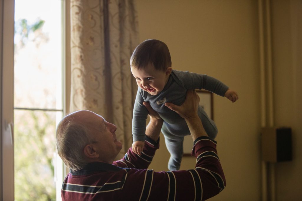
The soft, gentle murmurs of a baby’s first expressions, like little whispers of joy and wonder to doting parents, are actually signs that the baby’s heart is working rhythmically in concert with developing speech.

Jeremy I. Borjon, University of Houston assistant professor of psychology, reports in Proceedings of the National Academy of Sciences that a baby’s first sweet sounds and early attempts at forming words are directly linked to the baby’s heart rate. The findings have implications for understanding language development and potential early indicators of speech and communication disorders.

For infants, producing recognisable speech is more than a cognitive process. It is a motor skill that requires them to learn to coordinate multiple muscles of varying function across their body. This coordination is directly linked to ongoing fluctuations in heart rate.

Borjon investigated whether these fluctuations in heart rate coincide with vocal production and word production in 24-month-old babies. He found that heart rate fluctuations align with the timing of vocalizations and are associated with their duration and the likelihood of producing recognisable speech.

“Heart rate naturally fluctuates in all mammals, steadily increasing then decreasing in a rhythmic pattern. It turns out infants were most likely to make a vocalisation when their heart rate fluctuation had reached a local peak (maximum) or local trough (minimum),” reports Borjon.

“Vocalisations produced at the peak were longer than expected by chance. Vocalisations produced just before the trough, while heart rate is decelerating, were more likely to be recognised as a word by naïve listener,” he said.

Borjon and team measured a total of 2708 vocalisations emitted by 34 infants between 18 and 27 months of age while the babies played with a caregiver. Infants in this age group typically don’t speak whole words yet, and only a small subset of the vocalisations could be reliably identified as words by naïve listeners (10.3%). For the study, the team considered the heart rate dynamics of all sounds made by the baby’s mouth, be it a laugh, a babble or a coo.

“Every sound an infant makes helps their brain and body learn how to coordinate with each other, eventually leading to speech,” Borjon said.

As infants grow, their autonomic nervous system grows and develops. The first few years of life are marked by significant changes in how the heart and lungs function, and these changes continue throughout a person’s life.

The relationship between recognisable vocalisations and decelerating heart rate may imply that the successful development of speech partially depends on infants experiencing predictable ranges of autonomic activity through development.

“Understanding how the autonomic nervous system relates to infant vocalisations over development is a critical avenue of future research for understanding how language emerges, as well as risk factors for atypical language development,” said Borjon

Source: University of Houston

Men More Than Three Times as Likely to Die From a Brain Injury, New Study Shows

Photo by Anna Shvets

A new analysis of mortality data reveals the disproportionate impact of traumatic brain injuries (TBI) on older adults, males and certain racial and ethnic groups. The study, published in the peer-reviewed journal Brain Injury, provides a comprehensive analysis of TBI-related deaths across different population groups across the US in 2021.

The findings indicate that suicides remain the most common cause of TBI-related deaths, followed by unintentional falls, and specific groups are disproportionately affected by these tragedies.

Men, in particular, were found to be most likely to die from a TBI – more than three times the rate of women (30.5 versus 9.4). The reasons observed were multifactorial and could reflect differences in injury severity following a fall or motor vehicle crash, to the interaction of sex and age – with TBI outcomes in men worsening with age, while postmenopausal women fare better than men of similar age.

“While anyone is at risk for getting a TBI, some groups have a higher chance than others of dying from one. We identified specific populations who are most affected. In addition to men, older adults are especially at risk, with unintentional falls being a major cause of TBI-related death. American Indian or Alaska Native people also have higher rates of these fatal injuries,” says lead author Alexis Peterson PhD, of the National Center for Injury Prevention and Control at the Centers for Disease Control and Prevention.

“These findings highlight the importance of tailored prevention strategies to reach groups who may be at higher risk and the role healthcare providers can play in reducing TBI-related deaths through early intervention and culturally sensitive care.”

TBI remains a leading cause of injury-related death in the US In 2020, TBIs were associated with around a quarter of all injury-related deaths.

Using data from the National Vital Statistics System, the new analysis identified 69 473 TBI-related deaths among US residents during 2021. The age-adjusted TBI-related mortality rate was 19.5 per 100 000, representing an 8.8% increase from 2020.

Through statistical modeling, the researchers examined the simultaneous effect of multiple factors such as geographic region, sex, race and ethnicity, and age, on TBI-related mortality.

Key findings include:

  • Older adults (75+) had the highest rates of TBI-related deaths, with unintentional falls being the most common cause in this age group.
  • Non-Hispanic American Indian/Alaska Native individuals experienced the highest TBI-related death rate (31.5) compared to other racial and ethnic groups.
  • There were 37,635 TBI-related deaths categorised as unintentional injuries (ie, motor vehicle crashes, unintentional falls, unintentionally struck by or against an object, other).
  • 30,801 were categorized as intentional injuries (ie, all mechanisms of suicide and homicide).
  • Children aged from birth to 17 years accounted for around 4% of TBI-related deaths (2,977).

The authors emphasise the critical role of healthcare providers in preventing TBI-related deaths, particularly with groups at higher risk. “By assessing patients who may be at higher risk for TBI, especially due to falls or mental health challenges, healthcare providers can make timely referrals and recommend culturally tailored interventions to prevent further injury or death,” says Dr Peterson.

Public health efforts should focus on addressing the underlying causes of TBI-related deaths, such as unintentional falls and mental health crises, to help prevent further loss of life. “TBIs remain a significant public health concern, especially among older adults, men, and certain racial and ethnic groups,” says Peterson.  “CDC has proven resources that healthcare providers can use to not only reduce health disparities that increase the risk for TBI but also improve care for anyone affected by a TBI.”

The authors note the COVID-19 pandemic could have influenced TBI-related death trends in 2021. They also acknowledge several limitations of this analysis, including potential misclassification or incomplete documentation of causes on death certificates, which may lead to inaccuracies in estimating TBI-related deaths.

Source: Taylor & Francis Group

Sex Differences in Brain Structure Present at Birth

Photo by Chayene Rafaela on Unsplash

Sex differences in brain structure are present from birth, research from the Autism Research Centre at the University of Cambridge has shown.

While male brains tended to be greater in volume than female brains, when adjusted for total brain volume, female infants on average had significantly more grey matter, while male infants on average had significantly more white matter in their brains.

Grey matter is made up of neuron cell bodies and dendrites and is responsible for processing and interpreting information, such as sensation, perception, learning, speech, and cognition.  White matter is made up of axons, which are long nerve fibres that connect neurons together from different parts of the brain. 

Yumnah Khan, a PhD student at the Autism Research Centre, who led the study, said: “Our study settles an age-old question of whether male and female brains differ at birth. We know there are differences in the brains of older children and adults, but our findings show that they are already present in the earliest days of life.

“Because these sex differences are evident so soon after birth, they might in part reflect biological sex differences during prenatal brain development, which then interact with environmental experiences over time to shape further sex differences in the brain.”

One problem that has plagued past research in this area is sample size. The Cambridge team tackled this by analysing data from the Developing Human Connectome Project, where infants receive an MRI brain scan soon after birth. Having over 500 newborn babies in the study means that, statistically, the sample is ideal for detecting sex differences if they are present.

A second problem is whether any observed sex differences could be due to other factors, such as differences in body size.  The Cambridge team found that, on average, male infants had significantly larger brain volumes than did females, and this was true even after sex differences in birth weight were taken into account.

After taking this difference in total brain volume into account, at a regional level, females on average showed larger volumes in grey matter areas related to memory and emotional regulation, while males on average had larger volumes in grey matter areas involved in sensory processing and motor control.

The findings of the study, the largest to date to investigate this question, are published in the journal Biology of Sex Differences.

Dr Alex Tsompanidis who supervised the study, said: “This is the largest such study to date, and we took additional factors into account, such as birth weight, to ensure that these differences are specific to the brain and not due to general size differences between the sexes.

“To understand why males and females show differences in their relative grey and white matter volume, we are now studying the conditions of the prenatal environment, using population birth records, as well as in vitro cellular models of the developing brain. This will help us compare the progression of male and female pregnancies and determine if specific biological factors, such as hormones or the placenta, contribute to the differences we see in the brain.”

The researchers stress that the differences between males and females are average differences.

Dr Carrie Allison, Deputy Director of the Autism Research Centre, said: “The differences we see do not apply to all males or all females, but are only seen when you compare groups of males and females together. There is a lot a variation within, and a lot of overlap between, each group.”  

Professor Simon Baron-Cohen, Director of the Autism Research Centre, added: “These differences do not imply the brains of males and females are better or worse. It’s just one example of neurodiversity. This research may be helpful in understanding other kinds of neurodiversity, such as the brain in children who are later diagnosed as autistic, since this is diagnosed more often in males.”

The research was funded by Cambridge University Development and Research, Trinity College, Cambridge, the Cambridge Trust, and the Simons Foundation Autism Research Initiative.

Reference
Khan, Y.T., Tsompanidis, A., Radecki, M.A. et al. Sex differences in human brain structure at birth. Biol Sex Differ; 17 Oct 2024; DOI: 10.1186/s13293-024-00657-5

Republished under a Creative Commons Attribution-NonCommercial-ShareAlike 4.0 International License.

Source: University of Cambridge

Study Likely to Change Standard of Care for Deadly Vertebrobasilar Stroke

Ischaemic and haemorrhagic stroke. Credit: Scientific Animations CC4.0

Endovascular therapy (EVT), a minimally invasive surgery performed inside the blood vessels, is 2 ½ times more likely than standard medical management to achieve a positive outcome after vertebrobasilar stroke that affects the back of the brain, including the brain stem. A meta-analysis of four randomised clinical trials, published in The Lancet, was led by UPMC Stroke Institute director Raul Nogueira, MD.

Investigators from the US, Netherlands and China formed a multi-centre collaboration of all four randomised trials of EVT in vertebrobasilar occlusion with data that provides the strongest evidence to date of the benefits of EVT over alternative approaches for complicated vessel obstructions in life-sustaining areas of the brain.

Although vertebrobasilar artery occlusions interrupting blood flow in the back of the brain account for only a small fraction of all ischaemic strokes, they are especially deadly. Without an appropriate intervention, vertebrobasilar strokes lead to high rates of severe disability and mortality that may exceed 70%.

“While the overwhelming benefit of EVT for acute ischaemic strokes due to occlusions of large vessels that supply the anterior brain has been well established, the benefit of this therapy for vertebrobasilar artery occlusion, one of the most devastating forms of stroke, has been more controversial,” said Nogueira, endowed professor of neurology and neurosurgery at the University of Pittsburgh.

To address this uncertainty, the consortium of investigators, called VERITAS, focused on providing more precise, comprehensive and statistically powered estimates of the benefits of EVT with a particular focus on specific patient subgroups of clinical interest.

As the primary coordinating centre for the study, the Pitt team established common variables, definitions and trial specifications that laid the groundwork for a core pooled dataset from the four randomised controlled clinical trials ATTENTION, BAOCHE, BASICS and BEST of EVT for stroke due to vertebrobasilar artery occlusion.

Meta-analysis showed that at three months after the surgery, despite higher rates of brain bleeds with the procedure, EVT significantly reduced patient mortality and overall post-stroke disability, increasing patients’ functional independence. Notably, patients who underwent EVT were nearly 2 ½ times more likely to regain their ability to walk independently compared to patients who received the current medical standard of care, including intravenous thrombolytics.

“The results of the VERITAS collaboration are expected to influence treatment guidelines and impact stroke care globally,” Nogueira said. “We hope that this analysis sets the foundation for improved recovery after vertebrobasilar strokes and helps more people regain their independence after this catastrophic medical event.”

Source: University of Pittsburgh

Books Beat TV When it Comes to Brain Health

…but is that any surprise?

Photo by Nong on Unsplash

It’s that time of the year when most of us get the chance to sit back and enjoy some well-deserved down time. But whether you reach for the TV controller, or a favourite book, your choice could have implications for your long-term brain health, say researchers at the University of South Australia who published their research in the Journals of Gerontology.

Assessing the 24-hour activity patterns of 397 older adults (aged 60+), researchers found that the context or type of activity that you engage in, matters when it comes to brain health. And specifically, that some sedentary (or sitting) behaviours are better for cognitive function than others.

When looking at different sedentary behaviours, they found that social or mentally stimulating activities such as reading, listening to music, praying, crafting, playing a musical instrument, or chatting with others are beneficial for memory and thinking abilities. Yet watching TV or playing video games are detrimental.

Researchers believe that there is likely a hierarchy of how sedentary behaviours relate to cognitive function, in that some have positive effects while others have negative effects.

It’s a valuable insight that could help reduce risks of cognitive impairment, particularly when at least 45% of dementia cases could be prevented through modifiable lifestyle factors.

In Australia, about 411,100 people (or one in every 1000 people) are living with dementia. Nearly two-thirds are women. Globally, the World Health Organization estimates that more than 55 million people have dementia with nearly 10 million new cases each year.

UniSA researcher Dr Maddison Mellow says that not all sedentary behaviours are equal when it comes to memory and thinking ability.

“In this research, we found that the context of an activity alters how it relates to cognitive function, with different activities providing varying levels of cognitive stimulation and social engagement,” Dr Mellow says.

“We already know that physical activity is a strong protector against dementia risk, and this should certainly be prioritised if you are trying to improve your brain health. But until now, we hadn’t directly explored whether we can benefit our brain health by swapping one sedentary behaviour for another.

“We found that sedentary behaviours which promote mental stimulation or social engagement — such as reading or talking with friends — are beneficial for cognitive function, whereas others like watching TV or gaming have a negative effect. So, the type of activity is important.

“And, while the ‘move more, sit less’ message certainly holds true for cardiometabolic and brain health, our research shows that a more nuanced approach is needed when it comes to thinking about the link between sedentary behaviours and cognitive function.”

Now, as the Christmas holidays roll around, what advice do researchers have for those who really want to indulge in a myriad of Christmas movies or a marathon of Modern Family?

“To achieve the best brain health and physical health benefits, you should prioritise movement that’s enjoyable and gets the heart rate up, as this has benefits for all aspects of health,” Dr Mellow says.

“But even small five-minute time swaps can have benefits. So, if you’re dead set on having a Christmas movie marathon, try to break up that time with some physical activity or a more cognitively engaged seated activity, like reading, at some point. That way you can slowly build up healthier habits.”

Source: University of South Australia

T Cells could Ease Brain Injury after Cardiac Arrest

Photo by Mat Napo on Unsplash

Despite improvements in CPR and ambulance response times, only about one in 10 people ultimately survive after out-of-hospital cardiac arrest (OHCA). Most patients hospitalised with OCHA die of brain injury, and no medications are currently available to prevent this outcome. A team led by researchers from Mass General Brigham found that immune cells may play a key role.

Using samples from patients who have had an OHCA, the team uncovered changes in immune cells just six hours after cardiac arrest that can predict brain recovery 30 days later. They pinpointed a particular population of cells that may provide protection against brain injury and a drug that can activate these cells, which they tested in preclinical models. Their results are published in Science Translational Medicine.

“Cardiac arrest outcomes are grim, but I am optimistic about jumping into this field of study because, theoretically, we can treat a patient at the moment injury happens,” said co-senior and corresponding author Edy Kim, MD, PhD, of the Division of Pulmonary and Critical Care Medicine at Brigham and Women’s Hospital. “Immunology is a super powerful way of providing treatment. Our understanding of immunology has revolutionised cancer treatment, and now we have the opportunity to apply the power of immunology to cardiac arrest.”

As a resident physician in the Brigham’s cardiac intensive care unit, Kim noticed that some cardiac arrest patients would have high levels of inflammation on their first night in the hospital and then rapidly improve. Other patients would continue to decline and eventually die. In order to understand why some patients survive and others do not, Kim and colleagues began to build a biobank – a repository of cryopreserved cells donated by patients with consent from their families just hours after their cardiac arrest.

The researchers used a technique known as single-cell transcriptomics to look at the activity of genes in every cell in these samples. They found that one cell population – known as diverse natural killer T (dNKT) cells – increased in patients who would have a favourable outcome and neurological recovery. The cells appeared to be playing a protective role in preventing brain injury.

To further test this, Kim and colleagues used a mouse model, treating mice after cardiac arrest with sulfatide lipid antigen, a drug that activates the protective NKT cells. They observed that the mice had improved neurological outcomes.

The researchers note that there are many limitations to mouse models, but making observations from human samples first could increase the likelihood of successfully translating their findings into intervention that can help patients. Further studies in preclinical models are needed, but their long-term goal is to continue to clinical trials in people to see if the same drug can offer protection against brain injury if given shortly after cardiac arrest.

“This represents a completely new approach, activating T cells to improve neurological outcomes after cardiac arrest,” said Kim. “And a fresh approach could lead to life-changing outcomes for patients.”

Source: Mass General Brigham

The Heart has a ‘Brain’ of its Own

Human heart. Credit: Scientific Animations CC4.0

New research from Karolinska Institutet and Columbia University shows that the heart has a mini-brain – its own nervous system that controls the heartbeat. A better understanding of this system, which is much more diverse and complex than previously thought, could lead to new treatments for heart diseases. The study, conducted on zebrafish, is published in Nature Communications.

The heart has long been thought to be controlled solely by the autonomic nervous system, which transmits signals from the brain. The heart’s neural network, which is embedded in the superficial layers of the heart wall, has been considered a simple structure that relays the signals from the brain. However, recent research suggests that it has a more advanced function than that.

Controlling the heartbeat

Scientists have now discovered that the heart has its own complex nervous system that is crucial to controlling its rhythm.

“This ‘little brain’ has a key role in maintaining and controlling the heartbeat, similar to how the brain regulates rhythmic functions such as locomotion and breathing,” explains Konstantinos Ampatzis, principal researcher and docent at the Department of Neuroscience, Karolinska Institutet, Sweden, who led the study.

The researchers identified several types of neurons in the heart that have different functions, including a small group of neurons with pacemaker properties. The finding challenges the current view on how the heartbeat is controlled, which may have clinical implications.

Surprising complexity revealed

“We were surprised to see how complex the nervous system within the heart is,” says Konstantinos Ampatzis. “Understanding this system better could lead to new insights into heart diseases and help develop new treatments for diseases such as arrhythmias.” 

The study was conducted on zebrafish, an animal model that exhibits strong similarities to human heart rate and overall cardiac function. The researchers were able to map out the composition, organisation and function of neurons within the heart using a combination of methods such as single-cell RNA sequencing, anatomical studies and electrophysiological techniques.

New therapeutic targets

“We will now continue to investigate how the heart’s brain interacts with the actual brain to regulate heart functions under different conditions such as exercise, stress, or disease,” says Konstantinos Ampatzis. “We aim to identify new therapeutic targets by examining how disruptions in the heart’s neuronal network contribute to different heart disorders.”

Source: Karolinska Institutet

Scientists Identify a Type of Brain Cell That is a Master Controller of Urination

Photo by Jan Antonin Kolar on Unsplash

Researchers have identified a subset of brain cells in mice that act as the master regulators of urination.

The research, published as a Reviewed Preprint in eLife, is described by editors as an important study with convincing data showing that oestrogen receptor 1-expressing neurons (ESR1+) in the Barrington’s nucleus of the mouse brain coordinate both bladder contraction and relaxation of the external urethral sphincter.

Urination requires the coordinated function of two units of the lower urinary tract. The detrusor muscle of the bladder wall relaxes to allow the bladder to fill and empty, while the external sphincter opens when it’s appropriate to allow urine to flow out, but otherwise keeps tightly shut.

“Impairment of coordination between the bladder muscle and the sphincter leads to various urinary tract dysfunctions and can significantly degrade a person’s quality of life,” says first author Xing Li, Advanced Institute for Brain and Intelligence, School of Physical Science and Technology, Guangxi University, Nanning, China. “But although we know the individual nerve signalling pathways that control each of these urinary tract components, we don’t know which brain areas ensure they cooperate at the right time.”

To explore this, the authors used state-of-the-art live cell imaging to study the activity of brain cells in anaesthetised and awake mice during urination. They focused on a brain region called the pontine micturition centre (PMC), otherwise known as the Barrington’s nucleus, and compared the activity of different PMC nerve cell subtypes.

In their first experiments, they measured the activity of the cells as the bladder empties by measuring changes in levels of calcium. This revealed that the electrical firing rate of a subset of PMC cells expressing estrogen receptors (PMCESR1+ cells) was tightly linked to bladder emptying. When they combined this with monitoring bladder physiology, they found that it was not only the timing of PMCESR1+ cell activity that correlated with bladder emptying, but the strength of cell electrical activity, too.

Next, they tested what happened to urination if they blocked or triggered the PMCESR1+ cells. They found that when PMCESR1+ cell activity was blocked, the amount of urine the mice passed was significantly reduced and ongoing urination was suspended from the moment the cells were inactive. To understand the mechanism behind this, they measured the activity of the bladder muscle and sphincter. They discovered that both increase of bladder pressure and sphincter muscle bursting activity associated with bladder emptying both stopped when PMCESR1+ cell activity was blocked during an ongoing voiding even. Similarly, when PMCESR1+ cells were artificially activated using light, bladder emptying occurred 100% of the time. This suggests that PMCESR1+ cells work as a reliable master switch that either initiates or suspends bladder emptying.

To test whether PMCESR1+ cells can influence bladder emptying independently of controlling the sphincter, they disconnected either the nerve carrying messages from the brain to the sphincter, or the nerve carrying messages from the brain to the bladder. They found that PMCESR1+ cell control of the bladder was fully operational even when communication to the sphincter was blocked, and vice versa. This showed the cells could control the bladder and sphincter independently of one another, but the question remained: could they coordinate the action of the bladder muscle and sphincter together? That is, operate them in a controlled, perfectly timed manner, to trigger bladder emptying when appropriate?

To explore this, they simultaneously recorded bladder pressure and electromyography measurements of sphincter activity. The timing of bladder pressure changes immediately before sphincter bursting activity was consistent for both spontaneous bladder emptying and emptying caused by activating the PMCESR1+ cells, showing that these cells can coordinate the two steps in a precisely temporal sequence and controlled way.

“Our study shows that a subset of cells in the Barrington’s nucleus of the brain can initiate and suspend bladder emptying with 100% accuracy when needed, for example, to release only a small volume for landmarking by animals, or for a human to urinate into a small sample tube for a health check,” concludes senior author Xiaowei Chen, Third Military Medical University, and Chongqing Institute for Brain and Intelligence, China. “While other cells will no doubt be involved in perfect urination control, our pinpointing of PMCESR1+ cells’ crucial role in bladder–sphincter coordination will aid the development of targeted therapies for treating urination dysfunction caused by brain or spinal cord injury or peripheral nerve damage.”

Source: eLife

Soccer Headers may be More Damaging to Brain than Thought

Photo by Kenny Webster on Unsplash

Soccer heading may cause more damage to the brain than previously thought, according to a study presented at the annual meeting of the Radiological Society of North America (RSNA).

Heading is a widely used technique in soccer where the players control the direction of the ball by hitting it with their head. In recent years, research has been done that suggests a link between repeated head impacts and neurodegenerative diseases, such as chronic traumatic encephalopathy (CTE).

“The potential effects of repeated head impacts in sport are much more extensive than previously known and affect locations similar to where we’ve seen CTE pathology,” said study senior author Michael L. Lipton, MD, PhD, professor of radiology at Columbia University Irving Medical Center. “This raises concern for delayed adverse effects of head impacts.”

While prior studies have identified injuries to the brain’s white matter in soccer players, Dr Lipton and colleagues used a new approach in diffusion MRI to analyse microstructure close to the surface of the brain.

To identify how repeated head impacts affect the brain, the researchers compared brain MRIs of 352 male and female amateur soccer players, ranging in age from 18 to 53, to brain MRIs of 77 non-collision sport athletes, such as runners.

Soccer players who headed the ball at high levels showed abnormality of the brain’s white matter adjacent to sulci, which are deep grooves in the brain’s surface. Abnormalities in this region of the brain are known to occur in very severe traumatic brain injuries.

The abnormalities were most prominent in the frontal lobe of the brain, an area most susceptible to damage from trauma and frequently impacted during soccer heading. More repetitive head impacts were also associated with poorer verbal learning.

“Our analysis showed that the white matter abnormalities represent a mechanism by which heading leads to worse cognitive performance,” Dr Lipton said.

Most of the participants of the study had never sustained a concussion or been diagnosed with a traumatic brain injury. This suggests that repeated head impacts that don’t result in serious injury may still adversely affect the brain.

“The study identifies structural brain abnormalities from repeated head impacts among healthy athletes,” Dr Lipton said. “The abnormalities occur in the locations most characteristic of CTE, are associated with worse ability to learn a cognitive task and could affect function in the future.”

The results of this study are also relevant to head injuries from other contact sports. The researchers stress the importance of knowing the risks of repeated head impacts and their potential to harm brain health over time.

“Characterising the potential risks of repetitive head impacts can facilitate safer sport engagement to maximise benefits while minimising potential harms,” Dr Lipton said. “The next phase of the study is ongoing and examines the brain mechanisms underlying the MRI effects and potential protective factors.”

Source: Radiological Society of North America

Concussions from American Football Slow Brain Activity of High Schoolers

Photo by Jakob Rosen on Unsplash

A new study of high school American football players found that concussions affect an often-overlooked but important brain signal. The findings are presented at the annual meeting of the Radiological Society of North America (RSNA).

Reports have emerged in recent years warning about the potential harms of youth contact sports on developing brains. Contact sports, including high school football, carry a risk of concussion. Symptoms of concussion commonly include cognitive disturbances, such as difficulty with balancing, memory or concentration.

Many concussion studies focus on periodic brain signals. These signals appear in rhythmic patterns and contribute to brain functions such as attention, movement or sensory processing. Not much is known about how concussions affect other aspects of brain function, specifically, brain signals that are not rhythmic.

“Most previous neuroscience research has focused on rhythmic brain signaling, which is also called periodic neurophysiology,” said study lead author Kevin C. Yu, BS, a neuroscience student at Wake Forest University School of Medicine. “On the other hand, aperiodic neurophysiology refers to brain signals that are not rhythmic.”

Aperiodic activity is typically treated as ‘background noise’ on brain scans, but recent studies have shown that this background noise may play a key role in how the brain functions.

“While it’s often overlooked, aperiodic activity is important because it reflects brain cortical excitability,” said study senior author Christopher T. Whitlow, MD, PhD, MHA, radiology professor at Wake Forest University School of Medicine.

Cortical excitability is a vital part of brain function. It reflects how nerve cells, or neurons, in the brain’s cortex respond to stimulation and plays a key role in cognitive functions like learning and memory, information processing, decision making, motor control, wakefulness and sleep.

To gain a better understanding of brain rhythms and trauma, the researchers sought to identify the impacts of concussions on aperiodic activity.

Pre- and post-season resting-state magnetoencephalography (MEG) data was collected from 91 high school football players, of whom 10 were diagnosed with a concussion. MEG is a neuroimaging technique that measures the magnetic fields that the brain’s electrical currents produce.

A clinical evaluation tool for concussions called the Post-Concussive Symptom Inventory was correlated with pre- and post-season physical, cognitive and behavioral symptoms.

High school football players who sustained concussions displayed slowed aperiodic activity. Aperiodic slowing was strongly associated with worse post-concussion cognitive symptoms and test scores.

Slowed aperiodic activity was present in areas of the brain that contain chemicals linked with concussion symptoms like impaired concentration and memory.

“This study is important because it provides insight into both the mechanisms and the clinical implications of concussion in the maturing adolescent brain,” said co-lead author Alex I. Wiesman, PhD, assistant professor at Simon Fraser University. “Reduced excitability is conceptually a very different brain activity change than altered rhythms and means that a clear next step for this work is to see whether these changes are related to effects of concussion on the brain’s chemistry.”

The findings from the study may also influence tracking of post-concussion symptoms and aid in finding new treatments to improve recovery.

“Our study opens the door to new ways of understanding and diagnosing concussions, using this novel type of brain activity that is associated with concussion symptoms,” Dr Whitlow said. “It highlights the importance of monitoring kids carefully after any head injury and taking concussions seriously.”

Source: Radiological Society of North America