View of the spinal cord. Credit: Scientific Animations CC4.0
In a recent study published in Nature, researchers prevented T cells from causing the normal autoimmune damage that comes with spinal cord injury, sparing neurons and successfully aiding recovery in mouse models.
In spinal cord injury, the wound site attracts a whole host of peripheral immune cells, including T cells, which result in both beneficial and deleterious effects. Notably, antigen-presenting cells activate CD4+ T cells to release cytokines, ultimately leading to neuroinflammation and tissue destruction. This neuroinflammation is notably most pronounced during the acute phase of spinal cord injury. The problem is that these same T cells have a neuroprotective effect initially, only later developing autoimmunity and attacking the injury site.
Using single cell RNA sequencing, the researchers found that CD4+ T cell clones in mice showed antigen specificity towards self-peptides of myelin and neuronal proteins. Self-peptides have been implicated in a wide range of autoimmune conditions.
Using mRNA techniques, the researchers edited the T cell receptor, so that they shut off after a few days. In mouse models of spinal cord injury, they showed notable neuroprotective efficacy, partly as a result of modulating myeloid cells via interferon-γ.
Their findings provided insights into the mechanisms behind the neuroprotective function of injury-responsive T cells. This will help pave the way for the future development of T cell therapies for central nervous system injuries, and perhaps treatments for neurodegenerative diseases such as Alzheimer’s.
Researchers at Baylor College of Medicine and the Jan and Dan Duncan Neurological Research Institute at Texas Children’s Hospital have uncovered a new cell type in human brain cancers. Their study, published in Cancer Cell, reveals that a third of the cells in glioma, fire electrical impulses. Interestingly, the impulses, also called action potentials, originate from tumour cells that are part neuron and part glia, supporting the groundbreaking idea that neurons are not the only cells that can generate electric signals in the brain.
The scientists also discovered that cells with hybrid neuron-glia characteristics are present in the non-tumour human brain. The findings highlight the importance of further studying the role of these newly identified cells in both glioma and normal brain function.
“Previous studies have shown that patient survival outcomes are associated with tumour proliferation and invasiveness, which are influenced by tumour intrinsic and extrinsic factors, including communication between tumour cells and neurons that reside in the brain,” said Dr Benjamin Deneen, professor in the Department of Neurosurgery at Baylor.
Researchers have previously described that glioma and surrounding healthy neurons connect with each other and that neurons communicate with tumours in ways that drive tumour growth and invasiveness.
“We have known for some time now that tumour cells and neurons interact directly,” said first author Dr Rachel N. Curry, postdoctoral fellow in paediatrics – neuro oncology at Baylor, who was responsible for conceptualising the project. “But one question that always lingered in my mind was, ‘Are cancer cells electrically active?’ To answer this question correctly, we required human samples directly from the operating room. This ensured the biology of the cells as they would exist in the brain was preserved as much as possible.”
To study the ability of glioma cells to spike electrical signals and identify the cells that produce the signals, the team used Patch-sequencing, a combination of techniques that integrates whole-cell electrophysiological recordings to measure spiking signals with single-cell RNA-sequencing and analysis of the cellular structure to identify the type of cells.
The electrophysiology experiments were conducted by research associate and co-first author Dr Qianqian Ma in the lab of co-corresponding author associate professor of neuroscience Dr Xiaolong Jiang. This innovative approach has not been used before to study human brain tumour cells. “We were truly surprised to find these tumour cells had a unique combination of morphological and electrophysiological properties,” Ma said. “We had never seen anything like this in the mammalian brain before.”
“We conducted all these analyses on single cells. We analysed their individual electrophysiological activity. We extracted each cell’s content and sequenced the RNA to identify the genes that were active in the cell, which tells us what type of cell it is,” Deneen said. “We also stained each cell with dyes that would visualise its structural features.”
Integrating this vast amount of individual data required the researchers to develop a novel way to analyse it.
“To define the spiking cells and determine their identity, we developed a computational tool – Single Cell Rule Association Mining (SCRAM) – to annotate each cell individually,” said co-corresponding author, Dr Akdes Serin Harmanci, assistant professor of neurosurgery at Baylor.
“Finding that so many glioma cells are electrically active was a surprise because it goes against a strongly held concept in neuroscience that states that, of all the different types of cells in the brain, neurons are the only ones that fire electric impulses,” Curry said. “Others have proposed that some glia cells known as oligodendrocyte precursor cells (OPCs) may fire electrical impulses in the rodent brain, but confirming this in humans had proven a difficult task. Our findings show that human cells other than neurons can fire electrical impulses. Since there is an estimated 100 million of these OPCs in the adult brain, the electrical contributions of these cells should be further studied.”
“Moreover, the comprehensive data analyses revealed that the spiking hybrid cells in glioma tumours had properties of both neurons and OPC cells,” Harmanci said. “Interestingly, we found non-tumour cells that are neuron-glia hybrids, suggesting that this hybrid population not only plays a role in glioma growth but also contributes to healthy brain function.”
“The findings also suggest that the proportion of spiking hybrid cells in glioma may have a prognostic value,” said co-corresponding author Dr Ganesh Rao, professor and chair of neurosurgery at Baylor. “The data shows that the more of these spiking hybrid glioma cells a patient has, the better the survival outcome. This information is of great value to patients and their doctors.”
“This work is the result of extensive equal collaboration across multiple disciplines – neurosurgery, bioinformatics, neuroscience and cancer modelling – disciplines strongly supported by state-of-the-art groups at Baylor,” Deneen said. “The results offer an enhanced understanding of glioma tumours and normal brain function, a sophisticated bioinformatics pipeline to analyse complex cellular populations and potential prognostic implications for patients with this devastating disease.”
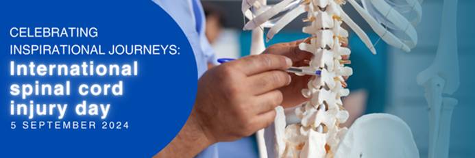
5 September 2024, International Spinal Cord Injury Day is commemorated on Thursday 5 September, drawing attention to the many ways people can be affected by spinal cord injury, creating awareness of prevention, and highlighting the possibilities for a fulfilling life after injury.
According to the World Health Organization, globally, over 15 million people are living with spinal cord injuries. Most of these cases are due to trauma, including falls, road traffic injuries or violence.
Jessica Morris, an occupational therapist at the Netcare Rehabilitation Hospital in Auckland Park, says one of the most critical aspects of care for those who’ve been impacted by spinal cord injuries is the importance of successful rehabilitation through a holistic, integrated approach from a multidisciplinary team.
“Many people just think it’s just about mobility. It’s so much more than that. Rehabilitation is complex because many different areas of our patients’ lives are affected.” Morris says they are fortunate that their team has so many different practitioners who can contribute to treating spinal cord injury patients, helping them regain a level of independence, which is vital to their self-confidence and sense of empowerment.
Dr Anrie Carstens, a general practitioner with a particular interest in physical medicine and rehabilitation who practises at the Netcare Rehabilitation Hospital, says the message of Spinal Cord Injury Awareness Day has relevance all year round, as people with spinal cord injuries need to be incorporated into society.
“It’s an opportunity to tell people not to be nervous to talk to someone in a wheelchair. They’re just like you or me, and they just have special ways of moving around and managing their pain and different aspects of their bodies. With the help of proper rehabilitation, the person can be better integrated as a functional, contributing member of society.”
Dr Carstens says people should also be aware that if they or their loved ones are ever impacted by a spinal cord injury, professional support is available. “Don’t just go straight home after your hospital stay and try to do everything on your own. Instead, come to a specialised spinal cord injury unit like ours, with therapists, doctors and nursing staff who are well versed in spinal cord injury and know the finer nuances necessary to optimally treat the person and show them how best to cope with their injury.
“In the multidisciplinary approach, every practitioner has a role in getting the person back into the real world, whether it means going back home, back to school, back to work or wherever they were before their injury occurred.”
From doctors and nurses with specialised skills to physiotherapists, occupational therapists, social workers and psychologists, speech therapists, a prosthetist and dieticians, the team provides a broad person focussed rehabilitation service to both adults and children. Their aim is to optimise their patients’ independence level using specialised equipment and teaching specific techniques to help overcome the obstacles a person may face.
Dr Carstens says it’s rewarding work for the staff at the hospital, who build up enduring relationships with those they care for. “One of the highlights is to compare and see what the patient was like when you admitted them and then see on discharge how much they’ve grown, how they’ve gained confidence and become more independent. What’s even better is to see them after they’ve been discharged and observe how well they’ve coped and how they’ve integrated and adjusted to their environment. We build a relationship with our patients because they stay with us for quite a while, and we usually have checkups every year after the person is discharged, often for life. We get to see them grow and thrive outside the healthcare setting, and we need more awareness about how much it is possible for people with spinal cord injuries to achieve.”
View of the spinal cord. Credit: Scientific Animations CC4.0
Injuries, infection and inflammatory diseases that damage the spinal cord can lead to intractable pain and disability but some degree of recovery may be possible. The question is, how best to stimulate the regrowth and healing of damaged nerves.
At the Vanderbilt University Institute of Imaging Science (VUIIS), scientists are focusing on a previously understudied part of the brain and spinal cord – white matter, which is made up of axons that relay signals. Their discoveries could lead to treatments that restore nerve activity through the targeted delivery of electromagnetic stimuli or drugs.
“In the spinal cord, the white matter signal is quite large and detectable, unlike in the brain, where it has less amplitude than the grey matter (signal),” said Sengupta, research instructor in Radiology and Radiological Sciences at Vanderbilt University Medical Center.
“This may be due to the larger volume of white matter in the spinal cord compared to the brain,” he added. Alternatively, the signal could represent “an intrinsic demand” in metabolism within the white matter, reflecting its critical role in supporting grey matter.
For several years, Gore, who directs the VUIIS, and his colleagues have used functional magnetic resonance imaging (fMRI) to detect blood oxygenation-level dependent (BOLD) signals, a key marker of nervous system activity, in white matter.
Last year, they reported that when participants undergoing fMRI perform a task, like wiggling their fingers, BOLD signals increase in white matter throughout the brain.
The current study monitored changes in BOLD signals in the white matter of the spinal cord at rest and in response to a vibrotactile stimulus applied to the fingers in an animal model. In response to stimulation, white matter activity was higher in “tracts” of ascending fibres that carry the signal from the spine to the brain.
This result is consistent with white matter’s known neurobiological function, the researchers noted. White matter contains non-neuronal glial cells that do not produce electrical impulses, but which regulate blood flow and neurotransmitters, the signaling molecules that transmit signals between nerve cells.
Much remains to be learned about the function of white matter in the spinal cord. But the findings from this research may help in improved understanding of diseases that affect white matter in the spinal cord, including multiple sclerosis, Sengupta said.
“We will be able to see how activity in the white matter changes in different stages of the disease,” he said. Researchers also may be able to monitor the effectiveness of therapeutic interventions, including neuromodulation, in promoting recovery following spinal cord injury.
The image represents a statistical average of how different types of love light up different regions of the brain. Photo: Pärttyli Rinne et al 2024, Aalto University.
We use the word ‘love’ in a bewildering range of contexts, from sexual adoration to parental love or the love of nature. Now, more comprehensive imaging of the brain may shed light on why we use the same word for such a diverse collection of human experiences.
“You see your newborn child for the first time. The baby is soft, healthy and hearty – your life’s greatest wonder. You feel love for the little one.”
The above statement was one of many simple scenarios presented to 55 parents, self-described as being in a loving relationship. Researchers from Aalto University utilised functional magnetic resonance imaging (fMRI) to measure brain activity while subjects mulled brief stories related to six different types of love.
“We now provide a more comprehensive picture of the brain activity associated with different types of love than previous research,” says Pärttyli Rinne, the philosopher and researcher who coordinated the study. “The activation pattern of love is generated in social situations in the basal ganglia, the midline of the forehead, the precuneus and the temporoparietal junction at the sides of the back of the head.”
Love for one’s children generated the most intense brain activity, closely followed by romantic love.
“In parental love, there was activation deep in the brain’s reward system in the striatum area while imagining love, and this was not seen for any other kind of love,” says Rinne. Love for romantic partners, friends, strangers, pets and nature were also part of the study, which was published in the journal Cerebral Cortex.
According to the research, brain activity is influenced not only by the closeness of the object of love, but also by whether it is a human being, another species or nature.
Unsurprisingly, compassionate love for strangers was less rewarding and caused less brain activation than love in close relationships. Meanwhile, love of nature activated the reward system and visual areas of the brain, but not the social brain areas.
Pet-owners identifiable by brain activity
The biggest surprise for the researchers was that the brain areas associated with love between people ended up being very similar, with differences lying primarily in the intensity of activation. All types of interpersonal love activated areas of the brain associated with social cognition, in contrast to love for pets or nature – with one exception.
Subjects’ brain responses to a statement like the following, on average, revealed whether or not they shared their life with a furry friend:
“You are home lolling on the couch and your pet cat pads over to you. The cat curls up next to you and purrs sleepily. You love your pet.”
“When looking at love for pets and the brain activity associated with it, brain areas associated with sociality statistically reveal whether or not the person is a pet owner. When it comes to the pet owners, these areas are more activated than with non-pet owners,” says Rinne.
Love activations were controlled for in the study with neutral stories in which very little happened. For example, looking out the bus window or absent-mindedly brushing your teeth. After hearing a professional actor’s rendition of each ‘love story’, participants were asked to imagine each emotion for 10 seconds.
This is not the first effort at finding love for Rinne and his team, which includes researchers Juha Lahnakoski, Heini Saarimäki, Mikke Tavast, Mikko Sams and Linda Henriksson. They have undertaken several studies seeking to deepen our scientific knowledge of human emotions. The group released research mapping subjects’ bodily experiences of love a year ago, with the earlier study also linking the strongest physical experiences of love with close interpersonal relationships.
Not only can understanding the neural mechanisms of love help guide philosophical discussions about the nature of love, consciousness, and human connection, but also, the researchers hope that their work will enhance mental health interventions in conditions like attachment disorders, depression or relationship issues.
An international study employing advanced measurements of brain ageing on a wide range of participants found that people from more disadvantaged countries and backgrounds had older biological ages for their brains compared to chronological ages. The results are published in Nature Medicine.
The pace at which the brain ages can vary significantly among individuals. This difference between biological and chronological ages may be affected by environmental factors like pollution and social factors like income or health inequalities, especially in older people and those with dementia. Until now, it was unclear how these combined factors could either accelerate or delay brain ageing across diverse geographical populations.
The study used advanced brain clocks based on deep learning of brain networks, involved a diverse dataset of 5306 participants from 15 countries. By analysing data from functional magnetic resonance imaging (fMRI) and electroencephalography (EEG), the researchers quantified brain age gaps in healthy individuals and those with neurodegenerative conditions such as mild cognitive impairment (MCI), Alzheimer’s disease, and frontotemporal lobe degeneration (FTLD).
Participants with a diagnosis of dementia, particularly Alzheimer’s disease, exhibited the most critical brain age gaps. The research also highlighted sex differences in brain ageing, with women in Latin American and Caribbean countries showing greater brain age gaps, particularly in those with Alzheimer’s disease. These differences were linked to biological sex and gender disparities in health and social conditions. Variations in signal quality, demographics, or acquisition methods did not explain the results. These findings underscore the role of environmental and social factors in brain health disparities.
The findings of this study have profound implications for neuroscience and brain health, particularly in understanding the interaction between macro factors (exposome) and the mechanisms that underlie brain ageing across diverse populations in healthy ageing and dementia. The study’s approach, which integrates multiple dimensions of diversity into brain health research, offers a new framework for personalised medicine. This framework could be crucial for identifying individuals at risk of neurodegenerative diseases and developing targeted interventions to mitigate these risks. Moreover, the study’s results highlight the importance of considering the biological embedding of environmental and social factors in public health policies. Policymakers can reduce brain age gaps and promote healthier ageing across populations by addressing issues such as socioeconomic inequality and environmental pollution.
Deep within either hemisphere of the brain is the “claustrum complex”, which contributes to consciousness and awareness. Many diseases known to be related to higher cognitive function, such as Alzheimer’s, schizophrenia, and ADD/ADHD, are also closely linked to abnormal function of this particular part of the brain. But how the different parts of the claustrum complex work or how its circuits and communication system are organised is not fully understood.
Researchers at Aarhus University have now uncovered this, and their results identify, down to the cellular level, which part of the claustrum complex controls our ability to discriminate familiar and novel things.
“Our study focuses on an area of the claustrum called the ‘endopiriform,’ which is a relatively unknown brain structure despite its unique brain network and cellular properties,” explains Asami Tanimura, an associate professor and the lead researcher of the study appearing as a preprint in eLife.
“For the first time, we have dissected the circuit of endopiriform to the hippocampus, and demonstrated how this pathway is crucial for recognition memory.”
In mouse models, researchers were able to observe how the mice’s behaviour changed when they respectively ‘turned on’ and ‘turned off’ the activity in this specific cell group.
Asami explains: “We observed that the cells in the endopiriform were active when the mice interacted with new conspecifics or objects, and when we inhibited this cell group, it reduced the mice’s ability to distinguish novel mouse or object from familiar ones.”
Based on this, the researchers concluded that this specific cell group in the claustrum seems to play a key role in sending memory-guided attention signal to the hippocampus.
“This is entirely new knowledge about this small but important part of the brain, and it gives us a unique understanding of the special circuit involved in recognition memory,” explains Asami.
What this knowledge might mean, and whether it could lead to the development of new treatment methods targeted at disorders in this part of the brain, remains to be seen. However, Asami and her colleagues are optimistic:
“To develop effective treatment methods, a very detailed understanding of the cells’ circuits is required. With our study, we have at least opened a door that has previously been closed in terms of specific role of the endopiriform-hippocampal circuit on higher cognitive function.”
A study published in Nature reveals the functional relevance of tumour-neuron interactions that regulate the growth of ependymoma brain tumours, one of the most common types in children. The study, conducted by researchers at Baylor College of Medicine and St. Jude Children’s Research Hospital, highlights how neuronal signalling, modifications in DNA-associated proteins and developmental programs are intertwined to drive malignancy in brain cancer.
“Ependymomas are the third most common type of paediatric brain tumours,” said co-corresponding author, Dr Benjamin Deneen professor in the Department of Neurosurgery. “These tumours are aggressive, resistant to chemotherapy and lack tumour-specific therapies, leading to poor survival.”
“We have not made an impact on patient survival in the last three decades. A major factor has been a poor understanding of the disease. The motivation of our collaborative work with the Deneen lab is to dissect the biology of these tumours as a basis for developing new therapies,” said co-corresponding author Dr Stephen Mack, associate member at St. Jude Children’s Research Hospital and member of the Department of Neurobiology, Neurobiology and Brain Tumor Program and Center of Excellence in Neuro-Oncology Sciences.
Previous studies have shown in other types of brain tumours that brain activity surrounding the tumour can influence its growth. “In the current study, we investigated whether brain activity played a role in ependymoma growth, specifically in a very aggressive type driven by a protein called ZFTA-RELA,” said first author Hsiao-Chi Chen, a graduate student in the Deneen lab. “In collaboration with the Mack lab, we developed an animal model to study this rare paediatric brain tumour and validated these findings in human tumour samples.”
The researchers discovered evidence of abnormal neuronal activity in ependymoma’s environment and investigated whether it affected ependymoma growth. They found that while hyperactivity of some neural circuits promoted tumour growth, hyperactivity of other neural circuits surprisingly reduced tumour growth, which had not been described before. Their study revealed a novel chain of events at play that regulates tumour growth, which may hold therapeutic applications.
“First, we found that normal neurons located in the brain region called dorsa raphe nucleus (dRN) project towards the cortex, where ependymoma grows. These neurons secrete serotonin, a brain chemical that carries messages between nerve cells, which surprisingly slows tumour growth,” Chen said.
Interestingly, ependymoma cells carry a serotonin transporter, a molecule that imports serotonin within the cell. “We were surprised to discover that serotonin enters ependymoma cells and binds to histone H3, a protein that is tightly associated with DNA,” Chen said. “Histone serotonylation, the addition of serotonin to histone, regulated tumour growth. Promoting it enhanced tumour growth while preventing it slowed down ependymoma growth in animal models.”
“Discovering histone serotonylation in ependymoma piqued our interest because a previous study from our lab had revealed that adding serotonin to histones affects which genes the cell turns on,” Deneen said.
The team discovered that histone serotonylation in ependymoma increases the expression of transcription factors, genes that regulate the expression of other genes,” Chen said. “We focused on transcription factor ETV5 whose overexpression accelerated tumour growth. But how does it do it?”
The next experiments showed that ETV5 expression triggers changes in the 3D structure of chromatin, the combination of DNA and proteins that forms chromosomes. The 3D changes prevent the activation of genes encoding neurotransmitters, molecules that mediate neural activity. The team focused on a neurotransmitter called neuropeptide Y (NPY) and found that growing tumours have little NPY. Restoring the levels of NPY in tumours slowed down tumour progression and tumour-associated neural hyperactivity through the remodeling of surrounding synapses or neuron-to-neuron communication.
“We knew that brain tumours release factors that remodel synapses towards hyperactivity. Here we found the opposite also can happen, that ependymoma tumours can release factors that suppress excitatory synaptic remodeling and that repressing this mechanism is essential for tumour progression,” Deneen said.
“I am excited that this work has redefined our understanding of how brain tumour cells grow, and how they take advantage of factors in their surrounding environment to initiate tumours,” Mack said. “I am equally excited that this work has revealed many new avenues for research that may in the future lead to new therapies, which is desperately needed for this devastating disease.”
More than half of us are carriers of chronic herpesvirus infections. But even though the herpes simplex virus can infect nerve cells, it rarely causes serious infection of the brain. Researchers from Aarhus University have now discovered a key element of the explanation.
The researchers have discovered a previously unknown defence mechanism in the body that is the reason why herpes infection causes a serious and potentially fatal brain inflammation in only one out of 250 000 cases. The study has recently been published in the scientific journal Nature.
“The study has exciting perspectives because it gives us a better understanding of how the brain defends itself against viral infections,” says Professor Søren Riis Paludan from the Department of Biomedicine at Aarhus University. He is the article’s last author, a Lundbeck Foundation Professor and centre director of the Excellence Centre CiViA.
“We’ve discovered how our body prevents herpesvirus from entering into the brain, even though 50–80% of us are chronically infected with this particular virus. The idea behind CiViA is that we want to understand how the body fights infections without harming itself at the same time. The mechanism we’ve found doesn’t cause inflammatory reactions,” he says.
The answer lies in the protective TMEFF1 gene.
The brain uses a novel mechanism to keep the virus out
Many years of experimenting with the genome-wide CRISPR screening technology and development of mice that lacked the critical gene have finally convinced the researchers that TMEFF1 produces a protein that prevents herpesvirus from entering into nerve cells.
The study in Nature is accompanied by another article describing two patients with brain inflammation caused by herpesvirus infection, called herpes encephalitic. In a collaborative study led by researchers in New York, the research group in Aarhus discovered that two children who developed herpes encephalitis were carrying a genetic defect that disabled the protective TMEFF1 gene.
“The new study is groundbreaking because it updates the basic understanding of immunity against viral infections,” explains Søren Riis Paludan.
“This is interesting for immunologists because it illustrates that there are still many immunological mechanisms in the brain that we don’t know about. “The study is also relevant for neuroscience because it sheds light on how the brain, so to say, prevents unwanted visitors from intruding without causing harm to the brain itself, i.e. the neuronal cells,” he says.
May provide a better understanding of Alzheimer’s
Søren Riis Paludan hopes that the study is the first step towards revealing a completely new range of brain defence mechanisms. One of the tracks that the researchers will now investigate is what the discovery may mean for the development of dementia.
Research has already demonstrated a correlation between infection with herpesviruses and later development of Alzheimer’s disease.
“Perhaps our discovery of a new antiviral mechanism in the brain can help to clarify whether individual differences in this particular mechanism or similar mechanisms can give the virus access to the brain and accelerate neurodegenerative processes,” says Søren Riis Paludan.
New research published in Developmental Medicine & Child Neurology reveals that children born preterm are more likely to screen positive for autism than full-term children.
For the study, 9725 toddlers were screened at 15-, 18-, or 24-month well child visits using a test called the Modified Checklist for Autism in Toddlers, Revised.
Screening results that were positive for autism were most common among children born extremely preterm (51.35%) and least common among those born full-term (6.95%). Subsequent evaluations after positive screening revealed the following rates of autism diagnoses: 16.05% of extremely preterm, 2.00% of very preterm, 2.89% of moderately preterm, and 1.49% of full-term births.
Utilising the screening test at ages unadjusted for early birth was effective for identifying autism, as only a small number of preterm children (1.90%) who screened positive with the test did not receive a diagnosis of autism or other developmental delay following evaluation.
“With this research, we are hoping to help dissipate doubts that clinicians might have about the utility of screening for autism in toddlers born preterm,” said corresponding author Georgina Perez Liz, MD, of the AJ Drexel Autism Institute. “Low-cost, universal public health strategies such as screening can lead to less disparity in autism detection and help children on the spectrum start specific intervention and supports earlier in life.”