Category: Neurology

How Molecules can ‘Remember’ and Contribute to Memory and Learning

Source: CC0

Researchers have discovered how an ion channel in the brain’s neurons has a kind of ‘molecular memory’, which contributes to the formation and preservation of lifelong memories. The researchers have identified a specific part of the ion channel at which new drugs for certain genetic diseases could be targeted.

Learning from past experiences and forming memories depend on the reshaping of connections between neurons in the brain. Synapses are strengthened or weakened throughout life in such a way that the brain is, in a certain sense, constantly being reshaped at the cellular level. This phenomenon is called synaptic plasticity.

There are several processes contributing to synaptic plasticity in the nervous system. One of these processes has to do with a type of molecules called calcium ion channels, which have long been of interest to researchers at Linköping University, LiU.

“I want to uncover the secret lives of these ion channel molecules. Calcium ion channels have very important functions in the body – by opening and closing, they regulate, among other things, nerve-to-nerve signalling. But beyond that, these molecules also have a kind of memory of their own, and can remember previous nerve signals,” says Antonios Pantazis, associate professor at the Department of Biomedical and Clinical Sciences at LiU, who led the study published in Nature Communications.

How can a molecule remember?

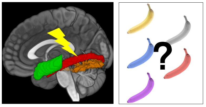
The focus of this study was on a specific type of ion channel, the CaV2.1 channel, which is the most common calcium ion channel in the brain. The ion channel is located at the synapse, at the very end of the neuron. When an electrical signal passes through the neuron, the ion channel open, setting in motion a process leading to neurotransmitter being released towards the receiving neuron in the synapse. In this way, CaV2.1 channels are the gatekeepers of synaptic, neuron-to-neuron communication.

Prolonged electrical activity reduces the number of CaV2.1 channels that can open, resulting in less transmitter release, so the receiving neuron receives a weaker message. It is as if the channels can ‘remember’ previous signalling, and in doing so, make themselves unavailable to open by subsequent signals. How this works at the molecular level has been unknown to scientists until now.

The Linköping researchers have now discovered a mechanism for how the ion channel can ‘remember’. The channel is a large molecule made up of several interconnected parts, which can move relative to each other in response to electrical signals. They discovered that the ion channel can take almost 200 different shapes depending on the strength and duration of an electrical signal; it is a very complex molecular machine.

“We believe that during sustained electrical nerve signalling, an important part of the molecule disconnects from the channel gate, similar to the way the clutch in a car breaks the connection between the engine and the wheels. The ion channel can then no longer be opened. When hundreds of signals occur over long enough time, they can convert most channels into this ‘declutched memory state’ for several seconds,” says Antonios Pantazis.

Target for future drugs

If the ion channel can ‘remember’ for just a few seconds, how does it contribute to lifelong learning? This type of collective memory in the ion channels can accumulate over time and reduce the communication between two neurons. This then leads to changes in the receiving neuron, lasting for hours or days. Eventually, it results in much longer-lived changes in the brain, such as the elimination of weakened synapses.

“In this way, a ‘memory’ that lasts for a few seconds in a single molecule can make a small contribution to a person’s memory that lasts for a lifetime,” says Antonios Pantazis.

Increased knowledge of how these calcium ion channels work can in the long term contribute to the treatment of certain diseases. There are many variants of the gene that produces the CaV2.1 channel, CACNA1A, that are linked to rare but serious neurological diseases, that often run in families. To develop drugs against these, it helps to know which part of the large ion channel you want to affect and in what way its activity should be changed.

“Our work pinpoints which part of the protein should be targeted when developing new drugs,” says Antonios Pantazis.

Source: Linköping University

Extending Ketamine’s Relieving Effect on Depression

Photo by Sydney Sims on Unsplash

For 30% of people with major depressive disorder (MDD), antidepressants don’t work. When infused at a low dose, ketamine shows remarkable efficacy as a rapidly acting antidepressant, with effects observed within hours even in patients who have been resistant to other antidepressant treatments. One drawback is that consistent infusions of ketamine are needed to maintain symptoms at bay, which could result in side effects, such as dissociative behaviours and the possibility of addiction, and stopping treatment can result in relapse.

In a new study published in Science, Lisa Monteggia’s and Ege Kavalali’s labs show that it is feasible to substantially extend the efficacy of a single dose of ketamine from its current duration of up to a week to a longer period of up to two months.

“The premise of this study, which was led by Zhenzhong Ma, a fantastic research assistant professor, was based on a testable mechanistic model that we developed that accounts for ketamine’s rapid antidepressant action,” Monteggia said.

Previously, researchers in the field had determined that ketamine’s antidepressant effect requires the activation of a key signalling pathway called ERK, but only ketamine’s long-term effects – not its rapid effects – are abolished when ERK is inhibited. As a fast-acting antidepressant, ketamine relies on ERK-dependent synaptic plasticity to produce its rapid behavioural effects. Ma and colleagues hypothesised that they could maintain ketamine’s effects for longer periods by enhancing ERK activity. 

In the recent paper, Ma discovered that ketamine’s antidepressant effects could be sustained for up to two months by using a drug called BCI, which inhibits a protein phosphatase and results in increased ERK activity. By inhibiting the phosphatase, the authors retained ERK’s activity and augmented the synaptic plasticity that drives ketamine’s prolonged antidepressant effects. 

lthough the use of BCI makes the application of these results to the clinic difficult, Monteggia said that the results provide a proof of principle that ketamine’s antidepressant action can be sustained by targeting intracellular signaling. She and Kavalali, the William Stokes Professor of Experimental Therapeutics and the chair of the Department of Pharmacology, have worked on the project since its inception and hope that it will foster other studies looking to identify specific molecules to enhance and sustain the action of a single dose of ketamine.

Ultimately, this work will be a stepping stone toward improving MDD patients’ lives by reducing the burden of treatment.

Source: Vanderbilt University

Glasgow Coma Scale Joined by New Measures to Assess TBI

Photo by Anna Shvets on Pexels

Trauma centres in the United States will begin to test a new approach for assessing traumatic brain injury (TBI) that is expected to lead to more accurate diagnoses and more appropriate treatment and follow-up for patients.

The new framework, which was developed by a coalition of experts and patients from 14 countries and spearheaded by the National Institutes of Health (NIH), expands the assessment beyond immediate clinical symptoms. Added criteria would include biomarkers, CT and MRI scans, and factors such as other medical conditions and how the trauma occurred.

The framework appears in the May 20 issue of Lancet Neurology.

For 51 years, trauma centres have used the Glasgow Coma Scale to assess patients with TBI, roughly dividing them into mild, moderate, and severe categories, based solely on their level of consciousness and a handful of other clinical symptoms.

That diagnosis determined the level of care patients received in the emergency department and afterward. For severe cases, it also influenced the guidance doctors gave the patients’ families, including recommendations around the removal of life support. Yet, doctors have long understood that those tests did not tell the whole story.

“There are patients diagnosed with concussion whose symptoms are dismissed and receive no follow-up because it’s ‘only’ concussion, and they go on to live with debilitating symptoms that destroy their quality of life,” said corresponding author Geoffrey Manley, MD, PhD, professor of neurosurgery at UC San Francisco and a member of the UCSF Weill Institute for Neurosciences. “On the other hand, there are patients diagnosed with ‘severe TBI’ who were eventually able to live full lives after their families were asked to consider removing life-sustaining treatment.”

In the US, TBI resulted in approximately 70 000 deaths in 2021 and accounts for about half-a-million permanent disabilities each year. Motor vehicle accidents, falls, and assault are the most common causes.

New system will better match patients to treatments

Known as CBI-M, the framework comprises four pillars – clinical, biomarker, imaging, and modifiers – that were developed by working groups of federal partners, TBI experts, scientists, and patients.

“The proposed framework marks a major step forward,” said co-senior author Michael McCrea, PhD, professor of neurosurgery and co-director of the Center for Neurotrauma Research at the Medical College of Wisconsin in Milwaukee. “We will be much better equipped to match patients to treatments that give them the best chance of survival, recovery, and return to normal life function.”

The framework was led by the NIH National Institute of Neurological Disorders and Stroke (NIH-NINDS), for which Manley, McCrea, and their co-first and co-senior authors are members of the steering committee on improving TBI characterisation.

The clinical pillar retains the Glasgow Coma Scale’s total score as a central element of the assessment, measuring consciousness and pupil reactivity as an indication of brain function. The framework recommends including the scale’s responses to eye, verbal, and motor commands or stimuli, presence of amnesia, and symptoms like headache, dizziness, and noise sensitivity.

“This pillar should be assessed as first priority in all patients,” said co-senior author Andrew Maas, MD, PhD, emeritus professor of neurosurgery at the Antwerp University Hospital and University of Antwerp, Belgium. “Research has shown that the elements of this pillar are highly predictive of injury severity and patient outcome.”

Biomarkers, imaging, modifiers offer critical clues to recovery

The second pillar uses biomarkers identified in blood tests to provide objective indicators of tissue damage, overcoming the limitations of clinical assessment that may inadvertently include symptoms unrelated to TBI.

Significantly, low levels of these biomarkers determine which patients do not require CT scans, reducing unnecessary radiation exposure and health care costs. These patients can then be discharged. In those with more severe injuries, CT and MRI imaging – the framework’s third pillar – are important in identifying blood clots, bleeding, and lesions that point to present and future symptoms.

Source: University of California – San Francisco

Study Sheds Light on How Autistic People Communicate

Photo by Peter Burdon on Unsplash

There is no significant difference in the effectiveness of how autistic and non-autistic people communicate, according to a new study, challenging the stereotype that autistic people struggle to connect with others.

The findings, published in Nature Human Behaviour, suggest that social difficulties often faced by autistic people are more about differences in how autistic and non-autistic people communicate, rather than a lack of social ability in autistic individuals, experts say. 

Researchers hope the results of the study will help reduce the stigma surrounding autism, and lead to more effective communication support for autistic people.  

Direct communication

Autism is a lifelong neurodivergence and disability, and influences how people experience and interact with the world. 

Autistic people often communicate more directly and may struggle with reading social cues and body language, leading to differences in how they engage in conversation compared to non-autistic people. 

Story sharing

The study, led by experts from the University of Edinburgh, tested how effectively information was passed between 311 autistic and non-autistic people. 

Participants were tested in groups where everyone was autistic, everyone was non-autistic, or a combination of both. 

The first person in the group heard a story from the researcher, then passed it along to the next person. Each person had to remember and repeat the story, and the last person in the chain recalled the story aloud. 

The amount of information passed on at each point in the chain was scored to discern how effective participants were at sharing the story. Researchers found there were no differences between autistic, non-autistic, and mixed groups.  

Increased awareness

After the task, participants rated how much they enjoyed the interaction with the other participants, based on how friendly, easy, or awkward the exchange was.  

Researchers found that non-autistic people preferred interacting with others like themselves, and autistic people preferred learning from fellow autistic individuals. This is likely down to the different ways that autistic and non-autistic people communicate, experts say.  

The findings confirm similar findings from a previous smaller study undertaken by the same researchers. They say the new evidence should lead to increased understanding of autistic communication styles as a difference, not a deficiency.   

Autism has often been associated with social impairments, both colloquially and in clinical criteria. Researchers have spent a lot of time trying to ‘fix’ autistic communication, but this study shows that despite autistic and non-autistic people communicating differently it is just as successful. With opportunities for autistic people often limited by misconceptions and misunderstandings, this new research could lead the way to bridging the communication gap and create more inclusive spaces for all.

 Dr Catherine Crompton, Chancellor’s Fellow at the University of Edinburgh’s Centre for Clinical Brain Sciences

Language Shapes how Sensory Experiences are Stored in the Brain

A study in stroke patients shows the brain’s vision-language connection shapes object knowledge

A schematic view of the main findings, adapted from a brain figure in the study. Image credit: Adapted from Liu Bet al., 2025, PLOS Biology, CC-BY 4.0

Our ability to store information about familiar objects depends on the connection between visual and language processing regions in the brain, according to a study published May 20th in the open-access journal PLOS Biology by Bo Liu from Beijing Normal University, China, and colleagues.

Seeing an object and knowing visual information about it, like its usual colour, activate the same parts of the brain. Seeing a yellow banana, for example, and knowing that the object represented by the word “banana” is usually yellow, both excite the ventral occipitotemporal cortex (VOTC). However, there’s evidence that parts of the brain involved in language, like the dorsal anterior temporal lobe (ATL), are also involved in this process – dementia patients with ATL damage, for example, struggle with object colour knowledge, despite having relatively normal visual processing areas. To understand whether communication between the brain’s language and sensory association systems is necessary for representing information about objects, the authors tested whether stroke-induced damage to the neural pathways connecting these two systems impacted patients’ ability to match objects to their typical colour. They compared colour-identification behaviour in 33 stroke patients to 35 demographically-matched controls, using fMRI to record brain activity and diffusion imaging to map the white matter connections between language regions and the VOTC.

The researchers found that stronger connections between language and visual processing regions correlated with stronger object color representations in the VOTC, and supported better performance on object color knowledge tasks. These effects couldn’t be explained by variations in patients’ stroke lesions, related cognitive processes (like simply recognizing a patch of color), or problems with earlier stages of visual processing. The authors suggest that these results highlight the sophisticated connection between vision and language in the human brain.

The authors add, “Our findings reveal that the brain’s ability to store and retrieve object perceptual knowledge – like the colour of a banana – relies on critical connections between visual and language systems. Damage to these connections disrupts both brain activity and behaviour, showing that language isn’t just for communication – it fundamentally shapes how sensory experiences are neurally structured into knowledge.”

Provided by PLOS

Could the Brain be Targeted to Treat Type 2 Diabetes?

Source: CC0

Successfully treating type 2 diabetes may involve focusing on brain neurons, rather than simply concentrating on obesity or insulin resistance, according to a study published in the Journal of Clinical Investigation.  

For several years, researchers have known that hyperactivity of a subset of neurons located in the hypothalamus, called AgRP neurons, is common in mice with diabetes. 

“These neurons are playing an outsized role in hyperglycaemia and type 2 diabetes,” said UW Medicine endocrinologist Dr Michael Schwartz, corresponding author of the paper.

To determine if these neurons contribute to elevated blood sugar in diabetic mice, researchers employed a widely used viral genetics approach to make AgRP neurons express tetanus toxin, which prevents the neurons from communicating with other neurons. 

Unexpectedly, this intervention normalised high blood sugar for months, despite having no effect on body weight or food consumption.   

Conventional wisdom is that diabetes, particularly type 2 diabetes, stems from a combination of genetic predisposition and lifestyle factors, including obesity, lack of physical activity and poor diet. This mix of factors leads to insulin resistance or insufficient insulin production.  

Until now, scientists have traditionally thought the brain doesn’t play a role in type 2 diabetes, according to Schwartz. 

The paper challenges this and is a “departure from the conventional wisdom of what causes diabetes,” he said. 

The new findings align with studies published by the same scientists showing that injection of a peptide called FGF1 directly into the brain also causes diabetes remission in mice. This effect was subsequently shown to involve sustained inhibition of AgRP neurons.

Together, the data suggest that, while these neurons are important for controlling blood sugar in diabetes, they don’t play a major role in causing obesity in these mice, the researchers noted in their report.  

In other words, targeting these neurons may not reverse obesity, even as it causes diabetes to go into remission, Schwartz explained. 

More research is needed on how to regulate activity in these neurons, and how they become hyperactive in the first place, he said. Once these questions are answered, Schwartz said that a therapeutic approach might then be developed to calm them down. 

This approach could represent a shift in how clinicians understand and treat this chronic disease, Schwartz said.  He noted, for instance, that semaglutide and other new drugs used to treat type 2 diabetes are also able to inhibit AgRP neurons.  

The extent to which this effect contributes to the antidiabetic action of these drugs is unknown. Further research might help scientists to better understand the role of AgRP neurons in how the body normally controls blood sugar, and to ultimately translate these findings into human clinical trials, he added.  

Source: University of Washington School of Medicine/UW Medicine

New Auditory Brainstem Implant Shows Early Promise

A new study co-led by Mass General Brigham researchers points to a promising new type of auditory brainstem implant (ABI) that could benefit people who are deaf due to Neurofibromatosis type 2 (NF2) and other severe inner ear abnormalities that prevent them from receiving cochlear implants. With further tests and trials, researchers hope it will provide a more effective treatment alternative than what is currently used.

In the new research, published in Nature Biomedical Engineering, scientists at Mass Eye and Ear, a member of the Mass General Brigham healthcare system, collaborated with scientists at the École Polytechnique Fédérale de Lausanne (EPFL) in Geneva, Switzerland, to report on a new class of soft, flexible ABIs that were designed to address the limitations of those currently used. These implants bypass damaged auditory structures and directly stimulate the brainstem’s sound-processing region to restore auditory function.

The new ABI was borne out of a decade-long collaboration between Mass Eye and Ear and EPFL scientists. It features an elastic, multilayer construct that includes ultra-thin platinum electrodes and silicone, a novel design that allows it to conform closely to the brainstem’s curved surface.

Conventional ABIs that are sometimes used in patients with NF2 rely on stiff electrodes that struggle to conform to the curved surface of the cochlear nucleus in the brainstem. That limits their effectiveness to modest benefits, typically providing only basic sound awareness to aid lip reading. The design can also cause side effects like discomfort that discourages long-term use.

The novel, soft electrode design was developed using advanced thin-film processing techniques, allowing for closer contact and more precise stimulation. In preclinical tests conducted in Switzerland, two macaques received the implants and underwent several months of behavioural testing. Results showed the animals could consistently distinguish between different patterns of stimulation – which indicated high-resolution auditory perception, a promising sign for eventual human use.

“While cochlear implants are life-changing for many, there remains a group of patients for whom current technology falls short,” said study co-senior author Daniel J. Lee, MD, FACS, Ansin Foundation Chair in Otolaryngology at Mass Eye and Ear. “Our research lays the groundwork for a future auditory brainstem implant that could improve hearing outcomes and reduce side effects in patients who are deaf and do not benefit from the cochlear implant.”

Source: Mass Eye and Ear

‘Sweet Spot’ for Focused Ultrasound Provides Relief from Essential Tremor

A surgical lesion site is highlighted in orange following MR-guided focused ultrasound treatment. Structural brain connections associated with optimal tremor response or side effects, as identified in the present study, are depicted in various colors. The background features an ultra-high resolution MRI image acquired at Massachusetts General Hospital. Image courtesy of Andreas Horn, Mass General Brigham.

Essential tremor, a common neurological movement disorder, causes uncontrollable shaking, most often in the hands, but it can also occur in the arms, legs, head, voice, or torso. Essential tremor impacts an estimated 1% of the worldwide population and around 5% of people over 60.

Investigators from Mass General Brigham identified a specific subregion of the brain’s thalamus that, when included during magnetic resonance-guided focused ultrasound (MRgFUS) treatment, can result in optimal and significant tremor improvements while reducing side effects. Their results are published in Science Advances.

“This one-time, noninvasive treatment can have immediate, long-lasting and lifechanging effects for patients and was pioneered here at Brigham and Women’s Hospital 30 years ago,” said co-senior author G. Rees Cosgrove, MD, FRCSC, director of functional neurosurgery at Brigham and Women’s Hospital. “The results of this study will help make the procedure even more safe and effective than it already is and will help other centres around the world improve their outcomes.”

MRgFUS treatment of essential tremor creates a small, permanent lesion in a specific nucleus in the thalamus that is thought to be part of the brain circuit mediating the disorder and disrupts the tremor-causing activity. The research team analysed data from 351 thalamotomy patients that were treated across three international hospitals, the largest cohort assessed to date, to identify the optimal location for this procedure and better understand its impacts on clinical improvements and side effects.

The study identified a set of optimal sites and brain connections to target, as well as locations and connections to avoid that lead to side effects. The team then tested whether this ‘sweet spot’ could be used as a model to predict the outcomes in a cohort of patients treated with the same procedure at another centre, which proved true. The more the ‘sweet spot’ was lesioned, the better the outcome was in all patients’ one-year, post-procedure comparison data. According to the researchers, when thalamotomy patients have good tremor control at one year, it is typically sustained over multiple years.

“Seeing how this procedure can make such a huge impact on patients’ lives is what motivated me to pursue this research,” said lead author Melissa Chua, MD, a senior resident in the Brigham’s Department of Neurosurgery. “It is very exciting to have such robust validation and to be moving toward this treatment becoming even more precise and personalized in the future.”

Next, the team plans to further analyse patient data for a more detailed picture of the evolution of this technology and how patient outcomes have improved, to fully understand the parameters that go into achieving long-term tremor control and minimise side effects.

“It is incredible when you can provide a patient with relief from these tremors,” Cosgrove said. “It is like a gift when patients who have not been able to sing, speak in public, write, or even drink from a cup for years can once again do so – we see it in case after case.”

Source: Mass General Brigham

Could a Transient Ischaemic Attack Leave Lasting Fatigue?

Photo by Karolina Grabowska on Pexels

A transient ischaemic attack (TIA) is typically defined as a temporary blockage of blood flow to the brain that causes symptoms that go away within a day, but a new study finds that people who have this type of stroke may also have prolonged fatigue lasting up to one year. The study is published in Neurology®, the medical journal of the American Academy of Neurology (AAN).

The study does not prove that TIAs, also known as mini-strokes, cause lasting fatigue; it only shows an association. “People with a transient ischaemic attack can have symptoms such as face drooping, arm weakness or slurred speech and these resolve within a day,” said study author Boris Modrau, MD, PhD, of Aalborg University Hospital in Denmark. “However, some have reported continued challenges including reduced quality of life, thinking problems, depression, anxiety and fatigue. Our study found that for some people, fatigue was a common symptom that lasted up to one year after the transient ischaemic attack.”

The study involved 354 people with an average age of 70 who had a mini-stroke. They were followed for a year.

Participants completed questionnaires about their level of fatigue within the first two weeks of the mini-stroke and again at three, six, and 12 months later. One questionnaire looked at five different types of fatigue, including overall tiredness, physical tiredness, reduced activity, reduced motivation and mental fatigue. Scores ranged from four to 20 with higher scores indicating more fatigue. Participants had an average score of 12.3 at the start of the study. At three months, the average score decreased slightly to 11.9, at six months to 11.4 and at twelve months to 11.1.

Researchers looked at how many participants experienced fatigue as defined as a score of 12 or higher. Of the participants, 61% experienced fatigue two weeks after the mini-stroke and 54% experienced fatigue at each of the three other testing time periods at three, six and 12 months.

Participants also had brain scans. Researchers found that the presence of a blot clot on a scan was equal between people with long term fatigue and those without it, so this did not explain the reason for the level of fatigue.

Researchers did find that previous anxiety or depression was twice as common in those participants who reported lasting fatigue.

“Long-term fatigue was common in our group of study participants, and we found if people experience fatigue within two weeks after leaving the hospital, it is likely they will continue to have fatigue for up to a year,” said Modrau. “For future studies, people diagnosed with a transient ischaemic attack should be followed in the weeks and months that follow to be assessed for lingering fatigue. This could help us better understand who might struggle with fatigue long-term and require further care.”

A limitation of the study was that while participants were asked to complete the questionnaires themselves, it is possible some responses may have been completed with assistance from relatives or caretakers and this may have influenced responses, including those around fatigue.

Source: American Academy of Neurology

Under Different Anaesthetics, Same Result: Unconsciousness by Shifting Brainwave Phase

MIT study finds that an easily measurable brain wave shift may be a universal marker of unconsciousness under anaesthesia

Photo by Anna Shvets on Pexels

At the level of molecules and cells, ketamine and dexmedetomidine work very differently, but in the operating room, they do the same exact thing: anaesthetise the patient. By demonstrating how these distinct drugs achieve the same result, a new study in animals by neuroscientists at The Picower Institute for Learning and Memory at MIT identifies a potential signature of unconsciousness that is readily measurable to improve anaesthesiology care.

What the two drugs have in common, the researchers discovered, is the way they push around brain waves, which are produced by the collective electrical activity of neurons. When brain waves are in phase, meaning the peaks and valleys of the waves are aligned, local groups of neurons in the brain’s cortex can share information to produce conscious cognitive functions such as attention, perception and reasoning, said Picower Professor Earl K. Miller, senior author of the new study in Cell Reports. When brain waves fall out of phase, local communications, and therefore functions, fall apart, producing unconsciousness.

The finding, led by graduate student Alexandra Bardon, not only adds to scientists’ understanding of the dividing line between consciousness and unconsciousness, Miller said, but also could provide a common new measure for anesthesiologists who use a variety of different anesthetics to maintain patients on the proper side of that line during surgery.

“If you look at the way phase is shifted in our recordings, you can barely tell which drug it was,” said Miller, a faculty member in The Picower Institute and MIT’s Department of Brain and Cognitive Sciences. “That’s valuable for medical practice.  Plus if unconsciousness has a universal signature, it could also reveal the mechanisms that generate consciousness.”

A figure from the paper summarises the main findings. Under either ketamine or dexmedetomidine general anaesthesia, brain waves become shifted out of phase within a hemisphere and more into phase across hemispheres.

If more anesthetic drugs are also shown to affect phase in the same way, then anaesthesiologists might be able to use brain wave phase alignment as a reliable marker of unconsciousness as they titrate doses of anesthetic drugs, Miller said, regardless of which particular mix of drugs they are using. That insight could aid efforts to build closed-loop systems that can aid anaesthesiologists by constantly adjusting drug dose based on brain wave measurements of the patient’s unconsciousness.

Miller has been collaborating with study co-author Emery N. Brown, an anaesthesiologist and Professor of Computational Neuroscience and Medical Engineering, on building such a system. In a recent clinical trial with colleagues in Japan, Brown demonstrated that monitoring brain wave power signals using EEG enabled an anaesthesiologist to use much less sevoflurane during surgery with young children. The reduced doses proved safe and were associated with many improved clinical outcomes, including a reduced incidence of post-operative delirium.

Phase findings

Neuroscientists studying anaesthesia have rarely paid attention to phase, but in the new study, Bardon, Brown and Miller’s team made a point of it as they anaesthetised two animals.

After the animals lost consciousness, the measurements indicated a substantial increase in “phase locking,” especially at low frequencies. Phase locking means that the relative differences in phase remained more stable. But what caught the researchers’ attention were the differences that became locked in: Within each hemisphere, regardless of which anesthetic they used, brain wave phase became misaligned between the dorsolateral and ventrolateral regions of the prefrontal cortex.

Surprisingly, brain wave phase across hemispheres became more aligned, not less. But Miller notes that case is still a big shift from the conscious state, in which brain hemispheres are typically not aligned well, so the finding is a further indication that major changes of phase alignment, albeit in different ways at different distances, are a correlate of unconsciousness compared to wakefulness.

“The increase in interhemispheric alignment of activity by anesthetics seems to reverse the pattern observed in the awake, cognitively engaged brain,” the Bardon and Miller team wrote in Cell Reports.

Determined by distance

Distance proved to be a major factor in determining the change in phase alignment. Even across the 2.5 millimetres of a single electrode array, low-frequency waves moved 20-30 degrees out of alignment. Across the 20 or so millimetres between arrays in the upper (dorsolateral) and lower (ventrolateral) regions within a hemisphere, that would mean a roughly 180-degree shift in phase alignment, which is a complete offset of the waves.

The dependence on distance is consistent with the idea of waves traveling across the cortex, Miller said. Indeed in a 2022 study, Miller and Brown’s labs showed that the anaesthetic propofol induced a powerful low-frequency traveling wave that swept straight across the cortex, overwhelming higher-frequency straight and rotating waves.

The new results raise many opportunities for follow-up studies, Miller said. Does propofol also produce this signature of changed phase alignment? What role do travelling waves play in the phenomenon? And given that sleep is also characterised by increased power in slow wave frequencies, but is definitely not the same state as anaesthesia-induced unconsciousness, could phase alignment explain the difference?

Source: Picower Institute