Older people with a genetic risk of Alzheimer’s disease did not experience the expected increase in cognitive decline and dementia risk if they consumed relatively large amounts of meat. This is shown in a new study from Karolinska Institutet published in JAMA Network Open. The results may contribute to the development of more individually tailored dietary advice.
APOE is a gene that affects the risk of Alzheimer’s disease. In Sweden, approximately 30 per cent of the population are carriers of the gene combinations APOE 3/4 or APOE 4/4. Among people with Alzheimer’s disease, those with these genotypes account for nearly 70 per cent.
When the Swedish Food Agency presented an overview of research on the link between diet and dementia last year, more research was requested to assess a possible link between meat consumption and the development of dementia.
‘This study tested the hypothesis that people with APOE 3/4 and 4/4 would have a reduced risk of cognitive decline and dementia with higher meat intake, based on the fact that APOE4 is the evolutionarily oldest variant of the APOE gene and may have arisen during a period when our evolutionary ancestors ate a more animal-based diet,’ says first author Jakob Norgren, researcher at the Department of Neurobiology, Care Sciences and Society, Karolinska Institutet.
The study followed more than 2100 participants in the Swedish National Study on Aging and Care, Kungsholmen (SNAC-K) for up to 15 years. All were aged 60 or older and had no diagnosis of dementia at the start of the study. The association between self-reported diet and cognitive health measures was analysed, adjusting for age, sex, education and lifestyle factors.
Twice the risk of dementia
At lower meat intake, the group with APOE 3/4 and 4/4 had more than twice the risk of dementia than people without these gene variants. However, the increased risk of cognitive decline and dementia in the risk groups was not seen in the fifth of participants who consumed the most meat. Their median consumption is estimated at approximately 870 grams of meat per week, standardised to a daily energy intake of 2,000 calories.
‘Those who ate more meat overall had significantly slower cognitive decline and a lower risk of dementia, but only if they had the APOE 3/4 or 4/4 gene variants,’ says Jakob Norgren. He continues:
‘There is a lack of dietary research into brain health, and our findings suggest that conventional dietary advice may be unfavourable to a genetically defined subgroup of the population. For those who are aware that they belong to this genetic risk group, the findings offer hope; the risk may be modifiable through lifestyle changes. ‘
The study also shows that the type of meat is important.
‘A lower proportion of processed meat in total meat consumption was associated with a lower risk of dementia regardless of APOE genotype,’ says Sara Garcia-Ptacek, assistant professor at the same department, who together with senior lecturer Erika J Laukka is the study’s last author.
The findings also extend beyond brain health. In a follow-up analysis, the researchers observed a significant reduction in all-cause-mortality in carriers of APOE 3/4 and 4/4 with higher consumption of unprocessed meat.
However, the study is observational and needs to be followed up with intervention studies that can better demonstrate causal relationships.
‘Clinical trials are now needed to develop dietary recommendations tailored to APOE genotype,’ says Jakob Norgren. He continues:
‘Since the prevalence of APOE4 is about twice as high in the Nordic countries as in the Mediterranean countries, we are particularly well suited to conduct research on tailored dietary recommendations for this risk group.’
The research was funded by, among others, the Swedish Alzheimer’s Foundation, the Swedish Dementia Foundation, the Emil and Wera Cornell Foundation, the Leif Lundblad family and other philanthropists, the Swedish Research Council and FORTE. The researchers state that they have no related conflicts of interest.
APOE Gene Facts:
Apolipoprotein E plays a central role in the transport of cholesterol and fats in the brain and blood. The protein is encoded by the APOE gene, which exists in three main variants: epsilon 2, 3 and 4. These variants affect the risk of developing Alzheimer’s disease and cardiovascular disease. Each person inherits two APOE genes, one from each parent, giving six possible combinations (genotypes): 2/2, 2/3, 2/4, 3/3, 3/4 and 4/4.
Compared to the most common genotype 3/3, one 4 variant increases the risk of Alzheimer’s disease by about three to four times and two 4 variants by about ten to fifteen times, while the 2 variant is associated with a lower risk. However, the increase in risk varies between different ethnic groups.
Researchers have discovered new diagnostic and prognostic markers for multiple sclerosis.
This is a pseudo-colored image of high-resolution gradient-echo MRI scan of a fixed cerebral hemisphere from a person with multiple sclerosis.
Credit: Govind Bhagavatheeshwaran, Daniel Reich, National Institute of Neurological Disorders and Stroke, National Institutes of Health
Researchers from the Max Planck Institute of Biochemistry and the Technical University of Munich (TUM) have discovered new diagnostic markers for multiple sclerosis (MS), a disease which affects 3 million people worldwide.
Using mass spectrometry, about 1500 proteins were analysed simultaneously per sample in the cerebrospinal fluid (CSF) of 5000 patients. The study uncovered a set of marker proteins that improve differentiation of MS from other inflammatory brain diseases where classical MS markers are negative. Additionally, the study identified changes in the CSF proteome that may potentially predict disease progression. This approach could also open up new avenues for the diagnosis of other diseases. The study was published in Cell.
Unspecific neurological symptoms can lead to lengthy or even inaccurate diagnoses of diseases, which is why improved protein markers are needed for swift and clear diagnosis
Using a new mass spectrometry method, approximately 1500 proteins were analyzed per cerebrospinal fluid sample across 5000 patients, and up to 2000 proteins in a further improved method
A new set of disease markers enable improved differentiation of multiple sclerosis (MS) from other inflammatory brain diseases, in particular for patients lacking the classical markers
The proteome of the cerebrospinal fluid (CSF) at diagnosis is informative for various aspects of disease evolution in a patient, such as long-term disability, risk of conversion from relapsing to progressive disease course and time to conversion
The method also has the potential to discover other proteins that could be used as markers for the diagnosis of other neurological diseases
Biomarker needs for multiple sclerosis
Imagine living with unexplained neurological symptoms: numbness, visual disturbances, fatigue, but not receiving a clear diagnosis for months or years. Non-specific neurological symptoms can make diagnosis difficult because, despite modern imaging techniques, there are no reliable molecular biomarkers for many neurological diseases.
Professor Bernhard Hemmer, head of the Department of Neurology at TUM University Hospital, explains: “The diagnosis of neurological diseases such as multiple sclerosis is based on a combination of imaging techniques using magnetic resonance imaging (MRI) and cerebrospinal fluid (CSF) analysis. While MRI reveals inflammatory changes in the brain and spinal cord, CSF shows chronic immune activity in the nervous system. In most cases, this combination enables a reliable diagnosis. In individual cases, however, differentiation can be challenging. This can lead to lengthy and less reliable diagnoses and is associated with uncertain and delayed treatment decisions. For this reason, we need new biomarkers to better diagnose the various diseases. In addition to diagnostic challenges, predicting disease progression, particularly disability accumulation, to guide optimal treatment, remains a major unmet need in MS”.
Proteomic study of cerebrospinal fluid across neurological diseases
In order to find new biomarkers, neurologists Bernhard Hemmer and Christiane Gasperi, both experts in MS research at TUM, have joined forces with Professor Matthias Mann, a world-leading expert in proteomics research. Matthias Mann, director at the MPI of Biochemistry, explains: “We have been developing the technology for measuring proteins using mass spectrometry in our laboratory together with colleagues for decades. Now we can reliably and accurately measure proteins in body fluids. However, for a long time researchers could only measure tens to hundreds of samples and only those proteins with the highest concentration in a body fluid. These proteins often turned out not to be the best markers for diseases. To go one step further, we combined the latest advances in mass spectrometry hardware, software, and sample preparation and adapted the workflow to cerebrospinal fluid.”
In this study, CSF samples from more than 5000 people with a wide range of neurological diseases were analysed. Jakob Bader, first author of the study and postdoctoral researcher in the field of proteomics research, explains: “Proteomics is a scientific discipline that aims to characterise a biological system by measuring all proteins, or at least as many as possible. For our study, it is essential to cover as many proteins as possible in order to increase the likelihood of measuring and later finding real disease markers in our analyses. The great advantage of this proteomic approach is that the identity of the markers does not have to be known beforehand. This saves years of research work in which individual candidates are examined one after the other.”
To avoid misinterpreting random differences between people as disease markers, it is essential to have a sufficient number of patients. Similarly, it is only possible to determine whether a marker is specific to a particular disease by considering the many other relevant diseases in parallel. “The breakthrough was achieving both objectives simultaneously: Analysing thousands of proteins while studying thousands of patients across many neurological diseases.” Jakob Bader adds.
A systematic analysis of disease effects and possible confounders
The 5,000 CSF samples came from a wide range of neurological disorders, including stroke, brain cancer, infections, autoimmune diseases such as MS, and others. Additionally, patient samples were analysed from individuals who had provided CSF samples for the diagnosis of severe headache disorders but in whom no neurological disease was found. This allowed the researchers to use these samples as controls. Systematic comparison of these disorders revealed shared and specific protein deviations from the controls.
For diagnostic use, an elevated protein concentration rarely points unambiguously to a single disorder. The study further revealed that disease-unspecific other effects like a person’s age, sex, and in particular degradation of the barriers insulating the brain from the CSF have a very large impact on the composition of this fluid, which complicates the quest for disease markers.
Biomarkers for a hard-to-identify form of multiple sclerosis
To showcase the potential of proteomic analysis for biomarker discovery, the researchers focused on the search for diagnostic markers for MS, a challenging task but with a direct medical need. Physician Christiane Gasperi says: “In approximately 10% of MS patients, diagnosis of the disease is particularly difficult because they lack the typical MS marker of so-called oligoclonal bands of antibodies that are specific to the CSF and not found in the blood. This complicates and potentially delays the diagnosis.”
She continues: “However, for our patients, a quick and clear diagnosis of the disease is of enormous importance. While current therapies cannot cure MS, they can slow its progression and reduce the long-term disability. That makes it crucial to start treatment early. At the same time, these therapies can have significant side effects, so treatment decisions require a high level of diagnostic certainty. When this confidence is not reached yet, therapy is often delayed. Thus, MS patients really benefit from an early intervention that depends on a clear and early diagnosis.”
To find better markers, the researchers applied an enhanced version of the proteomic method to measure about 2000 proteins in samples of MS and other inflammatory diseases of the CNS, which can mimic MS, and thus pose the greatest diagnostic challenges. This let them identify a set of 22 proteins that distinguishes MS from these inflammatory diseases with better accuracy than other parameters in the CSF that are currently measured in clinical practice. Christiane Gasperi comments: “It is particularly encouraging that we have found a combination of marker proteins that help in the diagnosis of this particularly difficult-to-identify form of MS.”
Predicting disease progression at diagnosis
Beyond improving diagnosis, the study also addressed a second major challenge: Some patients remain relatively stable for many years, while others accumulate disability more rapidly or transition from the relapsing disease course that is typical early on to a progressive course where disability accumulates persistently. At the time of diagnosis, it is very difficult to predict which trajectory a patient will follow. This uncertainty complicates treatment decisions and can be deeply unsettling for those newly diagnosed.
By analyzing hundreds of MS patient samples, the researchers showed that the CSF proteome at the time of diagnosis was associated with the level of disability years later. In addition, these patterns reflected a higher risk for patients to convert from the relapsing to the progressive disease course, as well as shorter times until such conversion occurred. Bernhard Hemmer explains: “Our findings suggest that important aspects of future disability and disease course are reflected in the proteome from the very beginning. This demonstrates that the biological information required for a prognostic test is already present at diagnosis.”
He summaries the study: “For diagnosis, we were able to define and validate a focused protein panel that improves differentiation in difficult cases. Additionally, we found that the overall protein pattern in the CSF at the time of diagnosis is linked to how the disease develops years later. Together, these findings bring us closer to more precise diagnosis and a more individualized treatment strategy from the very beginning.
An avenue for efficient biomarker discovery in neurology
Matthias Mann sees broader potential: “Proteins control almost all biological processes in the body and have long been the most important group of diagnostic markers. Nevertheless, we are probably only at the beginning here. With the methodology established here, we can now analyse the proteome in the CSF of many patients with an unprecedented number of proteins. This technological progress changes how we should search for biomarkers. Comprehensive proteome analysis of large patient collectives promise to be the most efficient path to new and better biomarkers. Beyond MS, this approach opens up prospects for many other diseases of the central nervous system – from Alzheimer’s and Parkinson’s to brain tumours and other neurological disorders.”
Cohort study finds people with stroke may be extra susceptible to air pollution’s impact on the brain
Photo by Kouji Tsuru on Pexels
People with greater exposure to air pollution face a higher risk of developing Alzheimer’s disease, according to a new study by Yanling Deng of Emory University, U.S.A., and colleagues, published February 17th in the open-access journal PLOS Medicine.
Alzheimer’s disease is the most common form of dementia, affecting about 57 million people worldwide. Exposure to air pollution is a known risk factor for Alzheimer’s disease, and for several common chronic health conditions, such as hypertension, stroke and depression. These chronic conditions are also linked to Alzheimer’s disease, but previously it was unclear whether air pollution causes these chronic conditions, which then lead to dementia, or if these conditions might amplify the effects of air pollution on brain health.
A team at Emory University studied more than 27.8 million U.S. Medicare recipients aged 65 years and older from 2000 to 2018. The researchers looked at individuals’ air pollution exposure level and whether they developed Alzheimer’s disease, while emphasizing the role of other chronic conditions. They found that greater exposure to air pollution was associated with an increased risk of Alzheimer’s disease, and that association was slightly stronger in individuals who had experienced a stroke. Hypertension and depression, however, had little additional impact.
Overall, the findings suggest that air pollution contributes to Alzheimer’s disease mostly through direct pathways rather than through other chronic health conditions. However, people with a history of stroke may be especially susceptible to the harmful effects of air pollution on brain health. The study indicates that improving air quality could be an important way to prevent dementia and protect older adults.
The authors add, “In this large national study of older adults, we found that long-term exposure to fine particulate air pollution was associated with a higher risk of Alzheimer’s disease, largely through direct effects on the brain rather than through common chronic conditions such as hypertension, stroke, or depression.”
“Our findings suggest that individuals with a history of stroke may be particularly vulnerable to the harmful effects of air pollution on brain health, highlighting an important intersection between environmental and vascular risk factors.”
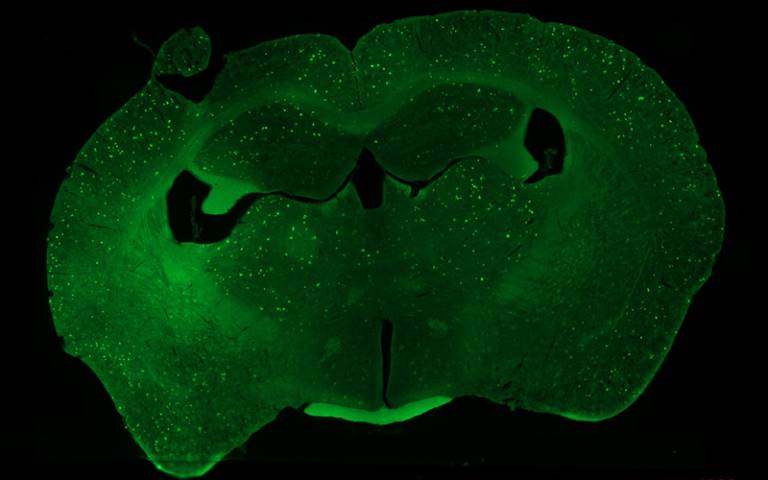
Mouse brain section highlights amyloid plaques, seen as bright green flecks (due to staining). Credit: Shipley et al.
Memory dysfunction in Alzheimer’s disease may be linked to impairment in how the brain replays our recent experiences while we are resting, according to a new study in mice by UCL scientists. The researchers say their findings, published in Current Biology, could help scientists develop drug treatments targeting this impaired brain function, or help design new tests for early diagnosis.
Co-lead author Dr Sarah Shipley (UCL Cell & Developmental Biology) said: “Alzheimer’s disease is caused by the build-up of harmful proteins and plaques in the brain, leading to symptoms such as memory loss and impaired navigation – but it’s not well understood exactly how these plaques disrupt normal brain processes.
“We wanted to understand how the function of brain cells changes as the disease develops, to identify what’s driving these symptoms.
“When we rest, our brains normally replay recent experiences – this is thought to be key to how memories are formed and maintained. We found this replay process is disrupted in mice engineered to develop the amyloid plaques characteristic of Alzheimer’s, and this disruption is associated with how badly animals perform on memory tasks.”
The replay process, which occurs in the brain’s hippocampus, involves place cells firing in rapid sequences during rest. Place cells – discovered by Nobel prize-winning UCL neuroscientist Professor John O’Keefe – are neurons (brain cells) that represent specific locations. When we visit somewhere, particular place cells fire, and as we move the cells fire in a sequence. Later, when we rest, these cells reactivate in the same sequence, helping memories become ingrained.
For the study, the researchers were testing how well mice performed in a simple maze task, while monitoring their brain activity with sets of electrodes that could simultaneously track roughly 100 individual place cells.
In mice with amyloid pathology, the replay process was fundamentally altered. Surprisingly, replay events occurred just as frequently as in healthy mice, but their structure was disorganised. The normal, coordinated patterns of place cell activity that should reinforce memories were scrambled. The researchers also found that place cells in affected mice became less stable over time, with individual neurons no longer reliably coding the same locations, particularly after rest periods – precisely when replay should be strengthening these representations.
This disruption had consequences on memory tasks: affected mice performed worse in the maze, appearing to forget where they had already been and revisiting corridors that led nowhere.
Co-lead author Professor Caswell Barry (UCL Cell & Developmental Biology) said: “We’ve uncovered a breakdown in how the brain consolidates memories, visible at the level of individual neurons. What’s striking is that replay events still occur – but they’ve lost their normal structure. It’s not that the brain stops trying to consolidate memories; the process itself has gone wrong.
“We hope our findings could help develop tests to detect Alzheimer’s early, before extensive damage has occurred, or lead to new treatments targeting this replay process. We’re now investigating whether we can manipulate replay through the neurotransmitter acetylcholine, which is already targeted by drugs used to treat Alzheimer’s symptoms. By understanding the mechanism better, we hope to make such treatments more effective.”
In research published in Developmental Medicine & Child Neurology, investigators developed a brief, reliable, and valid screening tool to help identify individuals with Duchenne muscular dystrophy (a neuromuscular disorder) who are at increased risk of brain-related comorbidities, such as language disorders, attention-deficit/hyperactivity disorder, and anxiety.
The research team developed the questionnaire-based screening tool, called the BIND (Brain Involvement iN Dystrophinopathies) screener, by reviewing the medical literature and incorporating expert consensus, and translated it into 11 languages. The questionnaire asks parents, caregivers, or patients to rate the impact of 18 cognitive, behavioural, and emotional items.
The BIND screener demonstrated strong accuracy in identifying individuals with Duchenne muscular dystrophy who had previously been diagnosed with neurodevelopmental or psychiatric conditions in an international sample of 835 participants. Additional validation was conducted in a subsample of 90 children and adolescents who underwent in-depth cognitive and clinical assessments.
“Families often tell us that cognitive and behavioural difficulties can be as challenging as the physical symptoms of Duchenne. This screener is designed to help identify those concerns earlier, so that children and adults can be referred for appropriate support,” said corresponding author Rubén Miranda, PhD, of Universidad Complutense de Madrid, Spain.
This is a pseudo-colored image of high-resolution gradient-echo MRI scan of a fixed cerebral hemisphere from a person with multiple sclerosis.
Credit: Govind Bhagavatheeshwaran, Daniel Reich, National Institute of Neurological Disorders and Stroke, National Institutes of Health
The immune system’s reaction to the common Epstein-Barr virus can ultimately damage the brain and contribute to multiple sclerosis (MS). This is shown by new research from Karolinska Institutet, published in Cell. The study provides new insight into the long-suspected link between Epstein-Barr virus (EBV) and MS.
Multiple sclerosis is a chronic inflammatory disease in which the immune system attacks the central nervous system and causes nerve damage. It has long been known that everyone who develops MS has had an infection with the Epstein-Barr virus (EBV) – a common virus that often infects young people, sometimes causing glandular fever but often without any obvious symptoms.. Exactly how this virus contributes to MS has long been unclear.
The new study shows that when the immune system fights EBV, certain T cells – which normally attack the virus – can also react to a protein in the brain called Anoctamin-2 (ANO2). This phenomenon is called molecular mimicry – immune cells mistaking the body’s own proteins for those of the virus.
The researchers found that these cross-reactive T cells are significantly more common in people with MS than in healthy controls. The study builds on previous research showing that misdirected antibodies after EBV infection may play a role.
“Our results provide mechanistic evidence that immune responses to EBV can directly damage the brain in MS. It is a complex neurological disease, and it may be that the molecular mechanisms vary between patients,” says the study’s first author, Olivia Thomas, assistant professor at the Department of Clinical Neuroscience at Karolinska Institutet.
The study is based on analyses of blood samples from people with MS and compared with healthy controls. The researchers were able to isolate T cells that react to both the EBV protein EBNA1 and ANO2 from people with MS. In addition, experiments in a mouse model showed that these cells can exacerbate MS-like symptoms and cause damage to the brain.
According to the researchers, the results may help explain why some people develop MS after an EBV infection while others do not.
“The discovery opens up new treatments that target these cross-reactive immune cells. Since several EBV vaccines and antiviral drugs are now being tested in clinical trials, the results may be of great importance for future preventive and therapeutic efforts,” says Professor Tomas Olsson, who led the study together with Associate Professor Andre Ortlieb Guerreiro-Cacais at the same institution.
A clinical trial from Wake Forest University School of Medicine shows that two widely available medications, the diabetes drug empagliflozin (Jardiance) and intranasal insulin, safely improve brain health in people with mild cognitive impairment and early Alzheimer’s disease. The study, published in Alzheimer’s & Dementia, marks the first time empagliflozin has been tested in non-diabetic patients with Alzheimer’s disease. The results show promising effects on memory, brain health and brain blood flow.
The research addresses a critical treatment gap for patients with Alzheimer’s disease. While recently approved anti-amyloid drugs represent progress, their benefits are modest, and they’re unavailable to many patients due to side effects and medical contraindications. They also don’t address the upstream metabolic and vascular problems that drive disease progression or help restore brain function after damage occurs.
“Our study suggests that targeting metabolism can change the course of Alzheimer’s disease,” said Suzanne Craft, PhD, lead investigator and professor of medicine and director of the Wake Forest Alzheimer’s Disease Research Center. “For the first time, we found that empagliflozin, an established diabetes and heart medication, reduced markers of brain injury while restoring blood flow in critical brain regions. We also confirmed that delivering insulin directly to the brain with a newly validated device enhances cognition, neurovascular health and immune function. Together, these findings highlight metabolism as a powerful new frontier in Alzheimer’s treatment.”
The four-week trial enrolled 47 older adults (average age 70) with mild cognitive impairment or early Alzheimer’s disease. Participants were randomly assigned to receive intranasal insulin alone, empagliflozin alone, both medications together or a placebo.
Both medications were safe and well-tolerated. Treatment-related side effects were mild and similar across all groups. Participants found the nasal insulin device highly feasible to use (4.6 out of 5.0), and compliance rates exceeded 97% for both medications throughout the study.
The results revealed different benefits for each medication. Intranasal insulin improved performance on sensitive cognitive tests that detect early memory and thinking changes. Brain imaging showed insulin treatment increased the structural integrity of white matter connections and changed blood flow patterns in memory-critical regions. The treatment also reduced plasma GFAP, a marker of astrocyte (support cells that maintain healthy connections between blood vessels and brain cells) dysfunction that’s elevated in Alzheimer’s disease.
Empagliflozin had different effects. The medication significantly lowered cerebrospinal fluid tau, a protein that forms toxic tangles in the brain in patients with Alzheimer’s disease. It also reduced neurogranin and vascular markers linked to disease progression and changed blood flow in key brain regions. Empagliflozin also increased HDL cholesterol, showing its beneficial metabolic effects work even in non-diabetic patients.
Both medications influenced multiple immune and inflammatory proteins in cerebrospinal fluid and blood. The changes suggest the drugs help activate protective immune responses while reducing harmful inflammation. Intranasal insulin particularly affected proteins involved in the nasal-olfactory plexus, a newly discovered pathway that connects the brain’s waste-clearance system to immune systems throughout the body.
The medications work differently but target overlapping problems. Empagliflozin, originally developed for diabetes, improves how the body processes glucose and sodium. That leads to better insulin sensitivity and vascular health throughout the body and brain. The drug also reduces oxidative stress and inflammation while improving how mitochondria produce energy in cells.
Intranasal insulin uses a precision delivery device to send insulin directly into the brain through the nose, bypassing the bloodstream. Once there, insulin activates receptors throughout the brain that keep synapses healthy, support blood vessel function, maintain white matter integrity, and regulate immune responses. Previous studies showed that lower doses of intranasal insulin preserved brain glucose metabolism and slowed white matter damage over 12 months.
The trial used higher insulin doses than previous studies (160 IU daily versus 40-80 IU) delivered through a cartridge pump system developed by Aptar Pharma and validated in earlier brain imaging studies. This device provides precise, reliable delivery to brain regions involved in memory and cognition. Empagliflozin was given at the standard 10 mg daily dose used for cardiovascular conditions in non-diabetic adults.
People with Alzheimer’s disease often have insulin resistance in the brain alongside vascular problems that reduce blood flow and nutrient delivery. These metabolic and vascular disruptions speed up the accumulation of amyloid plaques and tau tangles while preventing the brain from clearing these toxic proteins. Both medications tested in this trial target these upstream problems.
“We plan to build on these promising results with larger, longer studies in people with early and preclinical Alzheimer’s disease,” Craft said. “Because empagliflozin or intranasal insulin improved tau tangles, cognition, neurovascular health and immune function, we believe these treatments could offer real therapeutic potential, either on their own or in combination with other Alzheimer’s therapies.”
The complementary effects of the two medications could make them valuable additions to combination therapy approaches. Since both drugs are already FDA-approved for other conditions with well-established safety profiles, they could reach patients faster than entirely new medications would.
Research reveals Alzheimer’s disease-like DNA damage, hints at immune involvement
Photo by Hermes Rivera on Unsplash
Chronic traumatic encephalopathy (CTE), a neurodegenerative disease diagnosed after death, most often athletes of contact sports and military personnel, is not just caused by repeated head impact but also linked to DNA damage similar to that seen in Alzheimer’s disease, according to a new study led by researchers at Harvard Medical School, Boston Children’s Hospital, Mass General Brigham, and Boston University.
CTE is known to share characteristics with Alzheimer’s disease, namely the buildup of abnormal tau protein in the brain. CTE has also been associated with the development of dementia. The new research, published October 30 in Science, highlights the commonalities between CTE and Alzheimer’s at the genetic level and raises hopes that future treatments could target both diseases.
The findings also support recent work from study co-authors Jonathan Cherry and Ann McKee at Boston University in suggesting that immune system responses could help explain why only some people with repeated head impact go on to develop CTE.
“Our results suggest that CTE develops through some process in addition to head trauma,” said co-senior author Christopher A. Walsh, Professor of Pediatrics and Neurology and chief of the Division of Genetics and Genomics at Boston Children’s. “We suspect it involves immune activation in a way similar to Alzheimer’s disease, happening years after trauma.”
A new approach to studying CTE
The team used two types of single-cell genomic sequencing to identify somatic genetic mutations – non-inherited changes in DNA. This marked the first time scientists took such an approach to studying CTE.
Studying postmortem brain tissue samples, the researchers analysed hundreds of neurons from the prefrontal cortex of 15 individuals diagnosed with CTE after death and 4 individuals with repeated head impact but without CTE and compared them with 19 neurotypical controls and 7 individuals with Alzheimer’s.
The team found that neurons from individuals with postmortem CTE diagnoses had specific abnormal patterns of somatic genome damage that closely resemble those seen in Alzheimer’s. Individuals displaying signs of repeated head impact without postmortem CTE diagnoses didn’t have these changes.
“One of the most significant aspects of our work is the introduction of a new, single-cell genome approach to CTE,” said co-senior author Michael Miller, HMS assistant professor of pathology at Brigham and Women’s Hospital. “Our study provides further evidence that CTE is a bona fide neurodegenerative disease defined by its unique neuropathological features.”
The researchers also observed that the CTE brain samples showed signs of damage equivalent to more than 100 years of excess aging.
Clues to CTE’s origins
Repeated head impact most often occurs during contact sports such as American football, hockey, and rugby or during military service. CTE has been found postmortem in the brains of teenagers and young adults playing amateur sports as well as in older professional athletes.
Recent research in Nature from Cherry and McKee found that repeated head impact causes brain damage in young people even before tau deposition or symptoms indicative of CTE arise. That study also indicated that repeated head trauma induces immune activation in athletes’ brains, said Walsh, who is also an investigator of the Howard Hughes Medical Institute.
The October 30 paper adds to this growing evidence base by linking CTE with Alzheimer’s, which involves inflammation in microglial cells in the brain, despite the diseases’ differing risk factors, Walsh said.
Imagine knowing in your 20s or 30s that you carry a gene which will cause your mind and body to slowly unravel. Huntington’s disease is inherited, relentless and fatal, and there is no cure. Families live with the certainty of decline stretching across generations.
Now, a new treatment is being widely reported as a breakthrough.
Last week, gene therapy company uniQure announced that a one-time brain infusion appeared to slow the disease in a small clinical study.
If confirmed, this would not only be a landmark for Huntington’s disease but potentially the first time a gene therapy has shown promise in any adult-onset neurodegenerative disorder.
But the results, which were announced in a press release, are early, unreviewed and based on external comparisons. So, while these findings offer families hope after decades of failure, we need to remain cautious.
Symptoms usually start in mid-life. They include involuntary movements, depression, irritability and progressive decline in thinking and memory. People lose the ability to work, manage money, live independently and eventually care for themselves. Most die ten to 20 years after onset.
The disease is caused by an expanded stretch of certain DNA repeats (CAG) in the huntingtin gene. The number of repeats strongly influences when symptoms begin, with longer expansions usually linked to earlier onset.
Looking for a treatment
The gene that causes Huntington’s disease was identified in 1993, 32 years ago. Soon afterwards, mouse studies showed that switching off the mutant huntingtin protein even after symptoms had begun could reverse signs and improve behaviour.
This suggested lowering the toxic protein might slow or even partly reverse the disease. Yet for three decades, every attempt to develop a therapy for people has failed to show convincing clinical benefit. Trials of huntingtin-lowering drugs and other approaches did not slow progression.
What is the new treatment?
The one-time gene therapy, called AMT-130, involves brain surgery guided by MRI. Surgeons infuse an engineered virus directly into the caudate and putamen brain regions, which are heavily affected in Huntington’s.
The virus carries a short genetic “microRNA” designed to reduce production of the affected huntingtin protein.
By delivering it straight into the brain, the treatment bypasses the blood–brain barrier. This natural wall usually prevents medicines from entering the central nervous system. That barrier helps explain why so many brain-targeted drugs have failed.
What did they find?
Some 29 patients received treatment, with 12 in each group (one low-dose, and one high-dose) followed for three years. According to uniQure, those given the higher dose declined much slower than expected.
The study compared how much participants’ movement, thinking and daily function declined, compared to a matched external group from a global Huntington’s registry (meaning they weren’t part of the study). The company claimed those given the higher dose had a 75% slowing in their decline.
On a functional scale focused on independence, the company reported a 60% slowing in decline for the higher dose group.
Other tests of movement and thinking also favoured treatment. Nerve-cell damage in spinal fluid was lower for study participants than would be expected for untreated patients.
Why should we be cautious?
These findings are an early snapshot of results reported by the company, not yet peer-reviewed. The study compared treated patients to an external matched control group, not people randomised to placebo at the same time. This design can introduce bias. The numbers are also small – only 12 patients at the three-year mark – so we can’t draw solid conclusions.
The company reports the therapy was generally well tolerated, with no new serious adverse events related to the drug since late 2022. Most problems were related to the neurosurgical infusion itself, and resolved. But in a disease that already causes such severe symptoms, it is often hard to know what counts as a side effect.
The company uniQure has said it plans to seek regulatory approval in 2026 on the basis of this dataset.
Regulators will face difficult decisions: whether to allow access sooner before all the questions and uncertainties are addressed – based on the needs of a community with no effective options – and wait for further data while people are being treated, or to insist on larger trials that confirm results before approval.
What does it mean?
If upheld, these results represent the first convincing signs that a gene-targeted therapy can slow Huntington’s disease. They may also be the first evidence of benefit from a gene therapy in any adult-onset neurodegenerative disorder. That would be a milestone after decades of failure.
But these results do not prove success. Only larger, longer and fully peer-reviewed studies will show whether this treatment truly changes lives. Even if approved, a complex neurosurgical gene therapy may not be easily accessible to all patients.
The company has said the drug’s price would be similar to other gene therapies – which can cost over A$3 million per patient – and will have the added cost of brain surgery.
The takeaway
For families who carry this gene, the hope is profound. But caution is just as important.
We may be witnessing the first credible step toward slowing an inherited adult-onset neurodegenerative disease, or just an early signal that may not hold up.
Ultimately, only time and rigorous science will show whether this treatment delivers the benefits so urgently needed.
Mouse study reveals how females’ double X chromosomes drives brain inflammation and identifies diabetes drug as potential treatment
Photo by Karolina Grabowska on Pexels
New research by UCLA Health has identified a sex-chromosome linked gene that drives inflammation in the female brain, offering insight into why women are disproportionately affected by conditions such as Alzheimer’s disease and multiple sclerosis as well as offering a potential target for intervention.
The study, published in the journal Science Translational Medicine, used a mouse model of multiple sclerosis to identify a gene on the X chromosome that drives inflammation in brain immune cells, known as microglia. Because females have two X chromosomes, as opposed to only one in males, they get a “double dose” of inflammation, which plays a major role in ageing, Alzheimer’s disease and multiple sclerosis.
When the gene, known as Kdm6a, and its associated protein were deactivated, the multiple sclerosis-like disease and neuropathology were both ameliorated with high significance in female mice.
“It has long been known that there are sex differences in the brain. These can impact both health and neurological diseases,” said study lead author Dr Rhonda Voskuhl, director of the Multiple Sclerosis Program at UCLA Health and lead neurologist for the UCLA Comprehensive Menopause Program. “Multiple sclerosis and Alzheimer’s disease each affect women more often than men, about two to three times as often. Also, two-thirds of healthy women have ‘brain fog’ during menopause. These new findings explain why and point to a new treatment to target this.”
When first author Dr Yuichiro Itoh of the Voskuhl lab genetically “knocked out” the gene Kdm6a in brain immune cells, the inflammatory molecules shifted from being activated to a resting state. Additionally, the Voskuhl team performed a pharmacologic “knock down” of the protein made by this gene using metformin. Metformin is widely used as a treatment for diabetes, but is currently being researched for potential anti-ageing properties.
While these interventions were highly significant in female mice, their effect was almost undetectable in males, Voskuhl said.
“This is consistent with there being ‘more to block’ in females due to having two copies of the X-linked gene,” said Voskuhl, who is also a professor of neurology at UCLA Health. “It’s also why females are more likely to get MS and AD than males. This has implications for the clinic. Women may respond differently to metformin treatment than men.”
Voskuhl said the findings may also have implications for explaining a connection to brain fog in healthy women during menopause.
“Sex chromosomes and sex hormones achieve a balance through evolution,” Voskuhl said. “There is a selection bias to do so. Females have a balance between X chromosome-driven inflammation that can be good to fight infections at child-bearing ages. This is held in check by oestrogen, which is anti-inflammatory and neuroprotective. As women age, menopause causes loss of oestrogen, unleashing the proinflammatory and neurodegenerative effects of this X chromosome the brain immune cell.”
Voskuhl says together, these findings may support use of oestrogens that target the brain to keep the balance, and thereby protect the brain, during menopause.