In research published in the Journal of the American Geriatrics Society, investigators developed and validated models that can predict the risk of fall-related injuries (FRI) in nursing home residents based on routinely collected clinical data.
The researchers conducted retrospective cohort study of long-stay US nursing home residents (mean age 85 years, 69.6% female) between January 1, 2016 and December 31, 2017 (n = 733 427) using Medicare claims and Minimum Data Set v3.0 clinical assessments. Predictors of FRIs were selected through statistical methods, from an original set of 70 predictors. To come up with a useful clinical tool, they calculated a score using the five strongest predictors in the model.
Within 2 years of follow-up, 6% of residents experienced one or more FRI. The prediction models achieved good discrimination and excellent calibration for accurately estimating individuals’ six-month and two-year risk of fall-related injuries. In the clinical tool to predict 2-year risk, the five characteristics included independence in activities of daily living (ADLs) (HR 2.27; 95% CI 2.14–2.41) and a history of non-hip fracture (HR 2.02; 95% CI 1.94–2.12). Performance results were similar in the validation sample.
“These models can be used by researchers and clinicians to accurately determine patient risk for fall-related injuries using routinely collected clinical assessment data,” the authors wrote. “In nursing homes, these models should be used to target preventive strategies.”
Previous research has hinted at a possible link between head injury and increased rates of gliomas, rare but aggressive brain tumours. A University College London team has now identified a possible mechanism to explain this link, implicating genetic mutations acting in concert with brain tissue inflammation to change the behaviour of cells, making them more likely to become cancerous.
Publishing in Current Biology, the researchers have now identified a possible mechanism to explain this link, implicating genetic mutations acting in concert with brain tissue inflammation to change the behaviour of cells, making them more likely to become cancerous. Although this study was largely carried out in mice, it suggests that it would be important to explore the relevance of these findings to human gliomas.
The study was led by Professor Simona Parrinello (UCL Cancer Institute), Head of the Samantha Dickson Brain Cancer Unit and co-lead of the Cancer Research UK Brain Tumour Centre of Excellence. She said: “Our research suggests that a brain trauma may contribute to an increased risk of developing brain cancer in later life.”
Gliomas are brain tumours that often arise in neural stem cells. More mature types of brain cells, such as astrocytes, have been considered less likely to give rise to tumours. However, recent findings have demonstrated that after injury, astrocytes can exhibit stem cell behaviour again.
Professor Parrinello and her team therefore set out to investigate whether this property may make astrocytes able to form a tumour following brain trauma using a pre-clinical mouse model.
Young adult mice with brain injury were injected with a substance which permanently labelled astrocytes in red and knocked out the function of the p53 gene, known to have a vital role in suppressing many different cancers. A control group was treated the same way, but the p53 gene was left intact. A second group of mice was subjected to p53 inactivation in the absence of injury.
Professor Parrinello said: “Normally astrocytes are highly branched – they take their name from stars – but what we found was that without p53 and only after an injury the astrocytes had retracted their branches and become more rounded. They weren’t quite stem cell-like, but something had changed. So we let the mice age, then looked at the cells again and saw that they had completely reverted to a stem-like state with markers of early glioma cells that could divide.”
This suggested to Professor Parrinello and team that mutations in certain genes synergised with brain inflammation, which is induced by acute injury and then increases over time during the natural process of ageing to make astrocytes more likely to initiate a cancer. Indeed, this process of change to stem-cell like behaviour accelerated when they injected mice with a solution known to cause inflammation.
The team then looked for evidence to support their hypothesis in human populations. Working with Dr Alvina Lai in UCL’s Institute of Health Informatics, they consulted electronic medical records of over 20 000 people who had been diagnosed with head injuries, comparing the rate of brain cancer with a control group, matched for age, sex and socioeconomic status. They found that patients who experienced a head injury were nearly four times more likely to develop a brain cancer later in life, than those who had no head injury. It is important to keep in mind that the risk of developing a brain cancer is overall low, estimated at less than 1% over a lifetime, so even after an injury the risk remains modest.
Professor Parrinello said: “We know that normal tissues carry many mutations which seem to just sit there and not have any major effects. Our findings suggest that if on top of those mutations, an injury occurs, it creates a synergistic effect. In a young brain, basal inflammation is low so the mutations seem to be kept in check even after a serious brain injury. However, upon ageing, our mouse work suggests that inflammation increases throughout the brain but more intensely at the site of the earlier injury. This may reach a certain threshold after which the mutation now begins to manifest itself.”
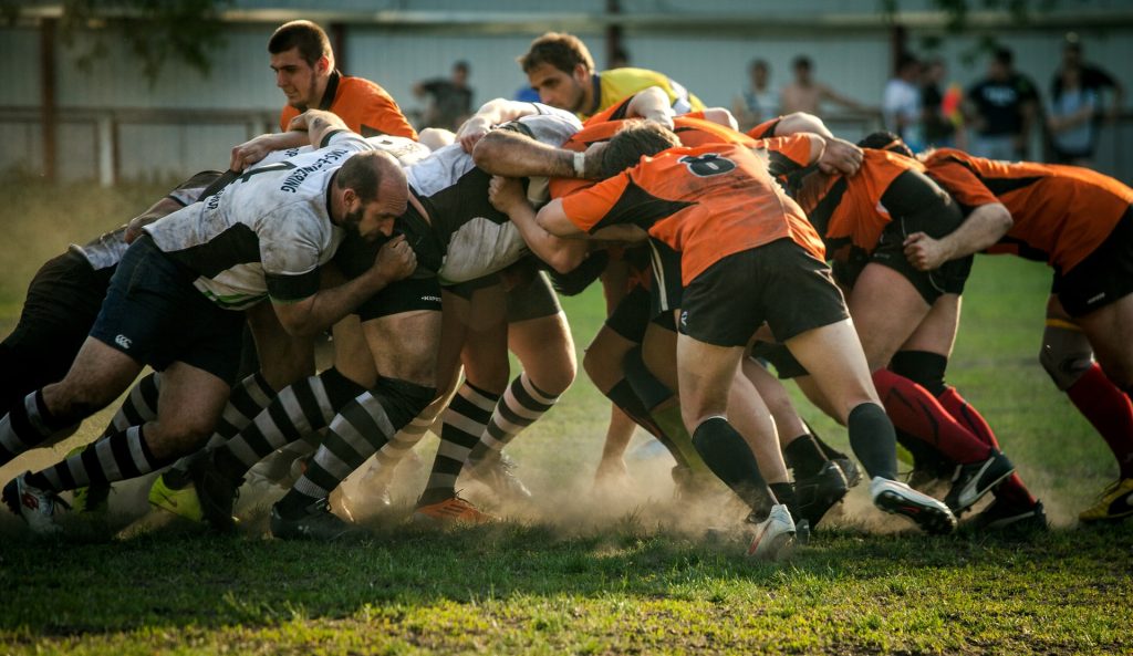
Over the past 17 years, evidence on chronic traumatic encephalopathy (CTE) has piled up. While some sports organisations like the National Hockey League and World Rugby still claim their sports do not cause CTE, a new review article in the journal Acta Neuropathologica strengthens the case that repetitive head impact (RHI) exposure is the chief risk factor for the condition.
CTE is characterised by a distinctive molecular structural configuration of p-tau fibrils that is unlike the changes observed with aging, Alzheimer’s disease, or any other diseases caused by tau protein.
Though CTE made US headlines in 2007, it wasn’t until 2016 that the National Institute of Neurological Disorders and Stroke/National Institute of Biomedical Imaging and Bioengineering (NINDS-NIBIB) criteria for the neuropathological diagnosis of CTE were published, and they were refined in 2021. Rare, isolated case studies reporting aberrant findings or using non-accepted diagnostic criteria have been disproportionately emphasised to cast doubt on the connection between RHI and CTE.
In the review, Ann McKee, MD, chief of neuropathology at VA Boston Healthcare System and director of the BU CTE Center, stresses that now over 600 CTE cases have been published in the literature from multiple international research groups. And of those over 600 cases, 97% have confirmed exposure to RHI, primarily through contact and collision sports. CTE has been diagnosed in amateur and professional athletes, including athletes from American, Canadian, and Australian football, rugby union, rugby league, soccer, ice hockey, bull-riding, wrestling, mixed-martial arts, and boxing.
What’s more, 82% (14 of the 17) of the purported CTE cases that occurred in the absence of RHI, where up-to-date criteria were used, the study authors disclosed that families were never asked what sports the decedent played.
According to the researchers, despite global efforts to find CTE in the absence of contact sport participation or RHI exposure, it appears to be extraordinarily rare, if it exists at all. “In studies of community brain banks, CTE has been seen in 0 to 3 percent of cases, and where the information is available, positive cases were exposed to brain injuries or RHI. In contrast, CTE is the most common neurodegenerative disease diagnosis in contact and collision sport athletes in brain banks around the world. A strong dose response relationship is perhaps the strongest evidence that RHI is causing CTE in athletes,” she added.
“The review presents the timeline for the development of neuropathological criteria for the diagnosis of CTE which was begun nearly 100 years ago by pathologist Harrison Martland who introduced the term “punch-drunk” to describe a neurological condition in prizefighters,” explained McKee, corresponding author of the study. The review chronologically describes the multiple studies conducted by independent, international groups investigating different populations that found CTE pathology in individuals with a history of RHI from various sources.”
New research published in the Journal of the American Geriatrics Society reports that motor vehicle collisions decreased after Japan implemented a mandatory cognitive screening test for older drivers when they renewed their drivers’ licences. For older pedestrians and cyclists, however, their number of collisions and injuries increased.
For the study, investigators analysed police-reported data on the number of collisions for drivers and injuries for pedestrians and cyclists among people aged 70 years or older in Japan from July 2012 to December 2019. As of March 2017, drivers aged 75 years or older who screen positive are required to see a physician before license renewal. If diagnosed with dementia, their licenses may be suspended or revoked.
From 2012 to 2019, there were 602 885 collisions for drivers and 196 889 injuries for pedestrians and cyclists among people aged 70 years or older. After the 2017 policy, collisions decreased among male drivers, and injuries increased among some age subgroups in both sexes. Cumulative estimated changes in the numbers of collisions and injuries from March 2017 to December 2019 were -3670 and 959, respectively.
“Safety measures need to be strengthened for older cyclists and pedestrians. We should also provide older people with necessary care to prepare for driving cessation and safe, alternative transport means,” said corresponding author Haruhiko Inada, PhD, a post-doctoral fellow at the Johns Hopkins Bloomberg School of Public Health.
Researchers have developed a handheld terahertz (THz) wave imaging device to assess burns faster and more accurately than current methods. The new device uses neural network model that uses terahertz time-domain spectroscopy (THz-TDS) data for non-invasive burn assessment.
“It is important for healthcare professionals to accurately assess the depth of a burn to provide the most appropriate treatment,” said research team leader M. Hassan Arbab from Stony Brook University. “However, current methods of burn depth evaluation, which rely on visual and tactile examination, have been shown to be unreliable, with accuracy rates hovering around 60–75%. Our new approach could potentially improve the accuracy of burn severity assessments and aid in treatment planning.”
THz-TDS uses short pulses of terahertz radiation, which lies between infrared and microwave wavelengths, to probe a sample. It is being examined for assessing burn injuries because physical changes caused by a burn will produce alterations in the skin’s terahertz reflectivity.
In the journal Biomedical Optics Express, the researchers reported that their artificial neural network classification algorithm was able to accurately predict the ultimate healing outcome of in vivo burns in an animal study with 93% accuracy. Their method needs much less training data, potentially making it more practical to process big data sets obtained over large clinical trials.
“In 2018, approximately 416 000 patients were treated for burn injuries in emergency departments in the United States alone,” said Arbab. “Our research has the potential to significantly improve burn healing outcomes by guiding surgical treatment plans, which could have a major impact on reducing the length of hospital stays and number of surgical procedures for skin grafting while also improving rehabilitation after injury.”
Better burn assessment
Various technologies have been developed to improve burn assessment, but they haven’t been widely adopted in the clinic due to drawbacks such as long acquisition times, high costs and limited penetration depth and field of view. Although THz-TDS looks promising for burn assessment, early demonstrations were limited to point spectroscopy measurements, which don’t account for burn heterogeneity and spatial variations. THz spectroscopy setups also tend to be bulky and difficult to set up.
“To address these challenges, we developed the portable handheld spectral reflection (PHASR) scanner, a user-friendly device for fast hyperspectral imaging of in vivo burn injuries using THz-TDS,” said Arbab. The device allows for “rapid imaging of a 37 x 27 mm2 field of view in just a few seconds.”
Previously, the researchers used numerical methods to extract features from the THz-TDS images and machine learning techniques to estimate the severity grade of in vivo burn injuries using measurements from the PHASR scanner. However, this approach did not consider the physical dynamics and macroscopic changes of the dielectric permittivity of burned skin tissue. Dielectric permittivity describes how a material responds to an electric field, and the researchers used Debye theory to explain how biological material interacts with THz waves.
The researchers tested their method by using the PHASR scanner to obtain spectroscopic images of skin burns and measure the permittivity of the burns. The researchers used this data to create a neural network model based on labelled biopsies. The model estimated the severity of the burns with an average accuracy rate of 84.5% and predicted the outcome of the wound healing process with an accuracy rate of 93%.
The researchers note that clinical testing of both the technique and the handheld imaging device are needed before this technique could be integrated into the existing workflow of clinical burn assessment.
Adults who suffered any head injury during a 30-year study period had two times the rate of mortality than those who did not have any head injury, and mortality rates among those with moderate or severe head injuries were nearly three times higher, according to new research published in JAMA Neurology.
Head injury can be attributed to a number of causes, from motor vehicle crashes, unintentional falls, or sports injuries. Furthermore, head injury has been linked with a number of long-term health conditions, including disability, late-onset epilepsy, dementia, and stroke.
Previous studies have shown increased short-term mortality among hospitalised patients with head injuries. This longitudinal study evaluated 30 years of data from over 13 000 community-dwelling participants (ie not hospitalised or in nursing homes) to determine if head injury has an impact on mortality rates in adults over the long term. Of these, 18.4% reported one or more head injuries during the study period, and of those who suffered a head injury, 12.4% were recorded as moderate or severe. The median period of time between a head injury and death was 4.7 years.
Death from all causes was recorded in 64.6% of those individuals who suffered a head injury, and in 54.6% of those without any head injury. Accounting for participant characteristics, investigators found that the mortality rate from all-causes among participants with a head injury was 2.21 times the mortality rate among those with no head injury. Further, the mortality rate among those with more severe head injuries was 2.87 times the mortality rate among those with no head injury.
“Our data reveals that head injury is associated with increased mortality rates even long-term. This is particularly the case for individuals with multiple or severe head injuries,” explained the study’s lead author, Holly Elser, MD, PhD, MPH a Neurology resident at Penn. “This highlights the importance of safety measures, like wearing helmets and seatbelts, to prevent head injuries.”
Investigators also evaluated the data for specific causes of death among all participants. Overall, the most common causes of death were cancers, cardiovascular disease, and neurologic disorders (which include dementia, epilepsy, and stroke). Among individuals with head injuries, deaths caused by neurologic disorders and unintentional injury or trauma (like falls) occurred more frequently.
When investigators evaluated specific neurologic causes of death among participants with head injury, they found that nearly two-thirds of neurologic causes of death were attributed to neurodegenerative diseases, like Alzheimer’s and Parkinson’s disease. These diseases composed a greater proportion of overall deaths among individuals with head injury (14.2%) versus those without (6.6%). Further research into this association is recommended.
Haemostatic microneedle technology can be applied like a typical adhesive bandage to quickly stop bleeding. The biocompatible and biodegradable microneedle arrays (MNAs) on the patch increase its surface contact with blood to accelerate the clotting process and also increase the adhesive properties of the patch via mechanical interlocking to promote wound closure. Credit: Designed by Amir Sheikhi and Reihaneh Haghniaz/Executed by Natan Barros. All Rights Reserved.
In the US, secondary, uncontrolled bleeding from traumatic injury is the leading cause of death from ages one to 46. Amir Sheikhi, assistant professor of chemical engineering and of biomedical engineering at Penn State, has a plan to change that with a novel microneedle patch that can immediately stop bleeding after injury.
He described his technology in a new paper in the journal Bioactive Materials.
“Excessive bleeding is a serious challenge for human health,” Sheikhi said. “With haemorrhaging injuries, it is often the loss of blood – not the injury itself – that causes death. There is an unmet medical need for ready-to-use biomaterials that promote rapid blood coagulation.”
Sheikhi’s haemostatic microneedle technology can be applied like a typical adhesive bandage to quickly stop bleeding. The biocompatible and biodegradable microneedle arrays (MNAs) on the patch increase its surface contact with blood, accelerating the clotting process. The needles also increase the adhesive properties of the patch via mechanical interlocking to promote wound closure.
“In vitro, the engineered MNAs reduced clotting time from 11.5 minutes to 1.3 minutes; and in a rat liver bleeding model, they reduced bleeding by more than 90%,” Sheikhi said. “Those 10 minutes could be the difference between life and death.”
The MNA patch is comparable hydrogel technology that is currently used to treat bleeding wounds in hospitals, but hydrogel applications require preparation and medical expertise. The microneedle patch is pre-engineered for immediate application that anyone can use to stop bleeding, Sheikhi said, much like a typical over-the-counter adhesive bandage.
Microneedles – which are already in use to deliver biologics, such as cells or drugs, through the skin or for cosmetic procedures to stimulate collagen production – are tiny, making their application pain-free, according to Sheikhi.
The researchers are now working to commercialise the patch, with more testing plans.
Contrary to popular belief, rest may not always be the best treatment after a concussion, according to the results of a large multi-centre study published in JAMA Network Open. In fact, an early return to school may be associated with a lower symptom burden after suffering a concussion and, ultimately, faster recovery.
“We know that absence from school can be detrimental to youth in many ways and for many reasons,” says study lead author Christopher Vaughan, PsyD, neuropsychologist at Children’s National Hospital. “The results of this study found that, in general, an earlier return to school after a concussion was associated with better outcomes. This helps us feel reassured that returning to some normal activities after a concussion – like going to school – is ultimately beneficial.”
In this cohort study, data from over 1600 youths aged five to 18 were collected across nine paediatric emergency departments in Canada. Because of the large sample size, many factors associated with greater symptom burden and prolonged recovery were first accounted for through the complex statistical approach used to examine the data. The authors found that an early return to school was associated with a lower symptom burden 14 days post-injury in the 8 to 12 and 13 to 18-year-old age groups.
“Clinicians can now confidently inform families that missing at least some school after a concussion is common, often between 2 and 5 days, with older kids typically missing more school,” Dr Vaughan says. “But the earlier a child can return to school with good symptom management strategies and with appropriate academic supports, the better that we think that their recovery will be.”
The results suggest a possible mechanism of therapeutic benefit to the early return to school. This could be due to:
Socialisation (or avoiding the deleterious effects of isolation).
Reduced stress from not missing too much school.
Maintaining or returning to a normal sleep/wake schedule.
Returning to light-to-moderate physical activity sooner (also consistent with previous literature).
Patients hospitalised with fractures typically receive low-molecular-weight heparin to prevent life-threatening blood clots. A new clinical trial, however, found that inexpensive over-the-counter aspirin is just as effective. The findings, published today in the New England Journal of Medicine, may lead surgeons to change their practice and administer aspirin to these patients.
With more than 12 000 patients, the multi-centre randomised clinical trial is the largest trial ever conducted on orthopaedic trauma patients. This multidisciplinary collaboration between orthopaedic surgeons and trauma surgeons points to the importance of evaluating techniques used to prevent post-surgical complications, like blood clots and infections, through high-quality, head-to-head comparison studies.
“Many patients with fractures will likely strongly prefer to take a daily aspirin over receiving injections after we found that both give them similar outcomes for prevention of the most serious outcomes from blood clots,” said the study’s principal investigator Robert V. O’Toole, MD. “We expect our findings from this large-scale trial to have an important impact on clinical practice that may even alter the standard of care.”
Patients who experience fractures that require surgery are at increased risk of developing blood clots, including life-threatening pulmonary embolisms. Current guidelines recommend prescribing low-molecular-weight heparin (enoxaparin) to prevent these clots, although smaller clinical trials in total joint replacement surgery suggested a potential benefit of aspirin as a less-expensive, widely available option.
The study enrolled 12 211 patients with leg or arm fractures that necessitated surgery or pelvic fractures regardless of the treatment. Half were randomised to 30mg of injectable enoxaparin twice daily. The other half received 81mg of aspirin twice daily. Patients were followed for 90 days to measure health outcomes from the two treatments.
The main finding of the study was that aspirin was “non-inferior,” or no worse than low molecular-weight heparin in preventing death from any cause – 47 patients in the aspirin group died, compared with 45 patients in the heparin group. For other important complications, the researchers also found no differences in pulmonary embolisms between the two groups. The incidence of bleeding complications, infection, wound problems, and other adverse events from the treatments was also similar in both groups.
Of all the outcomes studied, the only potential difference noted was in deep vein thrombosis. This condition was relatively uncommon in both groups as it occurred in 2.5% of patients in the aspirin group, and in 1.7% of patients in the heparin group.
“This relatively small difference was driven by clots lower in the leg, which are thought to be of less clinical significance and often do not require treatment,” said study co-principal investigator Deborah Stein, MD, MPH.
“Many patients don’t like giving themselves injections. It’s not fun in terms of giving the actual injection because it burns, and your stomach tends to bruise more easily compared to aspirin,” said Debra Marvel, a 53-year-old from Columbia, MD, who served as a patient advisor on the study. She received Lovenox (low-molecular-weight heparin) after her legs were crushed in a 2015 pedestrian accident, requiring multiple surgeries at the University of Maryland Shock Trauma Center. “Patients also prefer aspirin because Lovenox can be expensive based on insurance.”
“An estimated one million Americans are hospitalised each year with extremity fractures, and this new finding could help prevent potentially fatal blood clots in these patients using a medication that is cheaper and far easier to administer,” said Mark T. Gladwin, MD, Vice President for Medical Affairs, University of Maryland, Baltimore, and the John Z. and Akiko K. Bowers Distinguished Professor and Dean, University of Maryland School of Medicine. “Given these important results, we can expect the guidelines for the prevention of blood clots to be revised to include the option of aspirin for patients with traumatic bone fractures.”
Athletes who recover more slowly from concussion may be able to return to play with an additional month of recovery beyond the typical recovery time, according to a new study published in the journal Neurology. Slow recovery was defined as taking more than 14 days for symptoms to resolve or taking more than 24 days to return to play, both of which are considered the typical recovery times for about 80% of athletes with concussion.
“Although an athlete may experience a slow or delayed recovery, there is reason to believe recovery is achievable with additional time and injury management,” said study author Thomas W. McAllister, MD, of the Indiana University School of Medicine in Indianapolis. “This is an encouraging message that may help to relieve some of the discouragement that athletes can feel when trying to return to their sport. While some athletes took longer than 24 days to return to play, we found that three-quarters of them were able to return to sports if given just one more month to recover.”
The study looked at 1751 American college athletes diagnosed with a concussion by a team physician. Of Male athletes (63%) participated primarily in football, soccer and basketball. Female athletes (37%) participated primarily in soccer, volleyball and basketball.
Participants were evaluated five times: within six hours after their injury, one to two days later, once free of symptoms, once cleared to return to play and at six months.
Participants reported symptoms daily to medical staff, up to 14 days following injury and then weekly if they had not yet returned to play.
A total of 399 athletes, or 23%, had a slow recovery.
Researchers found that of the athletes who took longer than 24 days to return to play, more than three-fourths, or 78%, were able to return to play within 60 days of injury, and four-fifths, or 83%, were able to return to play within 90 days of injury. Only 11% had not returned to play six months after injury.
For the slow recovery group, the average time for returning to play was 35 days after injury, compared to 13 days in the overall group.
“The results of this study provide helpful information for athletes and medical teams to consider in evaluating expectations and making difficult decisions about medical disqualification and the value of continuing in their sport,” McAllister said.
A limitation of the study is that participants were all collegiate varsity athletes and may not be representative of other age groups or levels of sport, and the results may not apply to other types of mild brain injuries.